


Case Report - Year 2025 - Volume 40Issue 1
Amniotic Band Syndrome: Case Report
Síndrome da banda amniótica: Relato de caso
Augusto Ribeiro de Sousa Cardoso1 ; Jefferson Lessa Soares de Macedo1,; João Pedro Santana de Lacerda Mariz1; Jovita Maria Rafael Batista de Albuquerque Espíndola1; Thamara de Oliveira Vasconcelos1; Luís Felipe Rosa de Macedo2; Cecília Rosa de Macedo3; Simone Corrêa Rosa1
ABSTRACT
Amnioticbandsyndrome is a rare, congenital anomaly with several disfiguringanddisabling manifestations potentially leading to miscarriage. It often affects the distal extremities of limbs, resulting in lymphatic, vascular, and contour abnormalities. In our service, we followed a female patient with multiple amniotic bands in the upper limbs and the left lower limb. She underwent resection of an amniotic band at 10-months-old, in a single surgical time,withmultipleZ-plasties inthetotal planeof theleft leg,aswellas withreleaseof severedistal fusionsbetweenthe rightupper limbandthethorax. The rightupper limbband was not operated on as there would be little benefit. After surgery, the patient presented partial lymphedema regression and a vascular lesion in the dorsal region of the left foot, which underwent clinical treatment with dressing and topical medication. The evolution of the surgical scar was satisfactory, with complete resolution of the indentation of the amniotic band in the left leg and no signs of recurrence.
Keywords: amniotic band syndrome; leg; lower extremity; plastic surgery procedures; surgical flaps
RESUMO
A síndrome da banda amniótica é uma anomalia rara, congênita, com várias manifestações desfigurantes e incapacitantes podendo levar ao aborto espontâneo. Geralmente atinge as extremidades distais de membros, levando a alterações linfáticas, vasculares e de contorno. Em nosso serviço, acompanhamos uma paciente feminina com múltiplas bridas amnióticas, sendo estas em membros superiores e membro inferior esquerdo. Foi submetida à ressecção de brida amniótica aos 10 meses de idade em tempo único, com múltiplas zetaplastias na perna esquerda, assim como liberação de fusão distal grave entre o membro superior direito e o tórax. A brida de membro superior não foi operada, pois haveria pouco benefício. A paciente evoluiu com regressão parcial de linfedema no pós-operatório e lesão vascular em região dorsal do pé esquerdo, que foi tratada clinicamente com curativo e terapia tópica. A evolução da cicatriz cirúrgica foi satisfatória, com resolução completa da indentação da banda amniótica em perna esquerda e sem sinais de recidiva.
Palavras-chave: extremidade inferior; perna (membro); procedimentos de cirurgia plástica; retalhos cirúrgicos; síndrome de bandas amnióticas
Introduction
Amniotic band syndrome is a rare, congenital anomaly with several disfiguring and disabling manifestations that can even lead to spontaneous abortion. The incidence ranges from 1:1,200 and 1:15 thousand live births. Depending on severity, this spectrum can present diverse clinical manifestations, from mild soft tissue indentations to limb amputation. The most common presentations are acrosyndactyly, intrauterine amputations, and constrictions in distal limb rings. On average, the condition affects three different sites. The risk of functional loss may indicate surgical treatment to correct soft tissue deformities.1-3
Constriction band excision and repair in the neonatal period is indicated in patients with severe vascular insufficiency or lymphatic congestion resulting in signs of critical limb ischemia.2
There are several techniques for constrictive band excision and reconstruction, depending mainly on the affected site and the type of constriction resulting from the amniotic band (upper or lower limbs and fingers). The differences in surgical techniques refer to subcutaneous cellular tissue mobilization, fat grafting, or skin incision patterns (straight line, rectangular, and W- or Z-plasty). Incomplete amniotic band excision may hinder the full restoration of subcutaneous cellular tissue and lead to residual constrictions.2,4,5
Objective
The current study presents a case of amniotic band syndrome treated by the plastic surgery service from a public hospital, addressing the diagnosis, technique, postoperative followup, and outcomes.
Materials and Methods
We herein report to the case of a child with a constrictive band in the left leg treated by the plastic surgery team from a regional reference public hospital. We obtained the information for this study through a review of the patient’s medical records, an interview with the guardians, a photographic record of the therapeutic methods employed, and a literature review.
The study followed ethical research precepts by preserving the anonymity, privacy, and confidentiality of the information in the patient’s medical records. The present study was submitted tothe Research Ethics Committee of Fundação de Ensino e Pesquisa em Ciências da Saúde/Secretaria Estadual de Saúde do Distrito Federal (FEPECS/SES-DF, under CAAE 47391715.6.0000.5553, opinion number 1.167.841). We complied with all ethical requirements of the Resolution no. 466/2012 of the Brazilian National Health Council.
Case Report
A 10-months-old patient, female sex (JMNP), born by vaginal delivery at home with 38 weeks of gestational age, Appearance, Pulse, Grimace, Activity, and Respiration (Apgar) score of 8/9, weight of 3,070 g, head circumference of 33 cm, and length of 47 cm. There were no complications during pregnancy. The rapid tests for human immunodeficiency virus (HIV), syphilis, hepatitis B antigen (HBsAg), and hepatitis C (HCV) were negative. The severe acute respiratory syndrome coronavirus 2 (SARS-CoV-2) antigen test was nonreactive. The maternal and infant blood types were Oþ and O- respectively.
The mother denies consanguinity and maternal diseases, and the only family history was systemic arterial hypertension. The search for other congenital malformations was negative. There was a history of amniotic band syndrome diagnosed in the prenatal period by ultrasound.
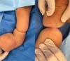
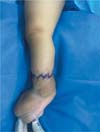
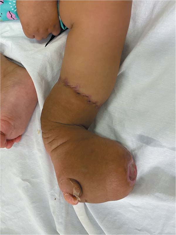
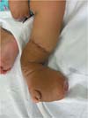
Our service admitted the patient after referral by the pediatric surgery team for evaluation and management (►Fig. 1). The patient presented with acrosyndactyly, shortening of the third, fourth, and fifth fingers, absence of the index finger of the right hand, a proximal constrictive band (Patterson grade III) in the middle third of the right arm, shortening of the forearm with severe distal bone fusion, and adhesion between the right hand and the thorax. There was no evidence of lymphedema of the right hand. A circumferential, deep amniotic band was evident in the distal third of the left leg, grade III, extending close to the bone region, with symbrachydactyly due to the shortening of the toes of the left foot. Additionally, the same left lower limb with the amniotic band presented a large distal lymphedema on the dorsum of the left foot but with no signs of vascular compromise.

Radiographs of the lower limbs did not show significant bone abnormalities. The vascular Doppler ultrasound evaluation of the left lower limb revealed arterial and venous perfusion distal to the amniotic band. There was no surgical planning for the right upper limb due to the severity of the congenital abnormalities in the hand (acrosyndactyly and finger amputation), with no lymphatic congestion and a good potential for limb rehabilitation.
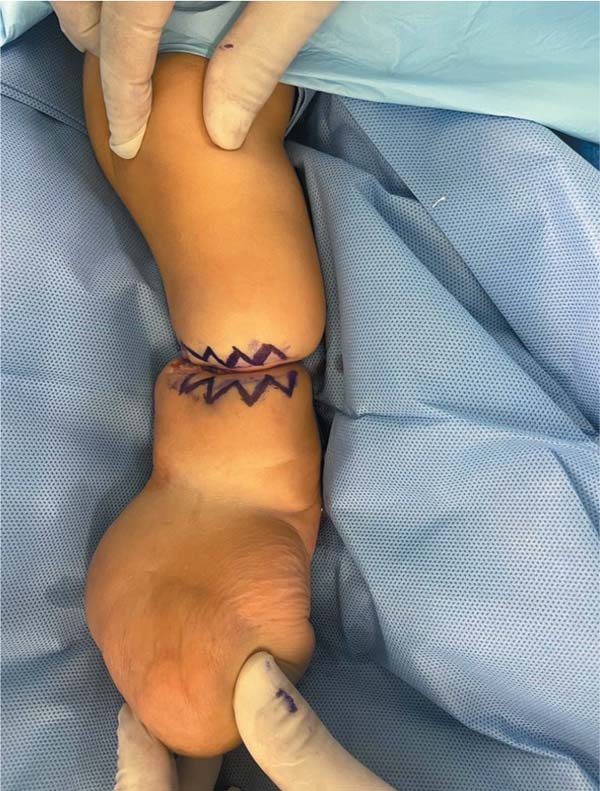
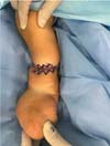
At 10-months-old, the child underwent surgery under general anesthesia to release adhesions between the chest and right hand with an incision, followed by closure with a continuous purse-string suture of the resulting chest wound. At the same procedure, we performed surgical treatment of the circumferential band of the left leg. This procedure involved the application of a cotton-cushioned Esmarch bandage at the level of the left thigh and infiltration of 10 mL of a 1% lidocaine solution with 1:120 thousand epinephrine throughout the entire region around the bandage. Next, we completely excised the abnormal fibrotic tissue (band) attaching the subcutaneous tissue to the deep fascial tissue of the left tibia. We performed multiple W-plasties involving the entire circumference of the limb (►Fig. 2), in the full plane at 60° angles, with no need for flaps (►Fig. 3). Due to the good mobility of the soft parts of the left leg, after releasing the fibrotic tissue from the band, it was easy to approximate the subcutaneous tissue and the dermis with a few absorbable sutures (polyglactin 4-0). Skin suture used nylon 5-0 and 6-0.
The operative time was 52 minutes, and we released the tourniquet on the left lower limb after finishing the dressing with petrolatum topical gauze (Adaptic, Systagenix), hydrophilic cotton, and bandaging. The total tourniquet time was 58 minutes. At the end of the procedure, we tested the distal perfusion, observing a capillary refill time lower than 2 seconds in the toes of the left foot. The patient remained hospitalized for 4 days with her leg elevated in bed. She presented significant lymphedema at the dorsum of the left foot, with peak edema at 48 hours. The patient was discharged after 4 days with analgesic treatment and no antimicrobials and scheduled a return visit within 3 days.
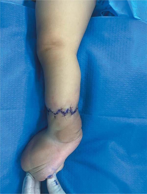
On the 7th postoperative day, we evaluated the patient in the outpatient clinic, revealing mild congestion, slight lymphedema reduction, and a good-looking surgical scar. On the 16th day, we removed the skin sutures (nylon). Finally, on the 32nd postoperative day, the patient developed an ischemic ulcer on the dorsum of the foot measuring 2 cm in diameter, treated on an outpatient basis with topical medication (healing oil of essential fatty acids AGE Dermaex) and dressings with calcium alginate fiber (Biatain, Coloplast), with resolution in 45 days (►Figs. 4,5).

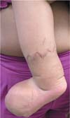
The patient showed significant improvement in vascular congestion, especially 4 months after surgery. There was no evidence of constriction or depression in the region of the amniotic band on the left leg, with total leveling of the skin and subcutaneous tissue with the rest of the limb. As moderate lymphedema persisted in the left foot, we recommended elastic stockings with mild compression up to the left knee and lymphatic drainage sessions. The surgical scar presented moderate hypertrophy in the first 6 months, and gradual improvement was noted with local massages and the use of silicone tapes.
Discussion
Amniotic band, or constriction ring, syndrome presents as a spectrum ranging from minor morphological abnormalities to severe constriction with limb amputation. It is not hereditary, and cases are sporadic. In our patient, we used a technique with multiple W-plasties in a single stage, which is a safe and effective method to release leg restrictions resulting from the amniotic band.2,5,6
Band excision can occur in a single or multiple stages, with intervals of 3 to 6 months depending on the depth of the lesion and the circulation status of the limb involved. Multistage reconstructions are indicated for patients with abnormal vascularization in the distal segment or presenting circumferential bands close to the bone, with a risk of distal flap necrosis.2,7
Patterson classified congenital constrictive bands according to their severity and clinical repercussions as follows: grade I, presence of simple constriction rings with normal extremity distal to these lesions; II, presence of a ring with distal atrophy and lymphedema; III, presence of ring with syndactyly in the affected extremities; and IV, presence of a ring leading to amputation.2,8
Among the lesions associated with congenital constriction bands that characterize amniotic band syndrome, syndactyly, finger amputations, brachydactyly, congenital hip dislocation, tibial pseudarthrosis, and congenital clubfoot stand out.9 Therefore, surgery for constriction band release is recommended before orthopedic procedures for any foot deformity at risk of toe necrosis.9
The case reported here presented a grade III constriction band according to the Patterson classification, and theband was virtually adhered to the left tibia, compromising the venous and lymphatic return of the affected limb. Likewise, the child presented a severe malformation associated with symbrachydactyly and amputation of a finger on the right hand. We did not release the constriction bands in the upper limb because the deformity was severe and there was no evidence of surgical benefit for the patient. However, the one-stage surgical procedure on the left leg had a satisfactory therapeutic response regarding the circumferential constrictive band.
Although surgeons widely accept W-plasty with subcutaneous tissue mobilization, insufficient tissue may result in incomplete correction of the limb contour. In contrast, linear circumferential closure has good immediate cosmetic outcomes but may result in secondary scar contraction and amniotic band recurrence, particularly in patients still in the growth phase.2,7
Mutaf and Sunay5 proposed a rectangular deepithelialized subcutaneous flap with a pedicle proximal to the lesion as an attempt to fill the associated band area, parallel to the skin tension lines for better limb contour. However, this flap is limited by the scarcity of subcutaneous cellular tissue in most cases. Other techniques using superficial Z-plasties with epidermal rearrangement alone have proven ineffective due to incomplete band excision, making limb contour restoration a challenge.1,7
The reported case did not present severe craniofacial or visceral abnormalities. Nonetheless, there are reports of craniofacial anomalies associated with amniotic band syndrome, including encephaloceles, cleft lip and palate, oralocular facial clefts, nasal-ocular facial clefts, and even the presence of a single oronasopharyngeal cavity. Visceral anomalies are rare in the syndrome, with gastroschisis as the most frequent. Omphalocele, bladder exstrophy, and ambiguous genitalia may also occur. Amniotic band syndrome may account for many unexplained fetal losses.10
Conclusion
This technique is a simple and reliable method for amniotic band excision in pediatric patients. When performed properly, it offers excellent outcomes for restoring the normal limb contour, with satisfactory evolution 1 year after surgery. The single-stage procedure was safe and effective even in a circumferential band of the lower limb.
References
1. Choulakian MY, Williams HB. Surgical correction of congenital constriction band syndrome in children: Replacing Z-plasty with direct closure. Can J Plast Surg 2008;16(04):221-223. Doi: 10.1177/229255030801600409
2. Chan AHW, Zeitlinger L, Little KJ. Multiple Continuous Y-to-VPlasties for Excision and Reconstruction of Constriction Band Syndrome: Case Series and Description of Surgical Technique. Plast Reconstr Surg 2022;149(04):774e-778e. Doi: 10.1097/ PRS.0000000000008954
3. Castro-Govea Y, Vela-Martinez A, Treviño-Garcia LA. Lipoinjection and Multiple Internal Cuts for Congenital Constriction Bands: A New Treatment Approach. Aesthetic Plast Surg 2017;41(02): 375-380. Doi: 10.1007/s00266-016-0744-4
4. Habenicht R, Hülsemann W, Lohmeyer JA, Mann M. Ten-year experience with one-step correction of constriction rings by complete circular resection and linear circumferential skin closure. J Plast Reconstr Aesthet Surg 2013;66(08):1117-1122. Doi: 10.1016/j.bjps.2013.04.042
5. Mutaf M, Sunay M. A new technique for correction of congenital constriction rings. Ann Plast Surg 2006;57(06):646-652. Doi: 10.1097/01.sap.0000235430.21875.55
6. Hung NN. Congenital constriction ring in children: sine plasty combined with removal of fibrous groove and fasciotomy. J Child Orthop 2012;6(03):189-197. Doi: 10.1007/s11832-012-0420-4
7. Moran SL, Jensen M, Bravo C. Amniotic band syndrome of the upper extremity: diagnosis and management. J Am Acad Orthop Surg 2007;15(07):397-407. Doi: 10.5435/00124635-200707000-00005
8. Cruz TBC, Freire IF, Marangoni AM, Tavares MTM, Aquino PLd, Pessoa SGdP. Cicatriz congênita circunferencial em membro inferior devido à síndrome da banda amniótica abordada com dablioplastia. Rev Bras Cir Plást 2024;39(03):e0912. Doi: 10.5935/2177-1235.2024RBCP0912-EN
9. Costa EN, Alves MdP, Fraga CEC, Silva JATd Jr, Daher O. Síndrome das bandas de constrição congênita: Estudo de 16 casos. Rev Bras Ortop 1996;31(04):341-346
10. Mayou BJ, Fenton OM. Oblique facial clefts caused by amniotic bands. Plast Reconstr Surg 1981;68(05):675-681. Doi: 10.1097/ 00006534-198111000-00001
1. Plastic Surgery Service, Hospital Regional da Asa Norte, Brasília, DF, Brazil
2. Universidade Católica de Brasília (UCB), Brasília, DF, Brazil
3. Centro Universitário de Brasília (UniCEUB), Brasília, DF, Brazil
Address for correspondence Jefferson Lessa Soares de Macedo, Plastic Surgery Service, Hospital Regional da Asa Norte, Brasília, DF, Brazil (e-mail: jlsmacedo@yahoo.com.br).
Artigo submetido: 04/12/2024.
Artigo aceito: 20/05/2025.
Conflict of Interests
The authors have no conflict of interests to declare.
 Read in Portuguese
 Read in English
 PDF PT
 Print
Print
 Send this article by email
 How to Cite
 Mendeley
 Pocket