




Case Report - Year 2026 - Volume 41Issue 1
Complex Marjolin's Ulcer of the Scalp - Case Report
Úlcera de Marjolin complexa de couro cabeludo - Relato de caso
Lucas Ruffo de Medeiros1 ; Frederico Alonso Sabino de Freitas1; Marcus Vinícius Jardini Barbosa1,
ABSTRACT
Marjolin's ulcer is a rare and aggressivemalignant neoplasm that arises from chronic wounds, the most common type being squamous cell carcinoma. The paper describes a 50-year-old womanwitha 30-year-old scalpulcer following traumatic avulsion,whichprogressed toinvasive carcinoma with intracranial extension. The patient presented with seizures, hemiparesis, and a large vegetating lesion. Imaging revealed extensive bone destruction and brain involvement. A wide surgical resection was done, followed by dura mater reconstruction with fascia lata, and microsurgical latissimus dorsi flap coverage. Despite initial recovery, the patient experienced local recurrence and died 2 months postoperatively. The case highlights the challenges of managingadvancedMarjolin'sulcer,particularly inthe scalp,wheredelayeddiagnosis anddeep invasion worsen prognosis. Early excision with adequate margins remains the gold standard, but intracranial extension necessitates multidisciplinary care. This report underscores the importance of prompt treatment for chronic wounds to prevent malignant transformation.
Keywords: scalp; squamous cell carcinoma; squamous cell carcinoma of head and neck; post-traumatic cancer; plastic surgery procedures
RESUMO
A úlcera de Marjolin é uma neoplasia maligna rara e agressiva que surge a partir de feridas crônicas, sendo o tipo mais comum o carcinoma de células escamosas. O artigo descreve o caso deumamulher de 50 anos comumaúlcera no couro cabeludo há 30 anos, decorrente de umaavulsão traumática,que evoluiuparaumcarcinoma invasivocomextensão intracraniana. A paciente apresentou convulsões, hemiparesia e uma grande lesão vegetante. Os exames de imagem revelaram extensa destruição óssea e comprometimento cerebral. Foi realizadauma ampla ressecção cirúrgica, seguida de reconstrução da dura-máter comfáscia lata e cobertura com retalho microcirúrgico de músculo latíssimo do dorso. Apesar da recuperação inicial, a paciente apresentou recidiva local e faleceu 2 meses após a cirurgia. O caso destaca os desafios no manejo da úlcera de Marjolin em estágio avançado, especialmente no couro cabeludo, onde o diagnóstico tardio e a invasão profunda pioram o prognóstico. A excisão precoce com margens adequadas continua sendo o padrão-ouro, mas a extensão intracraniana exige cuidados multidisciplinares. Este relato reforça a importância do tratamento imediato de feridas crônicas para prevenir a transformação maligna.
Palavras-chave: couro cabeludo; carcinoma de células escamosas; carcinoma de células escamosas de cabeça e pescoço; câncer pós-traumático; procedimentos de cirurgia plástica
Introduction
Marjolin’s ulcer is a rare and severe condition characterized by the malignant transformation of scars or chronic wounds into a squamous cell carcinoma. Skin lesions with impaired healing due to chronic irritation, repeated trauma, or other factors that persist for decades are more prone to malignancy.1–3
This condition is classified as acute if malignant transformation occurs within less than 12 months and chronic if it evolves over 12 months.1,4 The incidence of malignancy ranges from 2 to 6%, with an average latency of over 30 years between the initial injury and neoplasia. Epidemiologically, the extremities are affected in 58% of cases,5 while the scalp is less commonly involved.6
The pathophysiology involves repeated ulceration and prolonged stimulation of cell proliferation due to reepithelialization and possible spontaneous mutations.6 Early diagnosis is crucial, allowing for less complex reconstruction procedures. Clinical exams frequently show an ulcer with raised and hardened edges, foul odor, vegetating appearance, and purulent discharge. Histological exam confirms the diagnosis.7
Surgical excision is the most effective treatment, and lymph node dissection being suggested in some cases due to the risk of predominantly lymphatic spread of the tumor. Chemotherapy has shown limited efficacy, while radiotherapy is reserved for selected cases.1 Meningeal or brain involvement is extremely rare and associated with high morbidity and mortality.4 Considering the unpredictable progression, tissue damage, and poor prognosis, early diagnosis and excision are essential for better clinical outcomes;1 with a surgical margin of at least 3 cm being recommended.3
Therefore, this study aims to describe a rare case of Marjolin’s ulcer involving complete scalp infiltration and brain tissue invasion.
Case Report
A 50-year-old retired woman presented with a main complaint of “seizures for 1 month.” She reported focal seizures in the right upper limb (RUL) for 1 month and RUL paresis with decreased consciousness for 2 weeks associated with asthenia, adynamia, weight loss, and recent-onset fever. She reported a scalp wound of 30 years, due to a boat accident with scalp avulsion. The wound alternated between secondary healing and recurrent ulceration, with worsening progression over the past 2 years.
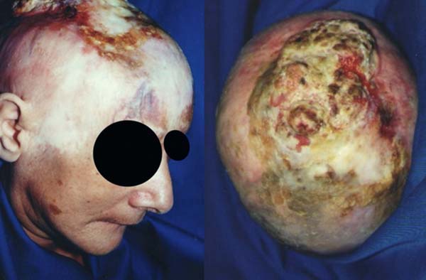
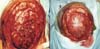
Physical exam revealed a debilitated, dehydrated, and apathetic patient, with right-sided hemiparesis and allopsychic disorientation. The scalp exhibited an extensive vegetating, infiltrative lesion with necrotic areas and foul odor, measuring 20 x 15 cm, located on the frontoparietal region (►Fig. 1 ).
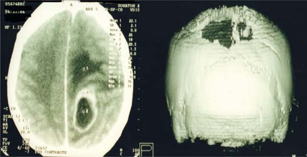
The diagnostic hypothesis was Marjolin’s ulcer, and a cranial computed tomography (CT) scan revealed a hypo-dense, extra-axial lesion in the left cerebral hemisphere with contrast enhancement, mass effect, sulcal effacement, midline shift, and an extensive frontoparietal bone destruction (►Fig. 2 ).

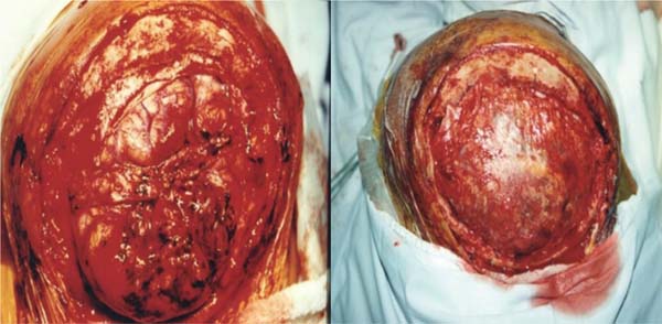
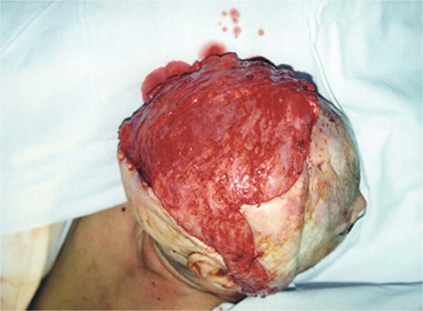
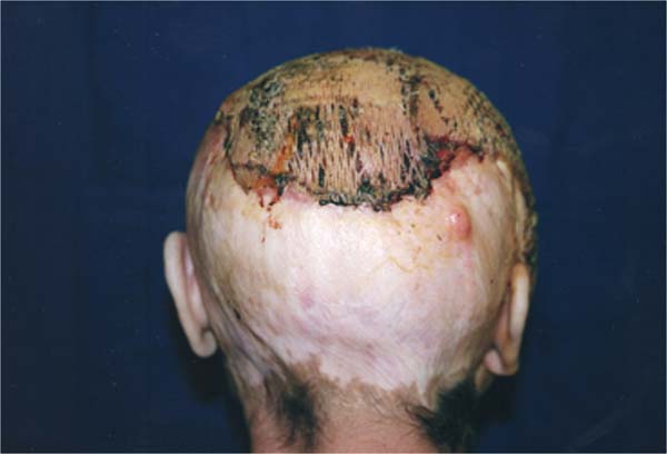
The patient underwent primary lesion resection involving all layers, with 5 cm safety margins. The procedure included: bone resection, resection of the dura mater, and affected brain tissue. The dura mater was reconstructed with a fascia lata graft (►Fig. 3 ); the latissimus dorsi muscle was transferred via microsurgery to cover the defect (►Fig. 4 ). Vascular anastomoses of the thoracodorsal vessels were performed on the facial artery (end-to-end) and external jugular vein (end-to-side), with a saphenous vein graft interposition. The patient showed postoperative improvement, with good reperfusion of the transferred muscle. A split-thickness skin graft was applied to the granulating area 2 weeks postoperatively (►Fig. 5 ).
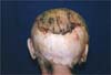
At the 40-day follow-up, local recurrence was detected, accompanied by worsening neurological symptoms and progressive decline. The patient died 2 months postoperatively.
Discussion
Marjolin’s ulcer is defined as a malignant transformation of chronic wounds that fail to heal properly or heal by secondary intention. It is commonly associated with burn scars and trauma, but can also arise from other conditions.6,8 The pathophysiology of malignant transformation remains unclear, but some authors suggested impaired immune response due to poor vascularization of scar tissue,9 and proto-oncogene overexpression.10 Scalp involvement is extremely rare;6 as such, the mechanisms of underlying scalp Marjolin’s ulcer require further elucidation due to limited case reports.6
This case involved a traumatic total scalp avulsion that evolved to a chronic wound. It is a rare report of infiltrative Marjolin’s ulcer with intracranial involvement. Scalp ulcers invading the skull and deep tissues pose significant challenges for resection and reconstruction, with low cure rates.6
Well-differentiated lesions have a better prognosis,8,10 and lymph node involvement is a crucial prognostic factor, as it represents the primary metastatic route. A study of 31 Marjolin’s ulcers reported a 3-year survival rate of 94% for well-differentiated tumors and 38% for poorly differentiated ones, with no survivors among patients with distant metastases.7–9 In this case, the tumor was undifferentiated, with no detectable nodal metastases. However, the patient presented with extensive cranioencephalic destruction, which certainly contributed to the poor prognosis.
Clinical treatment remains controversial and ineffective. Chemotherapy may be used as adjuvant therapy for metastatic cases but offers limited survival benefits. Radiotherapy is reserved for inoperable cases or postoperative consolidation, though its efficacy is debated due to poor vascularization of ulcerated tissue.1
Surgical excision with margins of at least 3 cm (extending to 5 cm in some cases) is the most effective treatment, reducing recurrence risk.1 In this case, a 5 cm margin was adopted but failed to prevent recurrence. Prophylactic lymph node dissection is suggested due to aggressive progression, with indications based on tumor grade or sentinel lymph node biopsy.1
After the resection, functional and aesthetic reconstruction should be considered to improve quality of life. Skin grafts are suitable for monitoring recurrence, while flaps are preferred for covering noble structures. Microsurgical flaps are indicated for extensive resections with limited local flap options or planned postoperative radiotherapy.1,9 In the presented case, the exposed brain tissue was covered with a fascia lata graft, and a microsurgical latissimus dorsi flap was used for extensive calvarial reconstruction. A meshed split-thickness skin graft was applied 15 days later. Despite no surgical complications, the advanced tumor stage and the patient’s clinical condition led to local recurrence, progressive decline, and death.
Conclusion
Concurrent Marjolin’s ulcer of the scalp and cranioencephalic involvement is rarely reported in the literature, which complicates the establishment of effective treatment protocols, leading to delayed diagnosis and reduced cure rates. Although case reports are considered low-level evidence,5 they provide valuable insights for managing rare conditions, which are often excluded from large multicenter studies.
REFERENCES
1. Rodovalho NN, Duarte LRA, Guimarães IS, Souza IH, Magalhães LO, Silva MP. Marjolin ulcer: The importance of early diagnosis and excision. Rev Bras Cir Plást 2022;38(02):1–6. Doi: 10.5935/21771235.2023RBCP0654-EN
2. Mousa AK, Elshenawy AA, Maklad SM, Bebars SMM, Burezq HA, Sayed SE. Post-burn scar malignancy: 5-yearmanagement review and experience. IntWound J 2022;19(04):895–909. Doi: 10.1111/iwj.13690
3. Machado AF, Fontinele DRS, Vieira SC. Marjolin’s ulcer in pressure injury scar: Case report. Rev Bras Cir Plást 2020;36(02):231–235. Doi: 10.5935/2177-1235.2021RBCP0073
4. Sengul G, Hadi-Kadioglu H. Penetrating Marjolin’s ulcer of scalp involving bone, dura mater and brain caused by blunt trauma to the burned area. Neurocirugia (Astur) 2009;20(05):474–477, discussion 477 . Doi: 10.1016/s1130-1473(09)70147-2
5. Simão TS, Almeida PCC, Faiwichow L. Úlcera de Marjolin: visão atualizada [Marjolin’s ulcer: Updated view]. Rev Bras Queimaduras 2012;11(04):251–253
6. Xiao H, Deng K, Liu R, et al. A review of 31 cases ofMarjolin’s ulcer on scalp: Is it necessary to preventively remove the scar? Int Wound J 2019;16(02):479–485. Doi: 10.1111/iwj.13058
7. Leonardi DF, Oliveira DS, Franzoi MA. Úlcera deMarjolin emcicatriz de queimadura: revisão de literatura [Marjolin’s ulcer in burn scar: Literature review]. Rev Bras Queimaduras 2013;12(01):49–52
8. Simman R, Caudil J. Marjolin Ulcers of the Scalp Post Trauma and of the Neck Post Radiation, Diagnosis, and Reconstruction. Eplasty 2022;22:ic10
9. Bostwick J III, Pendergrast WJ Jr, Vasconez LO. Marjolin’s ulcer: an immunologically privileged tumor? Plast Reconstr Surg 1976;57 (01):66–69
10. Smith J, Mello LF, Nogueira NC Neto, et al. Malignancy in chronic ulcers and scars of the leg (Marjolin’s ulcer): a study of 21 patients. Skeletal Radiol 2001;30(06):331–337 Doi: 10.1007/s002560100355
1. Medicine Program, Centro Universitário Municipal de Franca, Franca, SP, Brazil
Data Availability Data will be available upon request to the corresponding author.
Financial Support The authors declare that they did not receive financial support from agencies in the public, private, or non-profit sectors to conduct the present study.
Address for correspondence Marcus Vinícius Jardini Barbosa, MD, PhD, Alameda dos Flamboyants, 700–Morada do Verde, CEP: 14404409, Franca, SP, Brazil (e-mail: drmbarbosa@gmail.com.br ).
Article received: September 03, 2025.
Article accepted: November 24, 2025.
Conflict of Interests The authors have no conflict of interests to declare.
Editor-in-Chief: Dov Charles Goldenberg.
 Read in Portuguese
 Read in English
 PDF PT
 Print
Print
 Send this article by email
 How to Cite
 Mendeley
 Pocket
 Facebook
 Twitter