




Original Article - Year 2026 - Volume 41Issue 1
Traumatic Scalp Defects: Analysis of Management and a Treatment Algorithm in a Tertiary Hospital
Defeitos traumáticos do couro cabeludo: Análise e algoritmo de tratamento em um hospital terciário
Isabella Correa de Oliveira1, ; Pietro Maran Novais1; Roberto de Oliveira Basso1; Rodrigo Rezende Silva Cabral1; Maite Mateus1; Renato da Silva Freitas1
ABSTRACT
Background Scalp reconstruction following trauma presents unique challenges due to limited tissue extensibility and frequent association with severe injuries. The traditional "reconstructive ladder" may not always apply to emergency scenarios. This study aims to analyze the epidemiological profile and surgical outcomes of scalp trauma, proposing a treatment algorithm.
Methods A retrospective cohort study of 35 patients with scalp trauma treated at a Level I reference center between 2013 and 2023 was conducted. Demographic variables, injury characteristics, and surgical techniques were analyzed. Statistical analysis was performed using the chi-square test and Fisher's exact test (p<0.05).
Results The sample had a mean age of 35.4 years, with 28.6% pediatric patients. The profile indicated high severity: 91.4% emergency admissions and 77.1% with associated injuries. "Injury type" was the only statistically significant predictive factor for success (p<0.02). Anatomical location and the presence of polytrauma did not influence outcomes. Primary closure was successful only in wounds<3 cm; larger lesions required local flaps. A systematic failure was observed in the use of local flaps for total avulsions.
Conclusion The nature and size of the injury are the primary determinants of prognosis, regardless of location. This study proposes an algorithm based on injury type to optimize surgical outcomes in emergency settings.
Level of Evidence: IV.
Keywords: scalp; wounds and injuries; reconstructive surgical procedures; surgical flaps; microsurgery
RESUMO
Introdução A reconstrução do couro cabeludo após trauma apresenta desafios únicos devido à limitada extensibilidade tecidual e à frequente associação com lesões graves. A tradicional "escada reconstrutiva" nem sempre se aplica a cenários de emergência. Este estudo tem como objetivo analisar o perfil epidemiológico e os desfechos cirúrgicos dos traumas no couro cabeludo, bem como propor um algoritmo de tratamento.
Métodos Realizou-se umestudo de coorte retrospectivo de 35 pacientes comtrauma no couro cabeludo atendidos em um centro de referência Nível I entre 2013 e 2023. Foram analisadas variáveis demográficas, características da lesão e técnicas cirúrgicas. A análise estatística foi realizada por meio dos testes do qui-quadrado e do exato de Fisher (p<0,05).
Resultados A amostra apresentou média de idade de 35,4 anos, com 28,6% de pacientes pediátricos. O perfil indicou alta gravidade: 91,4% das internações foram de emergência e 77,1% com lesões associadas. O tipo de lesão foi o único fator preditivo estatisticamente significativo para o sucesso (p<0,02). A localização anatômica e o politrauma não influenciaram os desfechos. O fechamento primário foi bem-sucedido apenas em feridas<3 cm; lesões maiores exigiram retalhos locais. Observou-se falha sistemática no uso de retalhos locais em avulsões totais.
Conclusão A natureza e o tamanho da lesão são os principais determinantes do prognóstico, independentemente da localização. Este estudo propõe um algoritmo, baseado no tipo de lesão, para otimizar os desfechos cirúrgicos em ambientes de emergência.
Nível de evidência: IV.
Palavras-chave: couro cabeludo; ferimentos e lesões; procedimentos cirúrgicos de reconstrução; retalhos cirúrgicos; microcirurgia
Introduction
Scalp reconstruction poses a significant challenge for plastic surgeons due to unique anatomical features and limited local tissue elasticity. Surgical planning must prioritize the restoration of functionality, primarily protecting the cranial vault to prevent severe complications such as infections, bone sequestration, and intracranial involvement. Additionally, aesthetic restoration is crucial, as the scalp is an exposed area that serves as the foundation for hair. Issues such as hairline maintenance and scar positioning must be carefully considered.1 2 3 4 5 6 7 8 9 10
For reconstructive success, a mastery of anatomy is fundamental. The scalp is classically divided into five layers, represented by the mnemonic SCALP: Skin, Connective tissue (subcutaneous), Aponeurosis (galea), Loose connective tissue, and Pericranium. The skin is the thickest in the human body (3 mm to 8 mm), contributing to the region's low elasticity. The subcutaneous tissue contains vessels and nerves; therefore, flaps elevated too superficially in this plane carry a high risk of necrosis. The galea aponeurotica is a firm, inelastic structure. Beneath it, the loose connective tissue constitutes the ideal dissection plane, ensuring mobility and preserving vascularization. The deepest layer, the pericranium, lines the calvaria and, when preserved, serves as a recipient bed for skin grafts. Vascularization is extremely rich, with a robust collateral network formed by branches of the internal and external carotid arteries.1'3'4'6'8'10'11
While the etiology of scalp defects is varied, trauma represents a subcategory with unique challenges. Unlike elective lesions, traumatic causes are frequently managed in emergency settings, complex tissue loss, contamination, and associated injuries. This complexity requires a specific treatment approach and justifies the focus of this study exclusively on the analysis of reconstruction in trauma patients.1 2 3 4'6
The search for solutions to this challenge has a long history in medical literature. In 1696, French surgeon Augustin Belloste proposed trepanation of the skull when denuded of periosteum to stimulate local granulation and healing by secondary intention. The use of skin grafts was later advocated by Netolitzky in 1871 and Robinson in 1908.8'12 A significant advancement occurred in 1953, when Kazanjian demonstrated the benefits of galea relaxation incisions for greater advancement of local flaps. During the same period, Neumann, Radovan, and Manders explored the use of tissue expanders, and Uruguayan surgeon Orticochea described a three-flap technique for covering large defects. Since the 1970s, a major leap in technical complexity has been observed, driven by Miller's report of a microsurgical scalp replantation after complete avulsion, which opened new frontiers and stimulated further research in the field.8'12
Objective
The objective of this study was to analyze the clinical evolution of traumatic scalp reconstruction cases according to the surgical management adopted and to propose a treatment algorithm based on the observed results.
Methods
This retrospective cohort study was designed as a case series, and conducted at the Plastic and Reconstructive Surgery Service at a tertiary public hospital. The project followed the STROBE guidelines and was approved by the institutional Research Ethics Committee.
Inclusion criteria were: patients of all ages and both sexes, presenting scalp defects of traumatic etiology, treated between January 2013 and December 2023. Patients not managed by the Plastic Surgery team, injuries restricted to the frontal region without involvement of the hairline, or medical records with incomplete data (>25% missing information) were excluded.
Collected data included demographic variables, trauma mechanism, presence of associated injuries, and specific injury characteristics: injury type (partial or total avulsion, blunt-force trauma - BFT, scalping, or necrosis), topography, depth (classified by the deepest exposed structure), area (cm2), and number of surgical approaches. The primary outcome evaluated was the success of complete wound closure versus the occurrence of failures or complications (infection, hematoma, dehiscence, necrose).
Statistical analysis was performed using SPSS software v.29.0. Quantitative variables were analyzed by Student's t-test or Mann-Whitney U test, and qualitative variables by Chi-square or Fisher's Exact tests. Values of p< 0.05 were considered significant.
Results
A total of 35 patients were analyzed. The mean age was 35.4 years (SD±21.6), and 28.6% were pediatric patients (0-18 years). Males patients predominated (51.4%). The severity of the sample was evidenced by the high rate of emergency admissions (91.4%) and the presence of associated injuries in 77.1% of cases, primarily traumatic brain injury (TBI) (►Table 1).
| Variable | n (%) or mean ± SD |
|---|---|
| Age (years) | 35.4±23.6 |
| Gender | |
| Male | 18(51.4%) |
| Female | 17 (48.6%) |
| Comorbidities | |
| Absent | 28 (80.0%) |
| Present | 7 (20.0%) |
| Associated injuries | 27 (77.1%) |
| Eyelid BFT | 10(28.6%) |
| Facial BFT | 8 (22.9%) |
| Auricular BFT | 7 (20.0%) |
| Fractures (other sites) | 6(17.1%) |
| Facial fracture | 4(11.4%) |
| Skull fracture | 4(11.4%) |
| Cerebral hemorrhage | 3 (8.6%) |
| Ocular trauma | 2 (5.7%) |
| Multiple associated injuries | 12 (34.3%) |
| Length of hospital stay (days) | 33.5±98.4 |
| Follow-up time (days) | 206.9±326.2 |

Regarding the mechanism of injury, blunt-force trauma (BFT) was the most frequent (45.7%), followed by motor vehicle accidents (22.9%) and scalping/avulsions (20%). The most affected anatomical regions were the frontal (28.2%) and parietal (20.5%).
In surgical management, local flaps were the most used technique (46.2%), followed by primary closure (28.2%). The mean number of surgical procedures per patient was 2.03 (SD±1.9). Complete wound closure was achieved in 84.6% of patients. General complications occurred in 41.0% of cases, including infection, partial necrosis, and dehiscence (►Table 2).
| Variable | n(%) or meani ± SD |
|---|---|
| Type of injury | |
| BFT | 22 (56.4%) |
| Total avulsion | 7 (17.9%) |
| Scalping | 6(15.4%) |
| Partial avulsion | 3 (7.7%) |
| Necrosis | 1 (2.6%) |
| Depth (deepest exposed structure) | |
| Skull (exposed bone) | 9(23.1%) |
| Pericranium | 8 (20.5%) |
| Subcutaneous | 5 (12,8%) |
| Aponeurosis (galea) | 4(10.3%) |
| Meninges | 1 (2.6%) |
| Anatomical region | |
| Combinations (e.g., fronto-parietal) | 11 (28.2%) |
| Frontal | 10(25.6%) |
| Others (parietal, occipital, temporal) | 18(46.2%) |
| Surgical management | |
| Local flap | 18(46.2%) |
| Primary closure | 11 (28.2%) |
| Debridement | 4(10.3%) |
| Healing by secondary intention | 1 (2.6%) |
| Auxiliary measures | 5 (12.8%) |
| Delayed closure (3rd stage) | 4(10.3%) |
| Number of surgical procedures (mean) | 2.03 ±1.9 |
| Need for > 1 surgery | 12(30.8%) |
| Outcomes | |
| Complications (general) | 16(41.0%) |
| Complete wound closure | 33 (84.6%) |

In the statistical analysis, the "type of injury" was the only significant predictive factor for surgical success (p<0.02). Primary closure was successful only in small wounds (< 3 cm). For total avulsions, local flaps showed systematic failure, requiring the "inversion of the reconstructive ladder" with the use of grafts or more complex procedures. Variables such as anatomical location, age, and presence of polytrauma did not significantly influence the final success rate (p > 0.05).
Discussion
The reconstruction of the traumatized scalp represents a unique challenge, requiring a detailed analysis of the defect's characteristics. Our results demonstrate that, unlike elective reconstructions, the emergency scenario (91.4% of cases), the high incidence of associated injuries (77.1%), and the depth of the wounds (25.7% reaching the skull or meninges) are prominent factors in trauma epidemiology.
The traditional "reconstructive ladder" must be modulated in trauma.1 2 3 4'6 Rigorous statistical analysis of our data revealed that the "type of injury" was the only determinant variable for the outcome, showing robust statistical significance (p<0.02). These findings are clinically important, as they isolate the biological nature of the wound (total avulsion versus blunt-force trauma) as the main predictor of success or failure. It is fundamental to highlight that there was no statistically significant association related to anatomical location (frontal, parietal, occipital) or the presence of severe associated injuries. This allows for the inference that the plastic surgeon should not alter their reconstructive approach based solely on the affected region or polytrauma, but rather focus primarily on the diagnosis of the type and extent of the soft tissue injury.
Regarding defect size, our findings corroborate the 3 cm limit for primary closure.8 Above this cutoff, the rigidity of the galea aponeurotica made direct suturing unfeasible in our series, requiring immediate progression to local flaps. Skin grafting, in turn, remains a solution for extensive defects but critically depends on the presence of the pericranium. In the 23.1% of cases with bone exposure (loss of pericranium), grafting only became viable after preparing the bed with drilling or trepanation.8'12
Local flaps were the pillar of treatment (46.2%), offering the best aesthetic result in extensive lesions (above 3 cm).1'4'6'8'10 However, we identified a critical limitation: the use of local flaps for total avulsions resulted in systematic therapeutic failure. This suggests that the trauma zone in the avulsion compromises the vascularization of the adjacent tissue ("injury zone"), making local flap unreliable and prone to necrosis.
This failure of local flaps in total avulsion cases supports the concept of "inversion of the reconstructive ladder". In patients with this injury profile, the microsurgical free flap should no longer be considered the last resort but rather the preferred first-line option,14 ensuring stable coverage in a single stage and avoiding unnecessary re-interventions.6'13
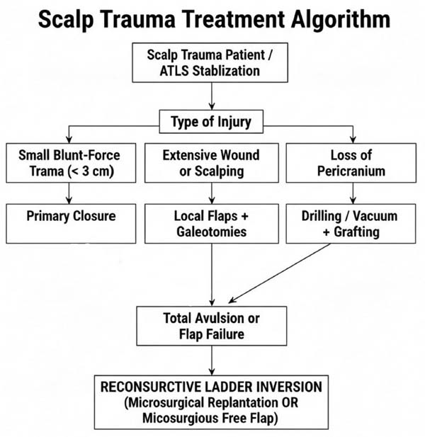
Based on these data, we propose a systematized treatment algorithm ( ►Fig. 1 ). The flow begins with patient stabilization (ATLS) and proceeds immediately to the decision based on the type of injury (a statistically validated factor). The algorithm branches into three initial paths: (1) small wounds (< 3 cm) are resolved with primary closure; (2) extensive wounds or scalping require local flaps with galeotomies; and (3) loss of pericranium demands bed preparation before grafting. The crucial point of the algorithm, however, is the identification of total avulsions or cases of local flap failure. In these specific scenarios, the protocol determines the interruption of further local reconstructive attempts and the immediate "Inversion of the Reconstructive Ladder," indicating replantation or microsurgical free flap as first-line therapy.
Conclusion
The analysis demonstrated that injury type and defect size were the main determinants of successful wound closure, regardless of anatomical location or associated trauma. The study supports the proposal of a systematized algorithm, an essential tool to guide surgical decision-making in emergency scenarios. The proposed protocol highlights the critical need to invert the reconstructive ladder in complex avulsions, prioritizing microsurgical techniques to avoid therapeutic failures and ensure definitive coverage.
References
1. Angelos PC, Downs BW. Options for the management of forehead and scalp defects. Facial Plast Surg Clin North Am 2009;17(03):379–393
2. Bas S, Oner C, Eren HI, Hacikerim Karsidag S, Yilmaz A. Reconstruction of complex scalp defects in different locations. Şişli Etfal Hastan Tıp Bül 2021;55(03):349–358
3. Berri DT, Ono MCC, Lopes MC, Groth AK, Silva ABD. Reconstrução de defeitos complexos da fronte e couro cabeludo. Rev Bras Cir Plást 2012;27(supl):45
4. Desai SC, Sand JP, Sharon JD, Branham G, Nussenbaum B. Scalp reconstruction: an algorithmic approach and systematic review. JAMA Facial Plast Surg 2015;17(01):56–66
5. Hofmann L, Rafizadeh Rashid M, Ludtka C, et al. Calvarial Reconstruction Following Massive Tissue Loss: A Feasible Treatment Strategy and Surgical Technique. Oper Neurosurg (Hagerstown) 2021;20(03):317–322
6. Leedy JE, Janis JE, Rohrich RJ. Reconstruction of acquired scalp defects: an algorithmic approach. Plast Reconstr Surg 2005;116(04):54e–72e
7. Ochs M, Chung W, Powers D. Trauma surgery. J Oral Maxillofac Surg 2017;75(8S):e151–e194
8. Seery GE. Scalp surgery: anatomic and biomechanical considerations. Dermatol Surg 2001;27(09):827–834
9. Iblher N, Ziegler MC, Penna V, Eisenhardt SU, Stark GB, Bannasch H. An algorithm for oncologic scalp reconstruction. Plast Reconstr Surg 2010;126(02):450–459
10. Zhang Y, Cheng K, Dong J, Li Q, Tremp M, Zhu L. Incidence and features of vertebral fractures after scalp avulsion injuries. J Craniofac Surg 2015;26(07):2217–2220
11. Fowler NM, Futran ND. Achievements in scalp reconstruction. Curr Opin Otolaryngol Head Neck Surg 2014;22(02):127–130
12. Vecchione TR, Griffith L. Closure of scalp defects by using multiple flaps in a pinwheel design. Plast Reconstr Surg 1978;62(01):74–77
13. Lutz BS, Wei FC, Chen HC, Lin CH, Wei CY. Reconstruction of scalp defects with free flaps in 30 cases. Br J Plast Surg 1998;51(03):186–190
14. Powers AK, Neal MT, Argenta LC, Wilson JA, DeFranzo AJ, Tatter SB. Vacuum-assisted closure for complex cranial wounds involving the loss of dura mater. J Neurosurg 2013;118(02):302–308
1. Plastic and Reconstructive Surgery Service, Hospital de Clínicas, Federal University
of Paraná (HC-UFPR), Curitiba, PR, Brazil
Address for correspondence Isabella Correa de Oliveira, MD, Rua Padre Agostinho, 2463, Apartment 1202, Bigorrilho, Curitiba, Paraná, 80710-000, Brazil (e-mail: isabella.oliveira21@gmail.com).
Article received: January 24, 2026.
Article accepted: June 01, 2026.
The authors have no conflict of interests to declare.
Editor-in-Chief: Dov Charles Goldenberg
 Read in Portuguese
 Read in English
 PDF PT
 Print
 Send this article by email
 How to Cite
 Mendeley
 Pocket
 Facebook
 Twitter