




Original Article - Year 2026 - Volume 41Issue 1
Extratemporal Facial Nerve Branching Patterns: Surgical Anatomy and Clinical Relevance
Padrões de ramificação do nervo facial extratemporal: Anatomia cirúrgica e relevância clínica

ABSTRACT
Background In facial plastic and reconstructive surgery, precise identification of the extratemporal facial nerve is critical to prevent iatrogenic injury. Surgical landmarks such as the tragal pointer, posterior belly of the digastric muscle, tympanomastoid suture, and retromandibular vein are commonly used to locate the facial nerve trunk. However, anatomical variations in branching patterns increase operative complexity.
Objective To evaluate the branching pattern and topographic relationships of the extratemporal facial nerve with clinically relevant surgical landmarks.
Methods The extratemporal segment of the facial nerve was dissected in 50 sides from 25 adult cadavers of both sexes (M: 21; F: 4) during routine dissections performed between 2016 and 2022. Length of the main trunk, division pattern within the parotid gland, relationship to the retromandibular vein, and interbranch communications were documented.
Results A single facial nerve trunk was observed at the stylomastoid foramen in all specimens. The trunk length ranged from 1.8 cm to 2.2 cm. In all cases, the nerve divided into temporofacial and cervicofacial divisions within the parotid gland. The facial nerve was located lateral to the retromandibular vein in 96% of sides. Multiple patterns of interbranch communications were identified, indicating anatomical variability relevant to surgical dissection planes.
Conclusion Knowledge of extratemporal facial nerve variations and its connection to operative landmarks is essential for improving surgical safety in parotidectomy, facial reconstruction, rhytidectomy, and aesthetic facial procedures. These findings support preoperative planning and careful intraoperative dissection to minimize nerve injury.
Keywords: facial nerve; surgical anatomy; parotid gland; facial nerve injuries; parotidectomy; anatomical variation
RESUMO
Introdução Na cirurgia plástica facial e reconstrutiva, a identificação precisa do nervo facial extratemporal é fundamental para prevenir lesões iatrogênicas. Pontos de referência cirúrgicos, como o ponteiro tragal, o ventre posterior domúsculo digástrico, a sutura timpanomastoidea e a veia retromandibular, são amplamente utilizados para localizar o tronco do nervo facial. Entretanto, variações anatômicas podem aumentar a complexidade cirúrgica.
Objetivo Avaliar o padrão de ramificação e as relações topográficas do nervo facial extratemporal com pontos anatômicos de importância cirúrgica.
Métodos O segmento extratemporal do nervo facial foi dissecado em 50 lados de 25 cadáveres adultos de ambos os sexos (M: 21; F: 4), durante dissecações de rotina realizadas entre 2016 e 2022. Foram analisados o comprimento do tronco principal, o padrão de divisão na glândula parótida, a relação com a veia retromandibular e as comunicações entre os ramos terminais.
Resultados Observou-se um único tronco do nervo facial na saída pelo forame estilomastoideo em todos os casos. O comprimento do tronco variou entre 1,8 cme 2,2 cm. Em todas as dissecações, o nervo dividiu-se em ramos temporofacial e cervicofacial. O nervo facial localizou-se lateralmente à veia retromandibular em 96% dos lados. Foram identificados diferentes padrões de comunicações entre os ramos terminais.
Conclusão O conhecimento das variações anatômicas do nervo facial extratemporal e de sua relação com pontos cirúrgicos é essencial para aumentar a segurança em parotidectomias, cirurgias reconstrutivas faciais, ritidoplastias e procedimentos estéticos.
Palavras-chave: nervo facial; anatomia cirúrgica; glândula parótida; lesões do nervo facial; parotidectomia; variação anatômica
Introduction
The facial nerve, which is the seventh cranial nerve, is at risk during facial plastic, reconstructive, and parotid surgeries. It has six segments: intracranial, meatal, labyrinthine, tympanic, mastoid and extratemporal. The extratemporal frees up the posterior auricular nerve and then innervates facial muscles through its terminal branches and is responsible for facial expression. The trunk of the facial nerve, which emerges from stylomastoid foramen, runs for 1 cm and then enters the posteromedial surface of the parotid gland, where it usually bifurcates into upper temporofacial and lower cervicofacial divisions. Both divisions give rise to five terminal branches: temporal, zygomatic, buccal, marginal mandibular, and cervical,1 which are frequently encountered during procedures such as parotidectomy, rhytidectomy, facial reconstruction, and cosmetic surgery.
Preserving these branches is essential to avoid postoperative facial paralysis and functional deficits. The branching of FN was first described by Davis et al, who described six types of facial nerve branching patterns (I, II, III, IV, V, and VI) based on the presence or absence of communications between its divisions and terminal branches.2 Another classification was described by Katz et al, with nine types of branching patterns (IA, IB, II, IIIA, IIIB, IIIC, IVA, IVB, and V) based on the number of trunks of facial nerve, the origin of the buccal branch, and communications between terminal branches.3 A new classification with three additional “double-trunk” types (VA, VB, and VC) was proposed by Kopuz et al.4 Several anatomical landmarks commonly used in plastic and head-and-neck surgery—including the stylomastoid foramen, posterior belly of the digastric muscle, tragal pointer, tympanomastoid suture, and retromandibular vein—serve as guides for identifying the facial nerve trunk intraoperatively. However, anatomical variations in branching patterns and interbranch communications increase the complexity of surgical dissection.
Methods
During routine educational dissections conducted between 2016 and 2022 in the Department of Anatomy, the extratemporal segment of the facial nerve was examined in 50 sides from 25 adult cadavers of both sexes (M: 21; F: 4). All cadavers had been embalmed using a 10% formaldehyde solution.
A preauricular surgical incision was employed, extending posteriorly behind the ear lobe over the mastoid process and inferiorly along the mandibular border, simulating operative exposure used in parotid and facial surgeries. The skin, subcutaneous tissue, and superficial fascia were carefully reflected to expose the parotid gland. The main trunk of the facial nerve was identified at the stylomastoid foramen using deep anatomical landmarks and traced through the gland to its terminal branching pattern.
The facial nerve trunk was followed meticulously along its intraparotid course, and its terminal branches were dissected and documented. All findings were recorded and photographed.
The study protocol included assessment of:
Number of facial nerve trunks emerging from the stylomastoid foramen.
Length of the main trunk from stylomastoid foramen to its intraparotid divisions.
Pattern of intraparotid divisions.
Relationship of the facial nerve to the retromandibular vein.
Terminal branching and interbranch communications based on Davis classification.2
Results
Number of Facial Nerve Trunks Exiting the Stylomastoid Foramen
In the present study, in all the dissections of 25 adult cadavers, there was only one trunk of facial nerve at the stylomastoid foramen. No double or triple trunks of facial nerve exiting through the stylomastoid foramen were observed in the present study.
Length of the Facial Nerve Trunk
In this study, length of the main trunk of facial nerve from stylomastoid foramen to its bifurcation point in the parotid gland ranged from 1.8 cm to 2.22 cm. The mean length was 1.9 cm and the SD for the mean length was 1.18 cm.
Number of Divisions of Facial Nerve Inside the Parotid Gland
The main trunk of the facial nerve on entering the parotid gland divided into upper temporofacial and lower cervico-facial divisions in all fifty sides. No other variations of divisions were observed in the present study.
Relation of Facial Nerve to Retromandibular Vein
The facial nerve was present lateral to the retromandibular vein in 96% of sides. In 4% of cases, facial nerve branches were found both lateral and medial to retromandibular vein.
Terminal Branches of Facial Nerve and Its Communications
The five branches—namely the temporal, zygomatic, buccal, marginal mandibular, and cervical—arose from the two divisions of the facial nerve in all sides. The temporal and zygomatic branches originated from the temporofacial division and marginal mandibular and cervical branches originated from the cervicofacial division in all specimens. The buccal branch of facial nerve arose from both divisions in 20 sides (►Fig. 1).
Communications of facial nerve were classified according to Davis et al:2
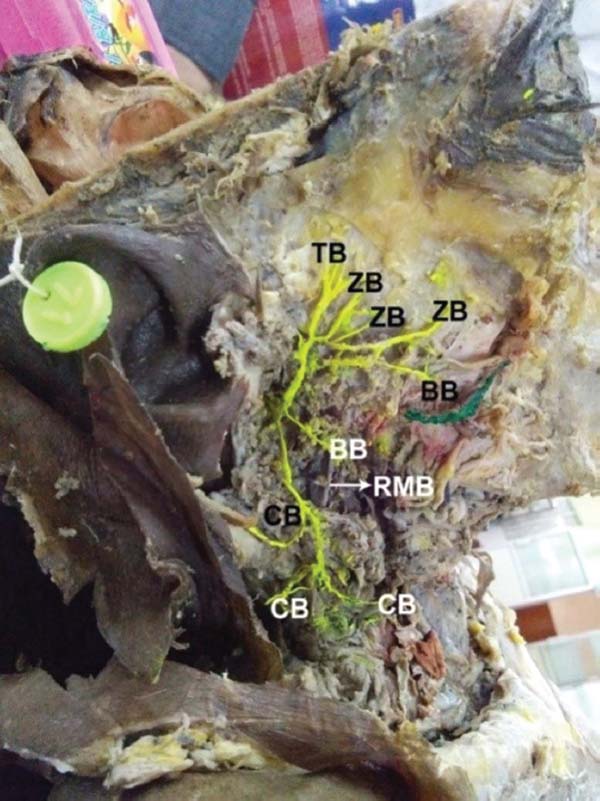
Type I: In seven sides (14%), there was no major communication between the divisions and branches of facial nerve (►Fig. 2).
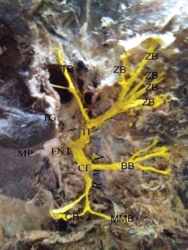
Type II: In nine sides (18%), communications between the branches of temporofacial divisions were observed (►Fig. 3).
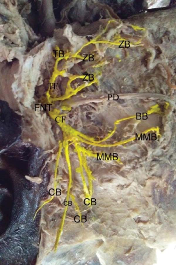
Type III: In 14 sides (28%), a single communication between the temporofacial and cervicofacial divisions was observed (►Fig. 4).
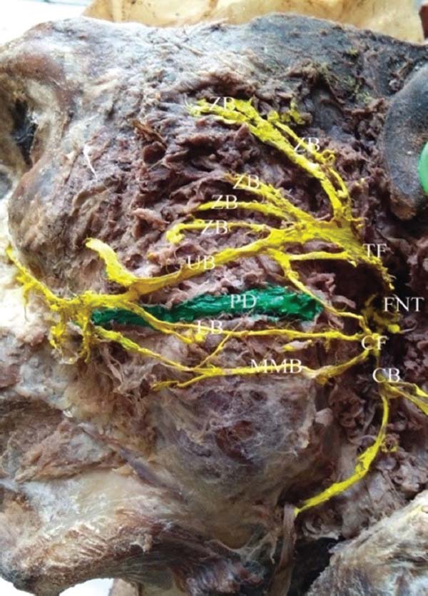
Type IV: In 12 sides (24%), a single communication between the temporofacial and cervicofacial divisions and communications among the temporofacial branches were observed (►Fig. 5).
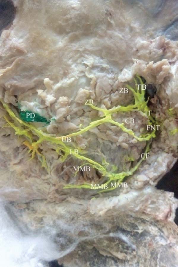
Type V: In four sides (8%), double communications between the temporofacial and cervicofacial divisions were observed.
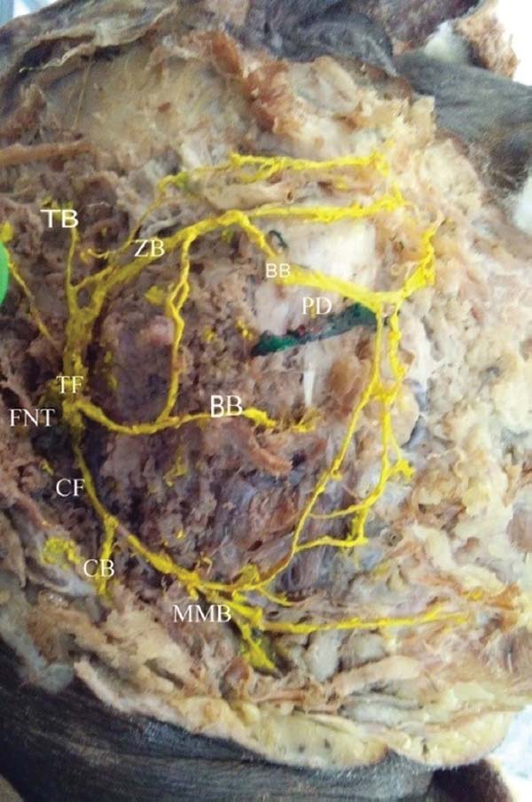
Type VI: In four sides (8%), complex communications between the branches of facial nerve were observed (►Fig. 6).
Discussion
A single facial nerve trunk exiting through the stylomastoid foramen has been described in the standard textbooks and in the previous literature.2, 5 Some authors have described that two facial nerve trunks exit from the stylomastoid foramen.6, 7 The presence of a single facial nerve trunk emerging from the stylomastoid foramen corresponds with classical anatomical descriptions and is consistent with previous studies. However, reports of multiple trunks emphasize the importance of cautious identification during surgical exposure. The mean distance between the emerging point of the facial nerve trunk from the stylomastoid foramen and its bifurcation point was 14.38 ± 3.04 mm according to Pascual et al.8 The measured trunk length in this study provides practical guidance for surgeons during deep parotid dissection, as excessive traction or blind dissection within this short segment increases the risk of nerve injury. Touré and Vacher classified the connection of the facial nerve to retromandibular vein in the parotid gland into six types, the most common type being that in which the facial nerve is lateral to retromandibular vein in 65.2% (Type I) of cases and medial to vein in 28% (Type II) of cases.9
Cruveilhier early on described six terminal branches, and Hovelacque later described eight terminal branches of facial nerve.10, 11 Davis et al simplified these earlier classifications of terminal branches of facial nerve and described five terminal branches of the facial nerve.2
Earlier classifications of communications among terminal branches of facial nerve were proposed by McCormack et al, Dargent and Duroux, and Pons-Tortella.12, 13, 14 Then, Davis et al in 1956 described a six-type classification based on the connections between the terminal branches.2
Type 1 (13%): No major communications between branches of facial nerve.
Type II (20%): Communications between the branches of temporofacial division only.
Type III (28%): Single communication between temporofacial and cervicofacial divisions.
Type IV (24%): Communications between branches of temporofacial division and also between temporofacial and cervicofacial divisions.
Type V (9%): Double communications between temporofacial and cervicofacial divisions.
Type VI (6%): Complex plexiform communications between the terminal branches.
Katz and Catalano proposed a new classification of communications based on the origin of buccal nerve and the number of trunks of the facial nerve.3 Anewer classification of communications of facial nerve into 12 types has been described by Pascual et al.8 It is well known that connections are more commonly found in the temporofacial division than in the cervicofacial division.2, 14 The observed communication patterns between branches highlight the complexity of intraparotid facial nerve anatomy. Such plexiform arrangements can obscure clear branch identification and increase the likelihood of inadvertent injury during facial lifting, parotidectomy, and reconstructive procedures.
Conclusion
Several variations in the branching pattern of facial nerve may increase the probability of a facial palsy after surgery if the surgeon is not aware of these anatomical variations.15 Variations in extratemporal facial nerve branching patterns and intraparotid communications significantly influence surgical safety. Detailed knowledge of these anatomical relationships and operative landmarks is essential to minimize postoperative facial nerve palsy during facial plastic surgery, parotid surgery, and reconstructive interventions.
References
1. Standring S. Gray’s anatomy: the anatomical basis of clinical practice. 39th ed. Edinburgh: Churchill Livingstone; 2005
2. Davis RA, Anson BJ, Budinger JM, Kurth LR. Surgical anatomy of the facial nerve and parotid gland based upon a study of 350 cervicofacial halves. Surg Gynecol Obstet 1956;102(04):385–412
3. Katz AD, Catalano P. The clinical significance of the various anastomotic branches of the facial nerve. Report of 100 patients. Arch Otolaryngol Head Neck Surg 1987;113(09):959–962
4. Kopuz C, Turgut S, Yavuz S, Ilgi S. Distribution of facial nerve in parotid gland: analysis of 50 cases. Okajimas Folia Anat Jpn 1994; 70(06):295–299
5. Gataa IS, Faris BJ. Patterns and surgical significance of facial nerve branching within the parotid gland in 43 cases. Oral Maxillofac Surg 2016;20(02):161–165
6. Kilic C, Kirici Y, Kocaoglu M. Double facial nerve trunk emerged from the stylomastoid foramen and petrotympanic fissure: a case report. J Korean Med Sci 2010;25(08):1228–1230
7. Kwak HH, Park HD, Youn KH, et al. Branching patterns of the facial nerve and its communication with the auriculotemporal nerve. Surg Radiol Anat 2004;26(06):494–500
8. Martínez Pascual P, Maranillo E, Vázquez T, Simon de Blas C, Lasso JM, Sañudo JR. Extracranial course of the facial nerve revisited. Anat Rec (Hoboken) 2019;302(04):599–608
9. Touré G, Vacher C. Relations of the facial nerve with the retromandibular vein: anatomic study of 132 parotid glands. Surg Radiol Anat 2010;32(10):957–961
10. Cruveilhier J. Traité d’anatomie descriptive. Paris: PAsselin; 1867. p. 544–48
11. Hovelacque A. Anatomie des nerfs craniens et rachidiens et du système grand sympathique chez l’homme. Paris: G Doin et Cie; 1927. p. 183–191
12. McCormack LJ, Cauldwell EW, Anson BJ. The surgical anatomy of the facial nerve with special reference to the parotid gland. Surg Gynecol Obstet 1945;80:620–630
13. Dargent M, Duroux PE. Données anatomiques concernant la morphologie et certains rapports du facial intra-parotidien. Presse Med 1946;37:523–524
14. Pons-Tortella E. El plexo parotídeo del nervio facial y las anastomosis periféricas entre el facial y el trigémino. Su importancia en la cirugía de la glándula parótida. Med Clin (Barc) 1947;9(01): 32–39
15. Lee DH, Yoon TM, Lee JK, Lim SC. Facial nerve anomaly in a patient with a parotid tumor: a case report. Medicine (Baltimore) 2016; 95(18):e3601
1. Department of Anatomy, Government Siddhartha Medical College, Vijayawada, Andhra
Pradesh, India
2. Department of Interventional Radiology, Miami Cardiac and Vascular Institute, Baptist
Health South Florida, Miami, Florida, United States
Address for correspondence Dr. Chitra Ramasamy, MD, DNB, Department of Anatomy, Government Siddhartha Medical College, Vijayawada 520008, Andhra Pradesh, India (e-mail: vjwchitra@gmail.com).
Article received: March 20, 2026.
Article accepted: June 01, 2026.
The authors have no conflict of interests to declare.
Editor-in-Chief: Dov Charles Goldenberg.
 Read in Portuguese
 Read in English
 PDF PT
 Print
 Send this article by email
 How to Cite
 Mendeley
 Pocket
 Facebook
 Twitter