




Original Article - Year 2026 - Volume 41Issue 1
Apert Syndrome: Evaluation of Patients Treated at A Public Institution
Síndrome de Apert: Avaliação de pacientes tratados em uma instituição pública
Renato da Silva Freitas1, ; Eduardo Angeli-Freitas1; Guilherme Augusto de Sá1; Julio Dallarmi Gil1; Igor Haddad Carvalho1; Mateus Vicente Andrade1; Isis Juliane Guarezi Nasser1; César Vinícius Grande1
ABSTRACT
Introduction Apert syndrome accounts for 4.5% of all cases of craniosynostosis, with brachycephaly being the most common presentation. It may lead to functional disturbances, including intracranial hypertension, hydrocephalus, respiratory difficulties, ocular abnormalities, and syndactyly. Although its treatment may employ different approaches, such as fronto-orbital advancement (FOA), posterior decompression, frontofacial advancement, Le Fort III (LFIII) osteotomy, and their variations, there is no well-defined therapeutic protocol. The current study evaluated the treatments for Apert syndrome performed at our institution.
Materials and Methods The present study evaluated the treatment of patients with Apert syndrome from 2004 to 2024 based on data on clinical features, previous treatments, complications, surgical courses, and specific outcomes.
Results We evaluated 30y patients with Apert syndrome, including 28 with brachycephaly. In total, 16 patients were younger than 6 months old, 5, between 6 and 13months old, 3, 1 to 4 years old, and 6, older than 4 years of age. Upon admission, 20 patients were previously untreated, and 10 had undergone prior surgical intervention. A total of 52 cranial surgeries were performed, including 18 FOAs, 5 monobloc procedures, 8 posterior decompressions, and 21 LFIII procedures.
Conclusion The management of the syndrome has benefited from technological advances and more refined surgical strategies, with an emphasis on multidisciplinary approaches and individualized treatment, requiring long-term follow-up and multistage interventions. Patients who presented at the ideal age initially underwent treatment with FOA and LFIII, and their current management includes posterior decompression and monobloc advancement.
Keywords: Apert syndrome; craniosynostosis; Le Fort III; obstructive sleep apnea; syndactyly
RESUMO
Introdução A síndrome de Apert é responsável por 4,5% de todas as ocorrências de cranioestenose, sendo a braquicefaliaa forma mais comum de sua apresentação. Pode levar a alterações funcionais, como hipertensão intracraniana, hidrocefalia, dificuldade respiratória, alterações oculares, além da sindactilia. Há diferentes abordagens, como avanço fronto-orbital (AFO), descompressão posterior, avanço frontofacial, osteotomia Le Fort III (LFIII), e suas variações. Ainda não há um protocolo bem definido para seu o tratamento. Avaliamos os tratamentos realizados em nossa instituição.
Materiais e Métodos Este estudo avaliou o tratamento de pacientes realizado de 2004 a 2024. Foram coletados dados relativos às características clínicas, aos tratamentos prévios, às complicações, à evolução cirúrgica e aos resultados específicos. Resultados Foram avaliados 30 pacientes com síndrome de Apert, e a braquicefalia esteve presente em 28 deles. Ao todo, 16 pacientes tinham menos de 6 meses de idade à admissão, 5, entre6e 12 meses, 3, entre 1e4 anos, e 6, mais de4 anos.À admissão, 20 pacientes jamais haviam sido submetidos a tratamento, e 10 haviam sido submetidos a intervenção cirúrgica prévia. Foram realizadas 52 de cirurgias cranianas, dentre elas: 18 procedimentos de avanço fronto-orbital, 5 de monobloco, 8 de descompressão posterior, e 21 de LFIII.
Conclusão O manejo da síndrome têm se beneficiado de avanços tecnológicos e estratégias cirúrgicas mais refinadas, com ênfase em abordagens multidisciplinares e tratamento individualizado, o que requer acompanhamento prolongado e intervenções em múltiplas etapas. Pacientes que chegaram em idade ideal foram tratados mediante AFO e LFIII em um primeiro momento, e atualmente estão sendo manejados por meio de descompressão posterior e avanço em monobloco.
Palavras-chave: síndrome de Apert; craniossinostose; Le Fort III; apneia obstrutiva do sono; sindactilia
Introduction
Craniosynostosis is a condition characterized by the premature closure of one or more cranial sutures, resulting in cranial or craniofacial deformity. Its classification depends on the shape of the calvaria: trigonocephaly (premature closure of the metopic suture); scaphocephaly (premature closure of the sagittal suture); brachycephaly (bilateral premature closure of the coronal sutures); plagiocephaly (unilateral premature closure of the coronal or lambdoid suture); and oxycephaly (premature closure of multiple sutures). This condition may or may not be associated with syndromes, and Apert syndrome, or type-I acrocephalosyndactyly, is a severe syndromic synostosis,1,2 is a rare genetic condition with a prevalence of 6 to 15 cases per 1 million live births. The condition follows an autosomal dominant pattern and is associated with mutations in the fibroblast growth factor receptor 2 (FGFR2) gene on chromosome 10q25–10q26 in 98% of the cases. Diagnosis is established in a proband with classic clinical features (multisuture craniosynostosis, midface retrusion, and complex syndactyly of the hands and feet) and/or by the identification of a heterozygous pathogenic variant in FGFR2 through molecular genetic testing, along with phenotypic features consistent with Apert syndrome.3 This condition may be associated with advanced paternal age, maternal infections, maternal exposure to medications, and inflammatory brain processes.
The premature closure of the bilateral coronal sutures leads to brachycephaly. Most subjects also present involvement of the sagittal and lambdoid sutures and are commonly associated with intracranial hypertension. Most subjects present with nonprogressive ventriculomegaly, and a subset exhibits true hydrocephalus. The cranial shape resembles a smahed tomato, with a normal head circumference. The biparietal diameter may be higher, resulting in a broad and high forehead and a tower-shaped skull (turribrachycephaly). In addition to this craniosynostosis manifestation, subjects with Apert syndrome may present with other phenotypes.1,2,4 The midface of patients with Apert syndrome is underdeveloped and presents marked retrusion. Cleft palate occurs in a subset of affected subjects. Multilevel airway obstruction may be present and result from nasal passage narrowing, tongue-based airway obstruction, and/or tracheal abnormalities. The hands of the subjects with Apert syndrome always show fusion of the three middle digits, potentially involving the thumb and fifth digit. In addition, feeding difficulties, dental abnormalities, hearing loss, hyperhidrosis, and progressive synostosis of multiple bones (such as in the hands, feet, and cervical vertebrae) are also common.3 Most subjects with Apert syndrome have normal intellectual development or mild intellectual disability, with reports of moderate and severe forms. A minority of affected subjects present with structural cardiac abnormalities, true gastrointestinal malformations, and genitourinary tract anomalies.3
Subjects with Apert syndrome require, in addition to cranial decompression and treatment of complex syndactyly, correction of sleep apnea, deepening of the orbits to improve exorbitism, and optimization of facial appearance. The treatment of synostosis may be subdivided into anterior frontoorbital advancement (FOA) and posterior decompression (PD), as surgical intervention in these different regions has distinct clinical implications.5 Early craniectomy was performed for many years to release synostotic cranial sutures, followed by a Le Fort III (LFIII) maxillary advancement at a later age.6 The traditional focus on anterior cranial fossa procedures should be reconsidered, as addressing the entire cranial vault leads to more consistent outcomes.7,8 Craniofacial approaches using monobloc (MB) osteotomy have been adopted by several centers, aiming at earlier improvement in esthetics and function, in spite of the higher surgical risk.
Despite the variety of approaches to treat Apert syndrome, there is no gold standard. Treatment centers partially or fully adopted the recent significant advances in techniques and technologies. The current study aims to evaluate our treatment approach, conducted at a referral center for subjects with craniofacial deformities.
Materials and Methods
The present study evaluated the treatment of subjects managed by the senior author (RSF) between 2004 and 2024 at the Comprehensive Care Center for Cleft Lip and Palate (Centro de Atendimento Integral ao Fissurado Lábio Palatal, CAIF, in Portuguese) at Hospital do Trabalhador and the Plastic Surgery Unit of Teaching Hospital at Universidade Federal do Paraná. The Ethics Committee approved the study under protocol number 87609425.7.0000.5225. Collected data included the type of synostosis, gender, age at presentation to the service, previous treatment, presence of sleep apnea, performance of tracheostomy, treatment of syndactyly (and the number of digits obtained), primary and secondary surgeries related to Apert treatment, and their surgical course. An extensive literature review in the PubMed database focused on treatment methods, classifications, and case reports of Apert syndrome, using the descriptors Apert syndrome and craniosynostosis.
Results
Thirty subjects with Apert syndrome were evaluated at CAIF. Among the craniosynostoses, brachycephaly was present in 28 subjects (93%), whereas 2 patients (6%) had plagiocephaly. Most subjects were female individuals, totaling 19 cases (63%). In total, 16 subjects (53%) were younger than 6 months upon admission, 5 (17%) were between 6 and 12 months, 3 (10%) were between 1 and 4 years, and 6 (20%) were older than 4 years. Upon admission, 20 subjects (67%) were previously untreated, and 10 (33%) had undergone prior surgical intervention. Among the 24 subjects younger than 4 years (80%), 19 (63%) were previously untreated, and 5 (17%) had already received treatment—2 (7%) underwent FOA with PD, 2 (7%) underwent isolated FOA, and 1 (3%) underwent gastrostomy.
Thus, ten subjects had already undergone surgical treatment. Two subjects had undergone procedures other than cranial surgeries, including one who had been submitted to a gastrostomy and another who had undergone hand reconstruction (►Table 1). The subject who presented to our service with gastrostomy as the only prior treatment underwent FOA and, after 3 years, LFIII with a rigid external distractor (RED). The subject previously treated for syndactyly underwent LFIII followed by Le Fort I (LFI).
| FOA 5 | FOA þ PD 3 | No cranioplasty 2 | |||||||
|---|---|---|---|---|---|---|---|---|---|
| LFIII | MB | PD | LFIII | No | LFIII | LFIII | LFI | FOA | LFIII |
| LFIII | LFIII | FOA | LFIII | FOA | LFIII | LFIII | LFI | ||
| LFIII | LFIII | LFI | |||||||
| LFIII | LFIII | ||||||||
Abbreviations: FOA, fronto-orbital advancement; LFI, Le Fort I; LFIII, Le Fort III; PD, posterior decompression.

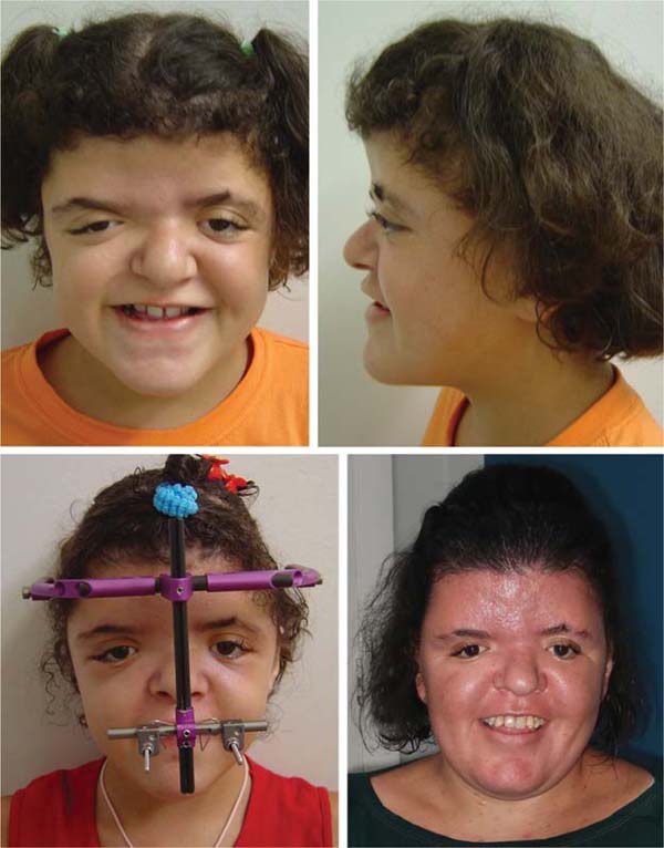
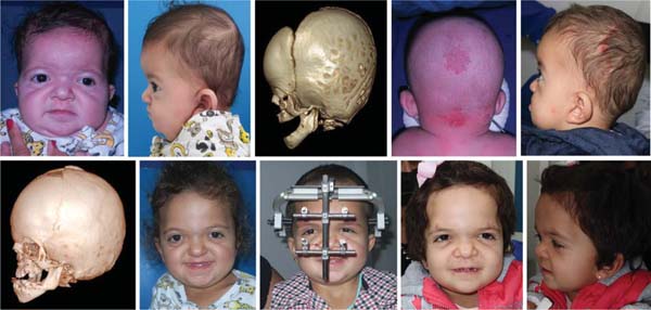
Eight subjects has undergone FOA at an outside institution, 3 of whom had also undergone PD (►Table 1). Four of these cases required, as the first cranial surgery at our service, LFIII with RED at a mean age of 93.25 months (►Fig. 1). Two of these 4 subjects underwent another LFIII as a second procedure at a mean age of 115.5 months. One of the subjects who did not undergo a repeat LFIII with RED underwent LFI after seven years. The other subject who did not undergo a repeat LFIII with RED returned to the original service for FOA. Later, they returned to our service to undergo 2 LFIII procedures at subsequent time points (1 after 5 years and a new advancement with RED after 7 years). One of the 8 subjects underwent MB osteotomy and, after 1 year and 3 months, LFIII with RED. One 27-year-old subject required LFI alone after orthodontic preparation. The last patient required no additional procedures beyond the previous FOA. They had been treated with FOA, underwent PD at our service, were submitted to another FOA procedure, and subsequently underwent 2 additional LFIII procedures at 6 and 14 years of age.
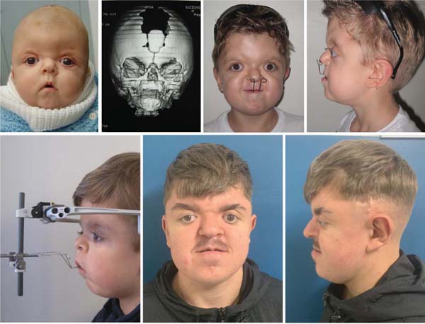
Twenty subjects were admitted to the service without having undergone any surgical treatment (►Table 2). One female subject with Apert syndrome presented at the age of 33 years, bringing her newborn child with the malformation for treatment. She had sleep apnea and underwent LFIII osteotomy with the use of a maxillary RED. Among the 19 previously-untreated subjects who presented at the ideal age, 11 (39%) initially underwent FOA, 6 (20%) underwent PD, 1 (3%) underwent MB advancement, and 1 (3%) remains untreated. Among the 11 previously-untreated subjects who underwent FOA, the mean age at the surgery was 7.5 months. Subsequently, eight of these subjects underwent additional procedures. Two subjects (7%) required repeat FOA at a mean age of 35.5 months, and 1 of them subsequently required LFIII with RED at 10 years of age (►Fig. 2). Three subjects (13%) underwent LFIII with RED at a mean age of 128.7 months. One subject (3%) underwent MB at 132 months of age. One subject (3%) underwent PD, a new FOA, and subsequently, LFIII with RED. One subject underwent LFI as a final procedure.
| FOA 11 patients |
LFIII 3 | ||
| FOA 2 | LFIII 1 | ||
| MB 1 | |||
| PD 1 | FOA 1 | LFIII 1 | |
| LFI 1 | |||
| NO 3 | |||
| PD 6 patients |
MB 2 | ||
| FOA 1 | |||
| NO 3 | |||
| MB 1 patient |
LFIII 1 | ||
| LFIII 1 patient |
|||
| No treatment 1 patient |
Abbreviations: FOA, fronto-orbital advancement; LFI, Le Fort I; LFIII, Le Fort III; MB, monobloc; NO, nothing; PD, posterior decompression.

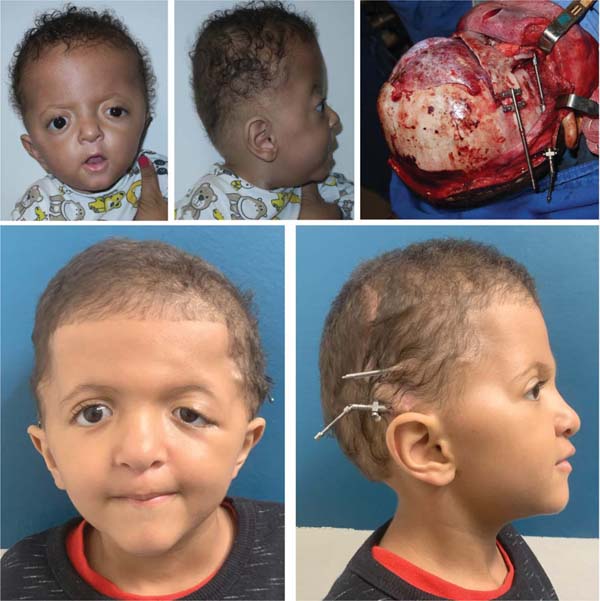
Among the 6 previously-untreated subjects who underwent PD as the initial surgical treatment, at a mean age of 13.3 months, 2 required MB advancement (►Fig. 3), and 1 required FOA, all of whom were operated on at 3 years of age. The only previously-untreated subject who underwent MB as the first-line procedure at age 3 required LFIII with RED at 10 years old.
Regarding the age at which subjects underwent the first surgical procedure at our center, regardless of whether they were previously untreated or not, 7 cases (23%) underwent PD at a mean age of 18.2 months; FOA was the most common procedure, performed 12 times (40%), at a mean age of 7.25 months; and 2 cases (7%) underwent MB advancement at a mean age of 97.5 months (►Fig. 4).
Among all 30 subjects analyzed, 17 (57%) presented with sleep apnea and required surgical treatment. Three subjects (10%) underwent only FOA, 3 (10%), only LFIII, and 1 (3%), only MB. Eight subjects (27%) underwent FOA followed by LFIII, 1 (3%) underwent FOA combined with MB, and 1 (3%) underwent MB combined with LFIII. In this group with respiratory complications, 12 FOA, 12 LFIII, and 3 MB procedures were performed, with some subjects undergoing a combination of 2 of these surgical therapies due to recurrence, which was observed in 10 cases (58%).
Regarding non-cranial surgeries, 25 subjects (83%) underwent a total of 53 secondary procedures. Hand reconstruction was the most frequent, performed in 25 subjects (83%). Six subjects (20%) underwent palatoplasty, including 4 (13%) who were younger than 5 years and 2 (7%) female patients who were older than 12 years. Eight subjects (27%) underwent canthopexy at ages ranging from 13 to 26 years. Other procedures included septoplasty (2 subjects [7%]) and turbinectomy (2 subjects [7%]). Isolated treatments included glabellar rhytidectomy, tonsillectomy, and tympanoplasty, each performed in 1 subject (3%). Lastly, only 2 subjects (6%) required tracheostomy.
Discussion
Apert syndrome is a severe craniofacial deformity with multiple repercussions for affected subjects. In recent decades, numerous techniques and devices have been developed for craniofacial surgery, such as absorbable plates and bone distractors. Patients with Apert syndrome have benefited from these advances, which enable procedures with improved outcomes, such as MB osteotomy. The objective of the current study was to evaluate the treatment adopted at our center for this malformation and the changes that have occurred with increased knowledge and the availability of these technologies. Thirty sequential subjects from different periods were studied, and the current series highlights these changes.
The most common finding in Apert syndrome is synostosis of the bicoronal suture. This was evident in the present study, in which 28 subjects (93%) presented with this type of craniosynostosis, and only 2 subjects (6%) presented with plagiocephaly. This anomalous fusion of the greater wing of the sphenoid bone with the temporal, frontal, and parietal bones forms the coronal ring, which restricts the growth of the anterior cranial fossa. As a result, the anteroposterior dimension is smaller, limiting the anterior displacement of the sphenoid. This restriction of anterior movement may lead to protrusion of the unfixed portion of the greater wing of the sphenoid, causing distortion rather than enabling homogeneous anterior displacement of the entire sphenoid structure.9 Forte et al.10 (2014) highlighted that, in subjects with Apert syndrome, the anterior cranial fossa is shorter and the sphenoid divergence angle is significantly more obtuse compared to controls. These anatomical findings are correlated with the high prevalence of brachycephaly observed in the current series. Furthermore, these authors10 concluded that midface retrusion is associated with abnormal sphenoid morphology and posterior rotation of the pterygoid plates. Cases with unilateral coronal synostosis present with a kyphotic cranial base, requiring specific treatments to correct this deformity. We observed two cases of unilateral plagiocephaly, both treated with FOA, with greater advancement on the affected side. Long-term follow-up demonstrated that these cases were less severe, with more consistent outcomes achieved using less extensive procedures.
Lu et al.9 (2018) analyzed preoperative computed tomography (CT) scans of 18 subjects with Apert syndrome and 36 CTs from control subjects to better understand orbital bone deformities. They concluded that these subjects are prone not only to acrocephaly, midface hypoplasia, and syndactyly, but also to visual impairment. The most significant orbital features identified in Apert syndrome were exorbitism, hypertelorism, and strabismus. In addition, the most common ophthalmological findings in these subjects were exposure keratitis and ocular ulceration resulting from exorbitism. Malposition of the lateral canthus, whether primary or secondary to multiple orbital and eyelid procedures, was addressed with lateral canthopexy in 8 subjects, aged between 13 and 26 years, to improve ocular protection and lateral canthal positioning. This orbital dysmorphology occurs before the age of 6 months. The authors9 observed that the zygomatic bone is the most severely-deformed facial structure in early childhood for subjects with Apert syndrome, both in its positional relationship and geometric shape. This deformity may act as a bridge, influencing and transmitting developmental stress forces to other facial and maxillary structures, caused by prematurely-fused coronal and perizygomatic sutures. Consequently, exophthalmos becomes more pronounced due to zygomatic hypoplasia. The FOA procedure advances the frontal bandeau, thereby protecting the eye. Improvement is more significant with MB advancement. Some cases may present a transition from exophthalmos to relative enophthalmos, which tends to improve with age.9
Le Fort III and MB osteotomies improve the positioning of the orbital bones, although they do not correct their dysmorphology. These procedures can reduce ocular protrusion and improve the esthetics of the orbitopalpebral region. They are frequently necessary and warranted, offering significant benefits such as improved psychosocial adjustment, reduction of exophthalmos, increased nasopharyngeal airway patency, and improved dental occlusion.6 In our service, two cases required LFIII with RED as the initial treatment.
The management of craniofacial synostoses has progressed continuously since the early days of craniofacial surgery in the 1950s. The FOA procedure consists of a craniotomy of the frontal region and orbital roof, followed by advancement of the orbital bandeau and remodeling of the frontal bone, resulting in improved frontal projection, greater ocular protection, and reduced intracranial pressure. Compression of the frontal brain tissue, the frontal subarachnoid space, and the frontal horns of the lateral ventricles is evident in Apert syndrome.10 This technique has been used as the first-line treatment in most centers, as it is less extensive than MB advancement. In an Australian study,11 94 subjects underwent 130 cranial surgeries, including 83 cases of FOA, 18 of PD, and 20 of MB. Subjects with bicoronal synostosis and significant airway compromise may require more aggressive and earlier interventions. This is consistent with the data from the present study, in which many subjects underwent FOA and PD to improve airway patency.12–14 One of the approaches for these cases is frontofacial MB advancement.15 Initially, this procedure was widely criticized due to high rates of surgical complications. Subsequently, a new approach was adopted, incorporating midface osteogenic distraction. Osteogenic distraction became the gold standard in the 2000s, as it enables frontofacial advancement in children, and longitudinal studies have demonstrated reductions in postoperative complications and surgical morbidity. This technique also reduces the recurrence of midface retrusion and the morbidity from intracranial procedures.16 Raposo-Amaral et al.17 (2020) reinforce the role of osteogenic distraction. These authors describe 69 subjects treated according to their institutional algorithm for Apert syndrome. The first important step in management is to distinguish between classic Apert syndrome and the cloverleaf skull deformity. Subjects with adequate bone thickness are referred for PD, whereas those without adequate bone thickness undergo venography to evaluate the supratentorial venous network and guide treatment planning. If a significant venous network is present, FOA is performed; if not, PD is indicated. Following these procedures, subjects undergo syndactyly treatment and, subsequently, palatal repair. Only later do subjects undergo LFIII or MB. These approaches are typically performed before 3 months of age in cases with intracranial hypertension and before 4 months of age in patients without it. Early intervention is carefully designed to use the brain’s malleability and rapid regenerative capacity during early childhood. This approach reduces the need for blood transfusions and minimizes the surgical risks associated with later interventions. Our center also prioritizes early treatment—among the 19 subjects younger than 6 months upon admission, 11 underwent cranial surgery before 9 months of age. The mean age at first surgical intervention in this group was 15.3 months. This higher mean age was due to surgical postponement secondary to clinical issues that delayed intervention.
Moreover, early correction of hand deformities enables the earlier development of basic motor skills, which can enhance children’s independence and quality of life.18
Osteogenic distraction is believed to provide greater skeletal stability during the facial-lengthening process, resulting in improved prognosis for subjects.12–14 Initially, external devices (RED system, KLS Martin SE & Co. KG) were used, enabling traction of the face and/or skull through fixation with pins placed posteriorly to the region to be advanced. This has been our primary method of traction. The use of RED for LFIII or MB provides considerable surgical flexibility, both in terms of device positioning and vector control and adjustment during the lengthening process. However, it requires adequate bone thickness to support fixation with transcutaneous pins. In the current series, RED was used in 20 LFIII procedures in 15 subjects and in 5 MB procedures (►Tables 1–2).
The use of internal distraction devices, in addition to being more convenient and discreet for patients, provides substantial skeletal stability during the distraction process and highly-efficient osteogenic consolidation. There is no risk of penetration of the distractor screws through the cranial bone table. However, this approach provides a unidirectional lengthening vector, which may limit the achievement of optimal outcomes, particularly in occlusion.12–14 A second surgical procedure is required for device removal after 3 to 6 months. This method was recently introduced in our service due to its high cost; nevertheless, it provided an additional option for synostosis treatment. In our series, only one patient with Apert syndrome underwent MB using an internal distraction device.
The PD procedure has been increasingly utilized. Although earlier approaches, such as those described by Marchac et al.19 (1994), did not include a specific treatment for the posterior cranial region, the technique described by Salyer and Bardach18 did. Some centers20 have combined PD in 21% of the cases as an adjunctive treatment before FOA to improve cranial shape. The PD procedure can be performed even in cases without lambdoid suture synostosis. It is now understood that remodeling of this cranial region is one of the most important aspects in the management of Apert syndrome, considering that brain growth tends to occur in areas with available space.18 Accordingly, some centers have adopted17 posterior treatment to delay intervention in the anterior region and postpone MB, rather than performing FOA initially. The options include lambdoid craniectomy, PD, or posterior remodeling. Our preference is PD, which was performed in eight cases and currently represents the initial treatment approach at our center.
Patients with Apert syndrome and bicoronal synostosis present with significant vertical compromise of the nasopharyngeal airway, whereas other subtypes present a more restricted oropharyngeal space.12–14 Xie et al.21 (2016) analyzed 25 subjects with Apert syndrome, 60% of whom presented nasal abnormalities, including bilateral choanal atresia (8%), bilateral (16%) or unilateral (4%) congenital bony nasal stenosis, and nasal septum deviation (32%). Moreover, 44% of the patients presented obstructive sleep apena (OSA); they were treated with nasal dilation procedures or surgical repair using devices such as nasopharyngeal airways, and a single patient required tracheostomy due to bilateral nasal obstruction. Adenoidectomy (12%), tonsillectomy (4%), and combined adenotonsillectomy (28%) were needed, along with noninvasive ventilation or long-term oxygen therapy in 12% of cases. Only one subject required midface advancement to treat OSA.21
In the current series of 30 cases, respiratory difficulty was evident, with 57% presenting with sleep apnea. In the literature,22 many cases require temporary tracheostomy. However, in the present study, only two patients underwent this procedure. The approach of our group aims at avoiding tracheostomy by performing additional procedures such as tonsillectomy and using continuous positive airway pressure (CPAP) while awaiting definitive midface advancement, either through LFIII or MB. The MB surgery (5 cases) or LFIII (15 cases) aimed to advance the midface, thereby enabling nasal breathing and improving apnea. In recent years, we have opted to perform PD initially, followed by MB from the age of 4, reserving LFIII for cases in which FOA had already been performed. Breik et al.20 (2016) recommended earlier midface advancement only in cases of OSA, although preferring to perform LFIII at a later stage.
The treatment of children with syndromic craniosynostosis involves several significant challenges due to severe deformities, limited physiological reserve, and substantial future craniofacial growth. Early intervention is advocated to facilitate corneal protection and the cranial expansion required for brain development. Subjects with intracranial hypertension have an indication for early surgery. On the other hand, procedures performed at a later stage tend to result in more stable skeletal correction and a lower need for subsequent revisions.
However, there is an ongoing debate regarding the relationship between cognitive development in subjects with Apert syndrome and their age at the timing of surgery. Some studies23 report delayed psychosocial development in cases of late surgery, while others24 have not found this association4 Early surgery, before 18 months of age, may lead to recurrence and the need for reoperation. Breik et al.20 reported that subjects operated after this age did not require a new FOA, whereas 63% of those operated earlier required an additional procedure.20 In the current series, 11 subjects underwent FOA at an early age, and 4 of them (36%) required a second FOA or MB. Accordingly, in our current approach, we have opted for PD as the initial procedure to allow more time before anterior surgery, whether FOA or MB, the latter being our current first-line option.
Variability in treatment approaches among craniofacial centers arises from differing perspectives on these challenges and the lack of robust outcome data. Over time, high-volume centers have reported high rates of reoperation for intracranial pressure and morphological issues following FOA and cranial vault remodeling. Some centers have shifted toward expansion of the midcranial vault or occipital region, while others have adopted less-invasive methods, such as regional craniectomy and spring-mediated remodeling, which reliably increase cranial volume in young subjects. Since 2009, posterior cranial vault distraction has become the initial procedure for many subjects with syndromic synostosis due to its effectiveness in expanding cranial volume, reliability in early childhood, and favorable perioperative morbidity profile. This method minimizes the risk of infection and preserves subsequent cranial growth potential, with reduced need for bone grafting. Its main limitation is the need for a second procedure to remove the distractor. Early posterior vault distraction has reduced the number of procedures in the first 5 years of life in subjects with Apert syndrome, decreasing exposure to general anesthesia and blood loss.25 However, many subjects still require FOA and cranial vault remodeling due to frontal and orbital retrusion. Fronto-orbital procedures performed in early years should advance the supraorbital bar, normalize the forehead, and reduce turribrachycephaly. Maintenance of correction requires the use of absorbable plates or bone grafts. Distraction associated with MB advancement may be considered in cases of severe exorbitism or OSA, with surgical timing adjusted according to functional needs, typically between the ages 5 and 8. Evidence25 suggests that there is limited vertical or sagittal maxillary growth following midface osteotomy.
The choice of osteotomy for midface advancement varies according to the patient’s anatomy. The LFIII method advances all midfacial elements as a single unit. Distraction facilitates greater advancement with control over the movement vector. The LFII procedure with zygomatic repositioning may be beneficial in subjects with Apert syndrome, improving ocular protection and facial proportions. Monobloc distraction with facial bipartition improves hypertelorism, corrects exorbitism, and adjusts facial width, although it is associated with relatively-high morbidity.25
Interestingly, we evaluated the age at presentation of syndromic subjects to our service. Subjects with Crouzon syndrome presented later, possibly due to less-severe facial involvement and the absence of hand deformities. In Apert syndrome, 50% of the subjects presented before 6 months of age; 16.7%, between 6 and 12 months; and 3.33%, between 12 and 24 months. Therefore, 70% of the cases presented within the period we consider most appropriate for treatment. This was also demonstrated by Renier et al.23 (2000), who conducted a retrospective observational study analyzing 2,137 cases of craniosynostosis treated over 23 years (1976–1999). These authors23 concluded that surgical correction should be performed as early as possible; to support this conclusion, they compared outcomes of procedures performed before and after 1 year of age, considering risks and outcomes. Regarding cognitive outcomes, children with Apert syndrome treated before age 1 demonstrated significantly-better results compared to those treated later. Among those evaluated before the age of 1 year, 44% had normal cognitive levels, whereas only 5% of those evaluated after this age achieved the same level. After surgical intervention, 17% of the children treated early achieved normal cognitive development, whereas none of those treated after 1 year did so. From a morphological standpoint, the outcomes were also more favorable in early interventions, with more satisfactory outcomes and fewer reoperations required. Additionally, the comparison of complication rates in the pre- and postoperative periods showed no significant differences between procedures performed before and after 1 year of age, reinforcing the safety of early intervention. Other cases presented between the ages of 2 and 4 years (6%) and above 4 years (20%). One previously-untreated female subject presented at 40 years old, bringing a 2-month-old child with Apert syndrome for treatment. After the child’s surgery, the mother underwent LFIII advancement using RED. Both had favorable outcomes, and the mother showed resolution of sleep apnea on polysomnography.
Correction of cranial deformity is essential to improve respiratory function and facial esthetics. In CAIF, the prevalence of FOA for brachycephaly correction demonstrated that previously-untreated cases were, for a long time, managed primarily using this technique. Many subjects previously treated with FOA at other centers sought CAIF for continuation of treatment and facial advancement. Craniofacial growth after FOA or even MB is often inadequate. It is worth emphasizing that midface advancement achieved with MB at 4 to 5 years of age tends to diminish over time, as the maxilla does not grow adequately. Therefore, some of these subjects (12 cases) required a subsequent LFIII to improve facial profile. In the study by Breik et al.20, 70% of the subjects required at least 2 transcranial procedures. In the study by Bachmayer and Ross6 horizontal maxillary growth after LFIII advancement was evaluated in subjects with Crouzon, Apert, or Pfeiffer syndromes. The authors6 concluded that horizontal maxillary growth invariably ceases after surgical midface advancement, leading to class-III malocclusion and maxillary deficiency that worsen over time due to continued mandibular growth. The LFIII procedure seems to inhibit further anteroposterior maxillary growth, although vertical growth of the maxilla is preserved and continues after surgery.
Due to severe maxillary hypoplasia, many subjects may also present with mandibular hypoplasia despite class-III occlusion. Changes in the spatial and morphological relationship between the mandible and the cranial base occur synchronously. Mandibular alterations are disproportionate in three dimensions—width, length, and height—with an initial shortening followed by reduced height. A narrow angle between the mandible and the posterior cranial base results in a more limited nasopharyngeal and oropharyngeal airway. These subjects may continue to present some degree of sleep apnea even after effective midface advancement. Thus, complementary orthognathic surgery at the end of facial growth, from ages 16 to 18, provides functional benefits for breathing and dental occlusion, as well as esthetic improvements. Orthognathic surgery was performed in four cases in our series. This pattern of deformity underscores the need for comprehensive and individualized surgical approaches to correct these complex anomalies.20 These findings are consistent with those of other studies.24,26
Therefore, the complex management of subjects with Apert syndrome demonstrates significant progress in recent years. Technological advances—including virtual surgical planning, prototyping, bone distractors, absorbable plates, CPAP, and other methods—have contributed substantially to improved outcomes. However, certain challenges persist. Long-term appearance may deteriorate despite adequate occlusion and good frontal projection, with the development of frontal wrinkles and insufficient nasal projection. Continued advances may enable better long-term restoration for these subjects.
Conclusion
The analysis of the results of the current study highlights the complexity of managing Apert syndrome and the importance of a multidisciplinary approach to achieve satisfactory clinical outcomes. The FOA procedure was the most frequently performed as the initial intervention, and it was often followed by additional craniofacial surgeries due to progression of the deformity or recurrence. The surgical procedures employed, such as MB osteotomy and LFIII maxillary advancement, demonstrated effectiveness in improving esthetic and functional outcomes. There was a high rate of requirement of multiple procedures among subjects with sleep apnea, reinforcing the complexity of respiratory management in these cases. In addition to cranial surgeries, secondary interventions, such as hand reconstruction (83% of the cases) and palatoplasty (20% of the cases), were frequently performed, highlighting the systemic impact of Apert syndrome. Furthermore, subjects who underwent early intervention, before the first year of life, demonstrated better cognitive and structural outcomes, emphasizing the importance of early treatment. Another relevant aspect discussed was the shift in the surgical approach adopted by the study team. The use of PD before anterior procedures (such as FOA and LFIII) was implemented to address one of the main challenges of Apert syndrome, intracranial hypertension, and it has shown promise in reducing the need for multiple interventions and improving long-term clinical outcomes. The variability of treatment protocols among specialized centers highlights the lack of consensus regarding the optimal surgical technique, underscoring the need for further comparative studies and long-term follow-up of subjects.
REFERENCES
1. Aloysio J. Neurologia Infantil – Capítulo 45: Cranioestenoses [Internet]. [citado 2024 out 10] Disponível em: http://josealoysio.com.br/site/wp-content/uploads/2016/08/Neurologia-Infantil-Cap-45-Cranioestenoses.pdf
2. Katouni K, Nikolaou A, Mariolis T, et al. Syndromic craniosynostosis: A comprehensive review. Cureus 2023;15(12):e50448. Doi: 10.7759/cureus.50448
3. Wenger TL, Hing AV, Evans KN. Apert Syndrome. In: Adam MP, Bick S, Mirzaa GM, et al., editores. GeneReviews® [Internet]. Seattle: University of Washington; 1993–2026 [citado 2024 out 10]. Disponível em: https://www.ncbi.nlm.nih.gov/books/NBK541728/
4. Kumari K, Saleh I, Taslim S, et al. Unraveling the complexity of Apert syndrome: Genetics, clinical insights, and future frontiers. Cureus 2023;15(10):e47281. Doi: 10.7759/cureus.47281
5. Lu X, Forte AJ, Wilson A, et al. Cranial fossa volume and morphology development in Apert syndrome. Plast Reconstr Surg 2020; 145(04):790e–802e
6. Bachmayer DI, Ross RB. Stability of Le Fort III advancement surgery in children with Crouzon’s, Apert’s, and Pfeiffer’s syndromes. Cleft Palate J 1986;23(Suppl 1):69–74
7. Mulliken JB, Bruneteau RJ. Surgical correction of the craniofacial anomalies in Apert syndrome. Clin Plast Surg 1991;18(02): 277–289
8. McCarthy JG, Glasberg SB, Cutting CB, et al. Twenty-year experience with early surgery for craniosynostosis: I. Isolated craniofacial synostosis–results and unsolved problems. Plast Reconstr Surg 1995;96(02):272–283. Doi: 10.1097/00006534-199508000-00004
9. Lu X, Forte AJ, Sawh-Martinez R, et al. Anterior convex lateral orbital wall: distinctive morphology in Apert syndrome. Br J Oral Maxillofac Surg 2018;56(09):864–869. Doi: 10.1016/j.bjoms. 2018.09.011
10. Forte AJ, Alonso N, Persing JA, Pfaff MJ, Brooks ED, Steinbacher DM. Analysis of midface retrusion in Crouzon and Apert syndromes. Plast Reconstr Surg 2014;134(02):285–293. Doi: 10.1097/PRS.0000000000000360
11. Moore MH, Hanieh A. Cerebrospinal fluid spaces before and after infant fronto-orbital advancement in unilateral coronal craniosynostosis. J Craniofac Surg 1996;7(02):102–105, discussion 106. Doi: 10.1097/00001665-199603000-00003
12. Lu X, Sawh-Martinez R, Forte AJ, et al. Classification of subtypes of Apert syndrome, based on the type of vault suture synostosis. Plast Reconstr Surg Glob Open 2019;7(03):e2158. Doi: 10.1097/GOX.0000000000002158
13. Lu X, Sawh-Martinez R, Forte AJ, et al. Mandibular spatial reorientation and morphological alteration of Crouzon and Apert syndrome. Ann Plast Surg 2019;83(05):568–582. Doi: 10.1097/SAP.0000000000001811
14. Lu X, Forte AJ, Sawh-Martinez R, et al. Spatial and temporal changes of midface in Apert’s syndrome. J Plast Surg Hand Surg 2019;53(03):130–137. Doi: 10.1080/2000656X.2018.1541324
15. Laure B, Joly A, Moret A, Travers N, Listrat A, Goga D. Frontofacial monobloc advancement with simultaneous frontal cranioplasty in adolescents with residual Apert syndrome deformations. J Craniofac Surg 2015;26(07):2059–2061. Doi: 10.1097/SCS.000 0000000001942
16. Rickart AJ, van de Lande LS, O’ Sullivan E, et al. Comparison of internal and external distraction in frontofacial monobloc advancement: A three-dimensional quantification. Plast Reconstr Surg 2023;152(03):612–622. Doi: 10.1097/PRS.0000000000010331
17. Raposo-Amaral CE, Denadai R, Oliveira YM, Ghizoni E, Raposo-Amaral CA. Apert syndrome management: Changing treatment algorithm. J Craniofac Surg 2020;31(03):648–652. Doi: 10.1097/SCS.0000000000006105
18. Salyer KE, Bardach J, editores. Salyer and Bardach’s Atlas of Craniofacial & Cleft Surgery: Volume I: Craniofacial Surgery. 1. ed. Filadélfia: Lippincott Williams & Wilkins; 2014
19. Marchac D, Renier D, Broumand S. Timing of treatment for craniosynostosis and facio-craniosynostosis: a 20-year experience. Br J Plast Surg 1994;47(04):211–222. Doi: 10.1016/0007-1226(94)90001-9
20. Breik O, Mahindu A, Moore MH, Molloy CJ, Santoreneos S, David DJ. Apert syndrome: Surgical outcomes and perspectives. J Craniomaxillofac Surg 2016;44(09):1238–1245. Doi: 10.1016/j.jcms.2016.06.001
21. Xie C, De S, Selby A. Management of the airway in Apert syndrome. J Craniofac Surg 2016;27(01):137–141. Doi: 10.1097/SCS.0000000000002333
22. Cuperus IE, Mathijssen IMJ, van Veelen MC, et al; Workgroup Craniosynostosis, European Reference Network CRANIO. A European multicenter study of perioperative airway management policies following midface surgery in syndromic craniosynostosis. Plast Reconstr Surg 2024;154(06):1281–1292. Doi: 10.1097/PRS.00000000000011317
23. Renier D, Lajeunie E, Arnaud E, Marchac D. Management of craniosynostoses. Childs Nerv Syst 2000;16(10-11):645–658. Doi: 10.1007/s003810000320
24. Fearon JA, Podner C. Apert syndrome: evaluation of a treatment algorithm. Plast Reconstr Surg 2013;131(01):132–142. Doi: 10.1097/PRS.0b013e3182729f42
25. Taylor JA, Bartlett SP. What’s new in syndromic craniosynostosis surgery? Plast Reconstr Surg 2017;140(01):82e–93e. Doi: 10.1097/PRS.0000000000003524
26. Nout E, Koudstaal MJ, Wolvius EB, Van der Wal KGH. Additional orthognathic surgery following Le Fort III and monobloc advancement. Int J Oral Maxillofac Implants 2011;40(07):679–684. Doi: 10.1016/j.ijom.2011.02.014
1. Unidade de Cirurgia Plástica, Complexo do Hospital de Clínicas da Universidade
Federal do Paraná, Curitiba, PR, Brazil
Data Availability Data will be available upon request to the corresponding author.
Financial Support The authors declare that they did not receive financial support from agencies in the public, private or non-profit sectors to conduct the present study.
Address for correspondence: Renato da Silva Freitas, Unidade de Cirurgia Plástica, Complexo do Hospital de Clínicas da Universidade Federal do Paraná, Rua General Carneiro 180, 9o andar, Curitiba, PR, 80060–150, Brazil (e-mail: dr.renato.freitas@gmail.com).
Article received: November 04, 2025.
Article accepted: December 16, 2025.
Conflict of Interests The authors have no conflict of interests to declare.
Editor-in-Chief: Dov Charles Goldenberg.
 Read in Portuguese
 Read in English
 PDF PT
 Print
Print
 Send this article by email
 How to Cite
 Mendeley
 Pocket
 Facebook
 Twitter