




Original Article - Year 2026 - Volume 41Issue 1
A Skin Fold Treated as Jowls
Uma dobra cutânea tratada como Jowls
Yuri Moresco de Oliveira1,
ABSTRACT
Introduction Loss of mandibular contour is one of the main reasons patients seek facial rejuvenation surgery. Jowls, which compromise this definition, result from accumulated fat and skin laxity and are influenced by factors such as displacement of the fat compartments and age-related changes in the fascial septa.
Methods This prospective study analyzed the treatment of the jowls as an anatomical unit in patients who underwent a deep plane face and neck lift with Perkins type III rhytidoplasty.
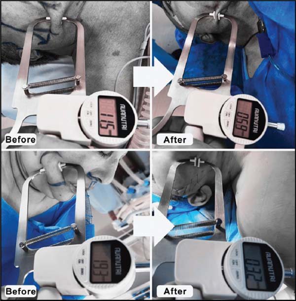
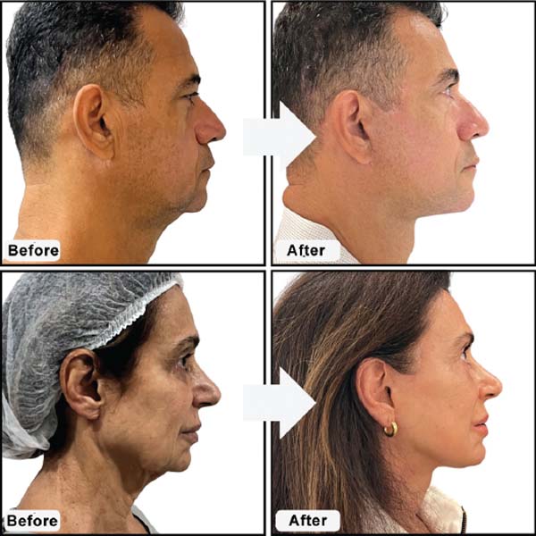
Objective assessment was performed using a skinfold caliper, demonstrating a reduction in the mean thickness of the jowls from 12.08mm to 4.69mm postoperatively.
Results Aesthetic improvement showed a significant correlation with patient satisfaction but not with age, bodymass index, or previous use of dermal fillers. The findings confirm that repositioning of the deep tissue layers, particularly the superficial musculoaponeurotic system (SMAS), is essential for restoring mandibular contour and enhancing facial rejuvenation. Redefinition of the jawline contributes decisively to the harmony of the lower third of the face.
Conclusion It is concluded that effective lifting techniques should consider reorientation of the soft tissues along a superolateral vector. A technique that unfolds, pulls, or elevates the tissues will produce a flattening effect on the structure being treated, and this effect is desirable for the jowls.
Keywords: superficial musculoaponeurotic system; rhytidoplasty; aging; rejuvenation; facelift
RESUMO
Introdução A perda do contorno mandibular é uma das principais razões pelas quais pacientes buscam cirurgia de rejuvenescimento facial. Os jowls, que comprometem essa definição, resultam de gordura acumulada e pele flácida, sendo influenciados por fatores como deslocamento dos compartimentos de gordura e alterações no septo fascial com o envelhecimento.
Métodos Este estudo prospectivo analisou o tratamento dos jowls como uma unidade anatômica em pacientes submetidos ao Deep Plane Face e Neck Lift, com ritidoplastia tipo III de Perkins. A avaliação objetiva foi feita com plicômetro, demonstrando redução média da espessura dos jowls de 12,08mm para 4,69mm no pósoperatório.
Resultados A melhora estética demonstrou correlação significativa com a satisfação dos pacientes, mas não com a idade, o IMC ou o uso prévio de preenchedores. Os achados confirmam que o reposicionamento das camadas profundas, especialmente do SMAS, é essencial para restaurar o contorno da mandíbula e melhorar o rejuvenescimento facial. A redefinição da jawline contribui de forma decisiva para a harmonia do terço inferior da face.
Conclusão Conclui-se que técnicas eficazes de lifting devem considerar a reorientação dos tecidos moles em vetor superolateral. Uma técnica que desdobre, puxe ou levante a região produzirá um efeito de achatamento da estrutura a ser tratada, efeito desejado no caso dos jowls.
Palavras-chave: sistema musculoaponeurótico superficial; ritidoplastia; envelhecimento; rejuvenescimento; lifting facial
Introduction
Loss of mandibular contour is one of the main reasons patients consider facial rejuvenation surgery.
Jowls, or the bulldog effect, arise from aging and change in the functional adaptations of the human mouth. A wide mandibular opening requires hypermobility of the tissues overlying the mandible immediately lateral to the level of oral commissure. This hypermobility over the mandibular insertion of the depressor muscles of the lower lip occurs entirely within the subcutaneous layer, allowing the mandible to move largely independently of the skin. The short, elastic subcutaneous connective tissue that permits this exceptional mobility without laxity during youth becomes elongated with aging, resulting in tissue laxity. The development of subcutaneous and dermal redundancy at this location constitutes the jowls.1
The zygomatic ligaments (McGregor’s patch) anchor the cheek skin to the inferior border of the zygoma, immediately posterior to the origin of the zygomaticus minor muscle. The mandibular ligaments attach the overlying skin to the mandible. Both ligaments constitute obstacles to surgical maneuvers intended to advance the overlying skin. They also restrict the facial skin against gravitational changes and delineate the anterior border of the jowl region.2
The presence of a mandibular ligament was first described by Furnas in his classic 1989 article, “The Retaining Ligaments of the Cheek.” More recent studies have described that the mandibular ligament extends up to 65 mm from the midline and 45 mm from the gonial angle, placing it much farther posteriorly than the anterior border of the jowl. Therefore, the labiomandibular fold is not formed by a ligament but by the direct insertion of the lower lip muscles into the dermis, in the same way that nasolabial fold is formed in the upper lip (►Fig. 1).1, 2, 3
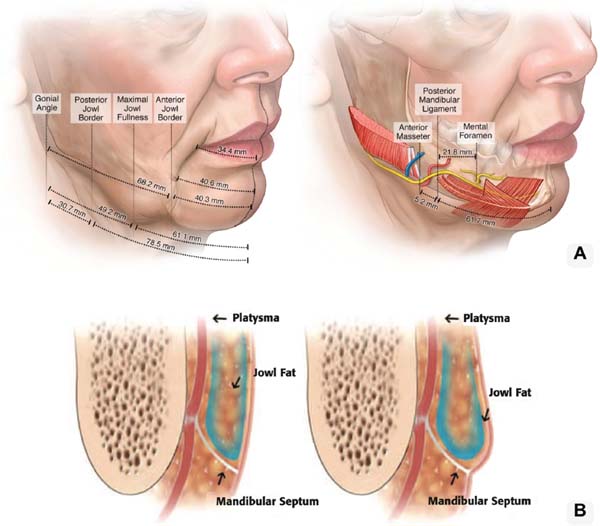
The jowls are formed by the combination of two fat compartments in the region and overlying skin. The interaction between these anatomical subunits and the dynamic forces of aging leads to the bulldog effect (►Fig. 1).1, 3
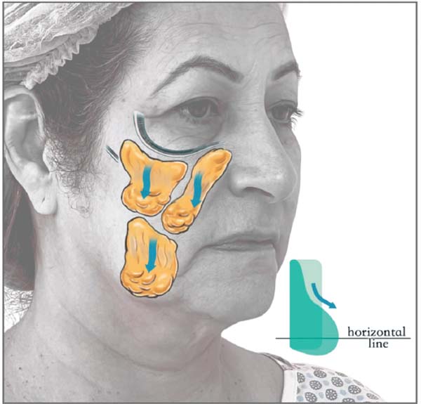
Facial aging is still poorly understood and is theorized to result from tissue deflation over time, displacement of the fat compartments, and septal dehiscence. These theoretical mechanisms may be applied to the jawline: atrophy of the superior and inferior jowl fat pads may reveal jowls present since an earlier age; displacement of the superior and inferior fat compartments and dehiscence of the mandibular septum may allow the development of the jowls and the migration of fat into the neck (►Fig. 2).3
Functional anatomy may explain how the formation of the jowls is related to the need for skin mobility. During youth, the subcutaneous connective tissue is elastic and shorter; with aging, it loses elasticity and becomes elongated, resulting in the secondary subcutaneous laxity that constitutes the jowl. The bulldog effect is greatest where it overlies the most posterior portion of the mandibular ligament because most skin movement over the underlying muscles occurs in this region.1
Several assumptions underlie the estimation of body fat percentage (% body fat) using skinfold calipers. These include: (1) the selection of number and location of the skinfold sites; (2) the relationship between internal and subcutaneous adipose tissue masses; (3) assumptions used in calibrating the technique against densitometry; and (4) the variability in the fat fraction of adipose tissue.4
The most common tool used to measure subcutaneous adipose tissue thickness is a skinfold caliper. In general, skinfold calipers are relatively inexpensive, portable, do not require extensive personnel training for accurate use, are painless, and are noninvasive (Borkan et al., 1982). Skinfold calipers are widely used in screening programs and large-sample studies, for which an extensive body of literature exists (Durnin and Rahaman, 1967; Fletcher, 1962; Keys and Brozek, 1953; Roche and Malina, 1983; Lohman, 1981).5
Interest in skinfold measurements, due to the easy accessibility of the subcutaneous layer and their noninvasive approach, has led to the widespread commercialization and use of skinfold calipers (Martin et al., 1985). Skinfold measurements and the variables derived from them are used in physiology, structural anatomy, endocrinology, kinanthropometry, nutrition, health and fitness, growth, sports, and exercise science. They have specificapplicationsin occupational biomechanics, human hydrodynamics, drug quantification, diabetes, coronary artery disease, hypertension, anorexia nervosa, and numerous epidemiological and human biology studies. The skinfold is a central parameter in the standardization of adipose tissue in studies of fat distribution.5, 6
The lower third of the face is crucial in shaping the overall facial appearance. The lower facial fat compartments tend to undergo selective atrophy or hypertrophy with aging, and apparently both processes may contribute to the development of tissue laxity. The lower facial fat, measured and treated as skinfold, will be evaluated and quantified preoperatively and postoperatively to study fat distribution in the jowl.
Objective
The present study aims to address one of the main concerns of patients seeking facial rejuvenation: the bulldog effect, or jowls. In this study, the jowl was considered a single anatomical unit, like a skin flap, with all its compartments unified to facilitate mobilization and management.
What is the actual postoperative improvement of the jowl? Does previous use of dermal fillers interfere with the outcome? Does body mass index make it more difficult to achieve a good result? Is the level of postoperative patient satisfaction related to previous use of dermal fillers?
Methods
This prospective, uncontrolled interventional study included 12 patients who underwent a combined deep plane face and neck lift during the same surgical procedure between September 2024 and April 2025. All procedures were performed by the author of this article. Patients of both sexes, aged 50 to 65 years, with a body mass index (BMI) ranging from 20.55 to 30.76, presenting with facial rhytides and facial and cervical laxity, and meeting the indication for Perkins type III rhytidoplasty7 (►Table 1), were included, following the jowl treatment algorithm proposed by Edward M. Reece, MD, and Rod J. Rohrich, MD (Scheme 1).3
| Perkins type III rhytidoplasty |
| Predominantly male patients |
| Thick cheeks |
| Marionette lines |
| Loss of mandibular contour |
| Heavy neck with lipoptosis |
| Significant platysmal bands |

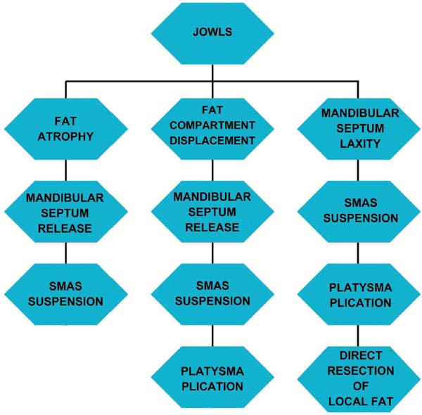
During the procedures, management of the jowl included release of the mandibular septum, unification of the superior and inferior jowl compartments, and release of the retaining ligaments in the jowl region.
Patients with an indication for Perkins type I or type II rhytidoplasty, those who underwent pre-jowl fat grafting (marionette line), those with a previous facelift, those younger than 40 years of age, and those without platysmal bands were excluded from the study.
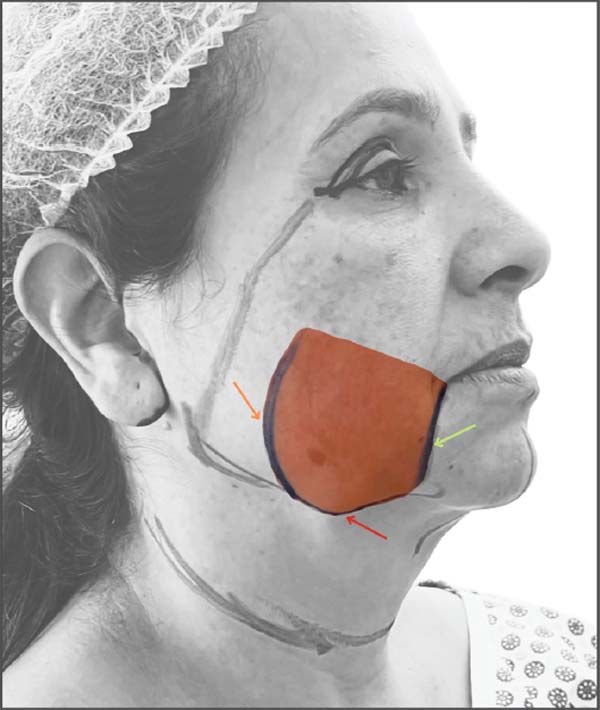
For the quantitative assessment of the jowls, measurements were obtained according to the reference points described by Minelli,1 namely the anterior border of the jowl (labiomandibular fold) and the posterior border of the jowl (anterior border of the masseter muscle). Measurements were taken at the midpoint between these two landmarks, at the lowest and most prominent point, with the patient in the supine position (when the jowl compartments assume their most craniocaudal position) (►Fig. 3). Measurements were obtained using an AVANUTRI Digital Scientific Skinfold Caliper (ANVISA Registration No. 81550810006), model AVA-450, with a sensitivity of 0.1 mm, a measurement range of 85 mm, and a pressure of 10g/mm2 (►Fig. 4).
The statistical analyses were based on standard descriptive statistics (mean, median, and standard deviation) and inferential analysis using the paired Student’s t-test for paired comparisons. Pearson’s correlation coefficient was used to assess the correlation between improvement of the jowl and the quantitative variables of age, BMI, and, in particular, patient-reported satisfaction. Finally, the effect of previous use of dermal fillers on jowl improvement was analyzed using Student’s t-test.
All participants were included after signing an informed consent form.
Surgical Technique
The procedure begins with infiltration of an anesthetic solution containing lidocaine, ropivacaine, epinephrine, tranexamic acid, and normal saline into the face, jowls, and neck. A 4-cm transverse incision is then made in the submental region along the cervical midline, through which dissection and release of the mandibular septum and mandibular ligament are performed, along with unification of the superior and inferior jowl compartments and release of the retaining ligaments defining the boundaries of the jowl. Using curved Metzenbaum scissors, subcutaneous dissection is performed in the labiomandibular fold, the jowl, and the region anterior to the masseter muscle, without extending to the entry point for the deep plane, thereby preserving a multilayered structure in this region to achieve the desired effect of the deep plane facelift (►Video 1).
Video 1
Releasing of the mandibular septum and mandibular ligament being performed
Online content including video sequences viewable at: https://www.thieme-connect.com/products/ejournals/html/10.1055/s-0046-1829145.
Following these maneuvers, the deep plane neck lift is completed with partial resection of the submandibular glands, resection and plication of the anterior digastric muscles, platysma plication, and the Crevasse maneuver. The deep plane facelift is then completed with release of the facial retaining ligaments.
Results
Asignificance level of 0.05 (5%) was established for this study. Therefore, results were considered statistically significant when the p-value was less than the adopted significance level.
The analyses began by comparing jowl measurements between the preoperative and postoperative periods using the paired Student’s t-test. These analyses were performed separately for each side (right and left) as well as for both sides combined, providing greater reliability of the results (►Table 2).
| Mean (mm) | Median (mm) | Standard deviation | CV | Min | Max | N | CI | P-value | ||
|---|---|---|---|---|---|---|---|---|---|---|
| Right jowl | Preop | 12.13 | 11.05 | 2.96 | 24% | 7.90 | 19.30 | 12 | 1.68 | <0.001 |
| Postop | 4.56 | 4.20 | 1.66 | 36% | 2.70 | 7.70 | 12 | 0.94 | ||
| Left jowl | Preop | 12.03 | 11.45 | 3.45 | 29% | 8.10 | 19.60 | 12 | 1.95 | <0.001 |
| Postop | 4.83 | 4.90 | 1.73 | 36% | 2.60 | 8.50 | 12 | 0.98 | ||
| Overall jowl | Preop | 12.08 | 11.25 | 3.15 | 26% | 7.90 | 19.60 | 24 | 1.26 | <0.001 |
| Postop | 4.69 | 4.35 | 1.66 | 35% | 2.60 | 8.50 | 24 | 0.67 | ||

In all three comparisons, there was a statistically significant difference in the mean jowl measurements between the two time points, with a consistent reduction in values. Overall, the mean decreased from 12.08 to 4.69 (p < 0.001) (►Fig. 5).
Because measurements were obtained at two different time points, it was possible to calculate the delta between them, representing the magnitude of the change. The delta was calculated by subtracting the preoperative value from the postoperative value. Positive values indicated an increase in jowl measurements, whereas negative values indicated a reduction. A complete descriptive analysis of the jowl delta was then performed (►Table 3).

Good variability was observed, as the coefficient of variation was below 50%, indicating data homogeneity. The mean reduction was -7.39 ± 1.40 points, indicating that, with 95% statistical confidence, the mean reduction may range from −8.79 to −5.99.
Based on the jowl delta, correlations were assessed with the quantitative variables of age, body mass index, and satisfaction to determine whether the reduction was influenced by any of these factors, using Pearson’s correlation coefficient (►Table 4).
| Jowl delta | ||
|---|---|---|
| Age | Correlation (r) | 0.046 |
| P-value | 0.831 | |
| BMI | Correlation (r) | −0.021 |
| P-value | 0.924 | |
| Satisfaction | Correlation (r) | 0.649 |
| P-value | 0.001 | |

The jowl delta showed a statistically significant correlation only with satisfaction (r = 0.649; p = 0.001). Because this was a positive correlation, higher satisfaction was associated with a greater jowl delta (values closer to zero, since the values were negative).
Finally, Student’s t-test was used to compare the mean jowl delta between patients who had and had not previously received dermal fillers (►Table 5).
| No filler | With filler | |
|---|---|---|
| Mean (mm) | −8.18 | −5.83 |
| Median (mm) | −6.35 | −6.00 |
| Standard deviation | 4.06 | 0.87 |
| Coefficient of variation | 50% | 15% |
| Minimum | −17.00 | −7.10 |
| Maximum | −3.70 | −4.60 |
| N | 16 | 8 |
| Confidence interval | 1.99 | 0.61 |
| P-value | 0.123 | |

No statistically significant effect of previous dermal filler use on the jowl delta was observed. The mean jowl delta was −8.18 among patients who had not received dermal fillers and -5.83 among those who had.
Discussion
Facial aging is a multifactorial process that results in a series of physiological and morphological changes in the facial tissues, including the bones, ligaments, muscles, fasciae, subcutaneous fat, and skin. The onset and rate of age-related changes in each structure vary among individuals, sexes, and ethnicities.8, 9
There is evidence that, with increasing age, total body fat is redistributed as subcutaneous fat shifts to the liver, bone marrow, and various other intra-abdominal and ectopic fat depots. Loss of subcutaneous fat in the face results in volume loss of the superficial and deep facial fat compartments, a concept summarized as the fat deflation theory.8, 9
A well-defined lower third of the face is crucial in shaping the overall facial appearance. Aging of this region is manifested primarily by the development of deformities in the lower cheeks around the oral commissures, the formation of redundant skin folds near the labiomandibular fold, and the loss of definition of the jawline.10
According to the volume restoration theory, the lower facial fat compartments tend to undergo selective atrophy or hypertrophy with aging, and, apparently, both processes may contribute to the development of tissue laxity. The lower facial fat has been divided into the jowl fat compartment, the labiomandibular fat compartment, and the chin fat compartment.10
The results demonstrate that resizing and redistributing the jowl compartments and layers restore the mandibular contour by compensating for deformities that have developed and for displaced and/or atrophied volumes.
Studies have shown statistically significant differences in the inferior segment of the superficial jowl fat compartment among young, middle-aged, and older individuals, demonstrating a tendency toward increased thickness with aging. However, no statistically significant differences in thickening were observed among the compartments across any age group. Similar changes have been observed in the deep jowl fat, which also showed a tendency toward increased thickness with aging.10
The same findings were observed in the present study, in which age and BMI showed no statistically significant association with the postoperative measurements.
Reece et al. and Braz et al. reported thickening of both the deep and superficial jowl compartments with aging. These findings are consistent with clinical observations and with the findings of Schenck et al., who suggested that descent and hypertrophy of the superficial and deep jowl fat compartments are the main causes of aging in the lower face.10 These findings suggest that the anterior and inferior portions of the jowls are key target areas for improvement in lower facial rejuvenation.
Facial attractiveness affects self-esteem and the way we are judged by others, and trait judgments based on facial appearance occur in as little as 100 ms. Dissatisfaction with facial appearance, particularly the appearance of the chin, neck, and jawline, increases with age. A well-defined mandibular border without prominent jowls is a hallmark of a youthful neck. Jowl fat atrophies with age, contributing to a square facial shape and loss of jawline definition. The jawline is therefore an essential component of neck rejuvenation, restoring a square face to a more youthful oval shape and enhancing facial definition.11
Atrophy of the subcutaneous tissue may reveal underlying jowls and is associated with thin, lax skin and reduced subcutaneous fat in the jowl region. Descent of the midface further worsens the appearance of the lower face secondary to ptosis of the skin and soft tissues cranial to the jowl. In addition, mild to moderate mandibular bone atrophy leads to deepening of the prejowl sulcus and ptosis of the overlying soft tissues, resulting in the appearance of prominent jowls.11
Facial aging is a dynamic process involving aging of both the soft tissues and the underlying bony structures. Much is known about how the face loses volume as the soft tissue structures age. Epidermal thinning and decreased collagen cause the skin to lose its elasticity. Fat loss, combined with gravity and muscle traction, leads to wrinkling and the formation of dynamic lines. Aging represents a transition from youth, in which there is an ideal relationship between the bony framework and the volume and surface area of the soft tissue envelope, to an imbalance between these components that results in the stigmata of the aging face. The anatomy of the facial soft tissues may be described as a series of layers: skin, subcutaneous fat (superficial and deep layers), superficial fascia and the muscles of facial expression, deep facial fascia (parotid-masseteric fascia), the facial nerve, and the parotid gland. As collagen and elastic fibers break down, the skin becomes laxer, and wrinkles and folds develop.12, 13
The effect of gravity on the lax skin envelope and atrophied soft tissues leads to the development of folds. This understanding has made soft tissue reconstruction with malar fat suspension and superficial musculoaponeurotic system (SMAS) tightening the predominant approach to rejuvenating the aging face. Restoration of the facial contour is significantly influenced by re-elevation of the descended facial fat through elevation and manipulation of the SMAS. The mandible forms the foundation of the lower face, and any change in its projection, width, or height may affect the overall facial aesthetics.12, 13
In current clinical practice, many patients have undergone multiple facial procedures and filler treatments to compensate for facial laxity and atrophy. Surprisingly, this study showed no statistically significant difference in jowl improvement between patients with and without previous facial fillers.
Facelifts generally redistribute skin laxity in two dimensions: vertically and laterally, or vertically and superolaterally. This two-dimensional facelift results from the combination of subperiosteal or supraperiosteal facelift techniques with subcutaneous rhytidectomy or SMAS elevation.14
Although many of these techniques can reposition lax soft tissues into a more favorable position, providing an apparent increase in anteroposterior (AP) projection, this is not achieved uniformly throughout the face, making additional techniques, such as adjunctive maneuvers, necessary to increase the AP dimension in specific areas. Conceptually, this three-dimensional approach has not been integrated into our routine way of thinking about restoring or enhancing the dimension lost during facial rejuvenation surgery.14
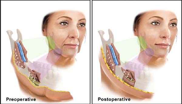
Soft tissue laxity during aging occurs in an inferior and inferomedial direction. The ideal reorientation of the soft tissues during facial rejuvenation is in the opposite direction: vertically and superolaterally. In principle, any technique that unfolds, pulls, or lifts the tissues will produce a flattening effect on the structure being treated. As previously mentioned, some treatments may increase the AP dimension, such as augmentation of cheek volume during a sub-SMAS or subperiosteal composite facelift, or when the malar fat is repositioned during a multiplane facelift. This occurs because the thickest portion of the advanced tissue is placed over a bony prominence. However, by definition, the absolute thickness of the soft tissue structure being advanced must decrease because it has been stretched (►Fig. 6).14
Conclusion
Rejuvenation of the facial contour is significantly influenced by re-elevation of the descended facial fat through elevation and manipulation of the SMAS. The mandible forms the foundation of the lower face, and any change in its projection, width, or height may affect the overall facial aesthetics. It is concluded that effective lifting techniques should consider reorientation of the soft tissues along a superolateral vector. A technique that unfolds, pulls, or lifts the tissues will produce a flattening effect on the structure being treated, an effect that is desirable for the jowls.
References
1. Minelli L, Yang HM, van der Lei B, Mendelson B. The surgical anatomy of the jowl and the mandibular ligament reassessed. Aesthetic Plast Surg 2023;47(01):170–180.. Doi: 10.1007/s00266-022-02996-3
2. Furnas DW. The retaining ligaments of the cheek. Plast Reconstr Surg 1989;83(01):11–16. Doi: 10.1097/00006534-198901000-00003
3. Reece EM, Rohrich RJ. The aesthetic jaw line: management of the aging jowl. Aesthet Surg J 2008;28(06):668–674. Doi: 10.1016/j.asj.2008.09.007
4. Martin AD, Drinkwater DT, Clarys JP, Daniel M, Ross WD. Effects of skin thickness and skinfold compressibility on skinfold thickness measurement. Am J Hum Biol 1992;4(04):453–460. Doi: 10.1002/ajhb.1310040404
5. Chumlea WC, Roche AF. Ultrasonic and skinfold caliper measures of subcutaneous adipose tissue thickness in elderly men and women. Am J Phys Anthropol 1986;71(03):351–357. Doi: 10.1002/ajpa.1330710310
6. Clarys JP, Martin AD, Drinkwater DT, Marfell-Jones MJ. The skinfold: myth and reality. J Sports Sci 1987;5(01):3–33. Doi: 10.1080/02640418708729760
7. Papel ID, et al. Facial plastic and reconstructive surgery. 4th ed. New York: Thieme; 2016. Chapter 14; p. 139–158
8. Eid L, Mao X, Zhao B, et al. Fat compartment gliding theory - a novel technique for the repositioning of superficial fat compartments for facial rejuvenation. Clin Cosmet Investig Dermatol 2023;16:3077–3090. Doi: 10.2147/CCID.S415467
9. Cotofana S, Koban KC, Konstantin F, et al. The Surface-volume coefficient of the superficial and deep facial fat compartments: a cadaveric three-dimensional volumetric analysis. Plast Reconstr Surg 2019;143(06):1605–1613. Doi: 10.1097/PRS.0000000000005524
10. Guo Y, Wen L, Wang X, et al. Analysis of age-related changes in lower facial fat compartments and of the course of blood vessels using computed tomography. Plast Reconstr Surg 2024;153(03):539e–548e. Doi: 10.1097/PRS.0000000000010506
11. Shridharani SM. Novel Surface Anatomic Landmarks ofthe Jowl to Guide Treatment with ATX-101. Plast Reconstr Surg Glob Open 2019;7(10):e2459. Doi: 10.1097/GOX.0000000000002459
12. Kahn DM, Shaw RB. Overview of current thoughts on facial volume and aging. Facial Plast Surg 2010;26(05):350–355. Doi: 10.1055/s-0030-1265024
13. Donofrio L, Weinkle S. The third dimension in facial rejuvenation: a review. J Cosmet Dermatol 2006;5(04):277–283. Doi: 10.1111/j.1473-2165.2006.00270.x
14. Ramirez OM. Full face rejuvenation in three dimensions: a “facelifting” for the new millennium. Aesthetic Plast Surg 2001;25(03): 152–164. Doi: 10.1007/s002660010114
1. Moresco Clinic, Curitiba, Paraná, Brazil
Address for correspondence Yuri Moresco de Oliveira, MD, Av. Visconde de Guarapuava, 4420, Batel, Curitiba, PR, 80240-010, Brazil (e-mail: dryuri@clinicamoresco.com.br).
Article received: January 29, 2026.
Article accepted: April 10, 2026.
The author has no conflict of interests to declare.
Editor-in-Chief: Dov Charles Goldenberg
 Read in Portuguese
 Read in English
 PDF PT
 Print
 Send this article by email
 How to Cite
 Mendeley
 Pocket
 Facebook
 Twitter