


Original Article - Year 2012 - Volume 27 -
Scalp reconstruction procedures
Métodos de reconstrução do couro cabeludo
ABSTRACT
Scalp avulsion is a devastating injury that affects the patient esthetically, functionally, and psychologically. The advent of microsurgery and the first reimplant performed by Miller in 1976 improved the treatment of scalp avulsion, making reimplant the first choice whenever possible. With modern techniques and the equipment available in specialized centers, scalp avulsion can be adequately treated using microsurgical reimplantation. This method achieves better results as compared to previously employed procedures. We report the reconstruction of the scalp of a 4-year-old boy after the avulsion of approximately two-thirds of the scalp area due to a dog bite. We focused on covering the skull with microsurgical latissimus dorsi and serratus muscles, and performing subsequent surgeries to completely cover the alopecic areas.
Keywords: Scalp/surgery. Microsurgery. Surgical flaps.
RESUMO
A avulsão de couro cabeludo apresenta-se como lesão devastadora aos pacientes acometidos, nas esferas tanto estética e funcional como psicológica. O advento da microcirurgia e a realização do primeiro reimplante por Miller, em 1976, revolucionaram o manejo da avulsão de couro cabeludo, tornando o reimplante o tratamento de escolha sempre que possível. Com as técnicas atuais e a estrutura disponível nos centros especializados, faz-se possível o adequado manejo das avulsões de couro cabeludo por meio do reimplante microcirúrgico, obtendo-se resultados adequados e superiores às opções oferecidas previamente. Os autores abordam a reconstrução do couro cabeludo após avulsão de aproximadamente dois terços de sua extensão, causado por mordedura de cão em uma criança de 4 anos de idade, enfocando a cobertura da calota craniana com retalho microcirúrgico dos músculos grande dorsal e serrátil e as sucessivas cirurgias até a cobertura total da área de alopecia.
Palavras-chave: Couro cabeludo/cirurgia. Microcirurgia. Retalhos cirúrgicos.
The coverage of the skull after scalp avulsion has always been a great challenge for plastic surgeons because reconstruction cannot be performed using local flaps. Furthermore, features including the age of the patient, head circumference, and skull thickness need to be evaluated.
Although scalp avulsion is rare, it can have serious repercussions depending on the mechanism of injury as well as psychological and social consequences1,2. These injuries mostly occur in work environments as a result of workers not wearing adequate protective gear; in female patients, this injury is often caused because of accidents related to their comparatively longer hair.
The affected area may be limited to the scalp or might involve other regions such as the forehead, eyebrows, ears, and lower parts of the face, thus rendering reconstruction more difficult.
Regarding the depth of the avulsion, the most common procedure is removal of the subgaleal region. However, in cases that involve great traction, subperiosteal damage and even the avulsion of skull parts have been observed, which are correlated with traumatic brain injuries1.
The definitive treatment for scalp avulsion underwent major changes after the improvement and standardization of microsurgical reimplantation techniques. In 1976, Miller et al.2 reported the first successful case of microsurgical reimplantation, thus encouraging the use of this procedure, which eventually became the treatment of choice. Other therapeutic options were reserved for cases in which treatment failed or as ancillary procedures.
Until now, the global experience with microsurgical scalp reimplants was confined to 3 or 4 dozen isolated case reports as well as small groups of studies. This has rendered it difficult to standardize the therapeutic response and success rates3,4.
Treatment success depends on several factors including ischemia time, the quality of the avulsion fragment, and the experience level of the surgeon5. The high probability of failure poses a challenging situation for the surgeon since there is often no remaining scalp and a large exposed area to be covered.
In this report, after an unsuccessful attempt at reimplantation, we performed a microsurgical transplant of the latissimus dorsi muscle with the first 4 digitations of the serratus to cover the skull.
CASE REPORT
The patient was a 4-year-old male who was a victim of a dog bite that resulted in the loss of approximately two-thirds of the scalp, including the whole pericranium.
The patient was admitted to the hospital with his scalp preserved under hypothermic conditions; there were many debris and many injuries derived due to lesions to the galea aponeurotica.
After clinical and neurological evaluation, the patient was taken to the surgical department 10 hours after the accident in an attempt to perform scalp reconstruction. Microanastomosis of the artery and superficial temporal vein was carried out, and perfect revascularization of the scalp was achieved. However, the flap became infected, resulting in arteriovenous thrombosis.
After stabilization of the general condition of the patient, regarding the debridement of devitalized tissue and antibiotic therapy, skull coverage by microsurgical transplantation of the latissimus dorsi muscle with the first 4 digitations of the serratus was proposed (Figure 1).

Figure 1 - Flap of the latissimus dorsi and the first digitation of the serratus.
The transplantation triggered venous thrombosis (Figure 2); 12 hours after surgery, the patient underwent a revision of the anastomosis with the realization of a bypass from the thoracodorsal vein to the external jugular vein.
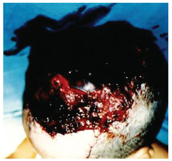
Figure 2 - Flap venous thrombosis.
The flap developed efficiently, although necrosis of approximately 30% was detected in its extension. The external wall of the skull was subjected to bone scraping (Figure 3). Furthermore, after the growth of granulation tissue, the entire wound was grafted with partial skin (Figure 4).

Figure 3 - Bone scraping of the external wall of the skull.

Figure 4 - Partial skin graft.
Six months after being discharged from the hospital, the patient underwent the first expansion of the remaining hairy area to cover one third of the alopecic area (Figure 5). After 5 years, 3 more expansions were carried out, and complete coverage of the alopecic area was ultimately achieved.

Figure 5 - Coverage of the skull after expansion of the hairy area.
DISCUSSION
Reimplantation is the treatment of choice for scalp lesions and should always be considered, even in cases of severe scalp damage. This procedure is potentially indicated in all cases due to its associated benefits. The specific procedures regarding pre-, intra-, and post-operative care are crucial for the success of the reimplantation, including the time of ischemia, scalp conservation, hemodynamic stabilization of the patient, cleaning, local debridement, and the use of antibiotics.
When reimplantation is not possible, the microsurgeon should strive not only for local coverage, but good esthetic results as well. The scalp tissue is the best replacement for the scalp; no other donor site possesses the same qualities of scalp tissue. A variety of techniques have been used to close these defects as follows:
Primary closure: This is the best option for small defects. Defects < 3 cm in diameter can be primarily closed; however, this varies by location. If primary closure is selected, any defect in the galea should be primarily closed with absorbable sutures and the skin edges should be re-approximated with sutures or staples. Skin graft and tissue expansion: Placing skin grafts can provide fast and effective closure of the defect. Skin grafts require properly vascularized tissue and are unsuccessful if applied directly to exposed bone. It is possible to perform bone trepanation and wait for local granulation. The intact pericranium is generally sufficient to support skin grafts. Tissue expansion generally provides broad tissue while preserving the sensitivity, color, and thickness of the scalp and hair; however, this ultimately requires a minimum of 2 surgical procedures. Patients should be told in advance that this procedure requires a commitment of at least 1-2 years2,3. Local flaps: These are preferred for the reconstruction of small- to medium-sized scalps. These flaps consist of skin, subcutaneous tissue, and the galea; however, small surface defects can occasionally be adequately reconstructed using the subcutaneous plane. For large flaps, the donor area is covered with skin graft6. Free flaps: Before the advent of microsurgery, the closing of scalp defects covering more than 15-20% of the total area was virtually impossible with a single procedure. However, as free flaps involve at least some donor site morbidity2, they should be reserved for appropriate situations when skin, local flaps, graft, and healing by secondary intention cannot be considered. They can also provide better results and scarring in the area corresponding to previous radiation exposure or infection.
There are several techniques for covering the skull. As a principle, more simple reconstruction procedures should be performed followed by more complex techniques. Each case must be evaluated individually, and the best method for reconstruction should be chosen after taking into account morbidity, available resources, and acceptable esthetic results.
CONCLUSIONS
Microsurgical reimplantation should be the preferred method for treatment of scalp avulsion because of its unquestionable superiority as compared to other therapeutic options as well as its esthetic and psychological benefits.
REFERENCES
1.Nahai F, Hester TR, Jurkiewicz MJ. Microsurgical replantation of the scalp. J Trauma. 1985;25(9):897-902.
2. Miller GD, Anstee EJ, Snell JA. Successful replantation of an avulsed scalp by microvascular anastomoses. Plast Reconstr Surg. 1976;58(2):133-6.
3.Van Beek AL, Zook EG. Scalp replantation by microsurgical revascularization: case report. Plast Reconstr Surg. 1978;61(5):774-7.
4. Buncke HJ, Rose EH, Brownstein MJ, Chater NL. Successful replantation of two avulsed scalps by microvascular anastomoses. Plast Reconstr Surg. 1978;61(5):666-72.
5. Cheng K, Zhou S, Jiang K, Wang S, Dong J, Huang W, et aI. Microsurgical replantation of the avulsed scalp: report of 20 cases. Plast Reconstr Surg. 1996;97(6):1099-106.
6. Svensson H, Njalsson T. Microsurgical replantation of partial avulsion of the scalp. Case report. Scand J Plast Reconstr Surg Hand Surg. 1995;29(2):177-80.
1. Plastic surgery resident medical doctor of the Santa Casa de Campo Grande, Campo Grande, MS, Brazil.
2. Plastic surgeon, full member of the Sociedade Brasileira de Cirurgia Plástica/Brazilian Society of Plastic Surgery (SBCP), preceptor at the Plastic Surgery Service of the Santa Casa de Campo Grande, Anatomy Assistant Professor at the Morphophysiology Department of the Universidade Federal de Mato Grosso do Sul (Federal University of Mato Grosso do Sul), Campo Grande, MS, Brazil.
3. Plastic surgeon, expert member of SBCP, member of the Plastic Surgery Service of the Santa Casa de Campo Grande, Campo Grande, MS, Brazil.
Rafael Anache Anbar
Av. Primeiro de Maio, 673 - Jardim São Bento
Campo Grande, MS, Brazil - CEP 79004-620
E-mail: raanbar@hotmail.com
Submitted to SGP (Sistema de Gestão de Publicações/Manager Publications System) of RBCP (Revista Brasileira de Cirurgia Plástica/Brazilian Journal of Plastic Surgery).
Article received: April 2nd, 2010
Article accepted: May 16, 2010
Study conducted at the Plastic Surgery Service of the Santa Casa de Campo Grande, Campo Grande, MS, Brazil.
 Read in Portuguese
 Read in English
 PDF PT
 Print
Print
 Send this article by email
 How to Cite
 Mendeley
 Pocket