


Case Reports - Year 2012 - Volume 27 -
Collumela reconstruction in a patient with necrosis resulting from nasogastric tube
Reconstrução de columela após necrose por uso de sonda nasogástrica
ABSTRACT
The present report describes the case of a patient with sequelae from a nasogastric tube used for a prior procedure performed during childhood. The reconstruction required 2 separate surgical procedures. The first surgery involved reconstruction of the columella with nasogenian flaps rotated upwards and the second procedure consisted of osteotomy and septoplasty.
Keywords: Rhinoplasty. Nose/surgery. Necrosis.
RESUMO
Os autores apresentam o caso de paciente portador de sequela de sonda nasogástrica utilizada para procedimento na infância. A reconstrução foi realizada em dois tempos cirúrgicos. No primeiro tempo cirúrgico, foi realizada reconstrução da neocolumela com retalhos nasogenianos rodados de baixo para cima. No segundo tempo, foram realizados osteotomia e ortosseptoplastia.
Palavras-chave: Rinoplastia. Nariz/cirurgia. Necrose.
"We restore, repair and make a whole with
parts that were created by nature and removed
by fate... not to delight the eyes, but to praise the
spirit and help the mind of the afflicted."
Gaspare Tagliacozzi
Historical
Nose reconstruction has been performed since ancient times. The first references appear among the Egyptians in 2200 BC and there are records of this surgery performed in India in 2000 BC (Indian flap).
Hippocrates, in 500 BC, described a procedure for nose reduction with immobilization of fractures. During the Renaissance, a major outbreak of leprosy and syphilis resulted in frequent nose defects, which led to the development of techniques for nose reconstruction with arm flaps (Tagliacozzi/Branca).
In 1845, Dieffenbach, in his book Operative Chirurgie, madeample references tonasalreconstruction. Morerecently, Gillies and Millard, in 1957, proposed the U-shaped frontal flap, which in 1959 was extended to the base of the nose by Converse.
Since then, several rhinoplasty techniques have been described18.
Surgical Anatomy
In nasal reconstruction, the vascular system of the face, and in particular the nose, is an important anatomical consideration9,10. Among the main vascular trunks, the most relevant are the sphenopalatine, the greater palatine arteries, the infraorbital and angular arteries, the upper lip, the lateral nasal artery, the anterior and posterior ethmoid arteries, and the dorsal artery.
CASE REPORT
A 24-year-old Caucasian male, resident of São Paulo, had a history of hospitalization in an intensive care unit (ICU) 23 years ago and treatment with a nasogastric tube, which resulted in the development of columella necrosis. The patient presented with absent columella and nasal tip and a short nose (Figure 1).
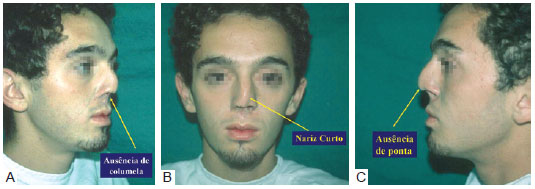
Figure 1 - Pre-operative appearance. In A, half right profile, absence of columella. In B, frontal view, short nose. In C, left profile, absence of the tip.
The reconstruction was performed in 2 separate surgical procedures. The first procedure used bilateral nasogenian rotating flaps with a superior pedicle rotated upwards (Figure 2).
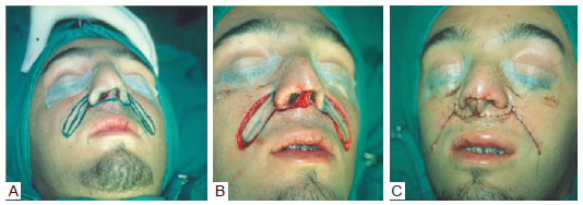
Figure 2 - Transoperative appearance. In A, delimitation. In B, flap production. In C, flap rotation.
Figure 3 shows the temporary result 3 days after surgery.

Figure 3 - Immediate post-operative appearance showing the temporary result after the first surgical procedure. In A, half right profile. In B, frontal view. In C, chin-nose.
In the second surgical procedure, osteotomy and septoplasty were performed to improve the dorsum/tip relationship.
Figure 4 shows the final result after the 2 surgeries.
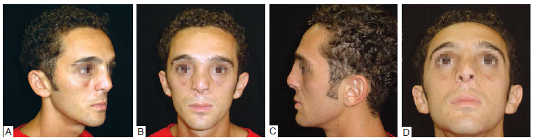
Figure 4 - Immediate post-operative appearance. In A, half right profile. In B, frontal view. In C, left profile. In D, chin-nose.
DISCUSSION
Several possibilities were considered for the reconstruction of the nose; this included nasal base and frontal flaps as well as composite grafts. After considering all options, the recommendation was to reconstruct the nose in 2 separate surgical procedures. The first surgery consisted of the use of nasogenian flaps for the reconstruction of the columella and the second procedure included a rhinoplasty with osteotomy and orthopositioning of the nasal pyramid.
CONCLUSIONS
The surgical result obtained was aesthetically satisfactory and the physiological function of the nose was preserved.
REFERENCES
1. Schaupp H. Submental hair-bearing skin flap to upper lip. In: Strauch B, Vasconez LO, Hall-Findlay EJ, eds. Grabb's encyclopedia of flaps. Boston: Little, Brown and Company; 1990. p. 647-8.
2. Tobin GR. Functional lower lip and oral sphincter reconstruction with innervated depressor anguli oris flaps. In: Strauch B, Vasconez LO, Hall-Findlay EJ, eds. Grabb's encyclopedia of flaps. Boston: Little, Brown and Company; 1990. p. 665.
3. Kernahan DA. Reconstruction of the nose. In: Grabb W, Smiths J, eds. Plastic surgery: a concise guide to clinical practice. 2nd ed. Boston: Little, Brown and Company; 1973.
4. Metzenbaum M. Replacement of the lower end of the dislocated septal cartilage versus submucous resection of the dislocated end of the septal cartilage. Arch Otolaryngol HNS. 1929;9:282-311.
5. Seltzer AP. The nasal septum: plastic repair of the deviated septum associated with a deflected tip. Arch Otolaryngol. 1944;40:443-4.
6. Cottle MH, Loring RM. Surgery of the nasal septum: new operative procedures and indications. Ann Otol Rhinol Laryngol. 1948;57(3):705-13.
7. Cottle MH, Loring RM, Fischer GG, Gaynon IE. Themaxila-premaxila approach to extensive nasal septum surgery.AMA Arch Otolaryngol. 1958;68(3):301-13.
8. Cutting C, Grayson B, Brecht L, Santiago P, Wood R, Kwon S. Presurgical columellar elongation and primary retrograde nasal reconstruction in one-stage bilateral cleft lip and nose repair. Plast Reconstr Surg. 1998;101(3):630-9.
9. Lockhart RD. Anatomia humana. Cidade do México: Editorial Interamericana; 1965.
10. Sabino Neto M. Anatomia do músculo depressor do ângulo da boca [dissertação de mestrado]. São Paulo: Universidade Federal de São Paulo, Escola Paulista de Medicina; 1995. p. 71.
1. Chief physician of the Serviços Oficiais de Ensino pós-graduado, MEC-SBCP, Hospital Ipiranga (Integrated Services of Plastic Surgery, Official Services of Post-Graduate Education, MEC-SBCP, Ipiranga Hospital), São Paulo, SP, Brazil.
2. Regent of the Serviços Oficiais de Ensino pós-graduado, MEC-SBCP, Hospital Ipiranga (Integrated Services of Plastic Surgery, Official Services of Post-Graduate Education, MEC-SBCP, Ipiranga Hospital), São Paulo, SP, Brazil.
3. Training supervisor of the Serviços Oficiais de Ensino pós-graduado, MEC-SBCP, Hospital Ipiranga (Integrated Services of Plastic Surgery, Official Services of Post-Graduate Education, MEC-SBCP, Ipiranga Hospital), São Paulo, SP, Brazil.
Correspondence to:
Aymar Sperli
Avenida Açocê, 174 - Moema
São Paulo, SP, Brazil - CEP 04075-020
E-mail: aymar.sperli@uol.com.br
Article submitted to SGP (Sistema de Gestão de Publicações/Manager Publications System) of RBCP (Revista Brasileira de Cirurgia Plástica/Brazilian Journal of Plastic Surgery).
Article received: January 22, 2010
Article accepted: May 20, 2010
This work was performed at the Serviços Integrados de Cirurgia Plástica, Serviços Oficiais de Ensino pós-graduado, MEC-SBCP, Hospital Ipiranga (Integrated Services of Plastic Surgery, Official Services of Post-Graduate Education, MEC-SBCP, Ipiranga Hospital), São Paulo, SP, Brazil.
 Read in Portuguese
 Read in English
 PDF PT
 Print
Print
 Send this article by email
 How to Cite
 Mendeley
 Pocket