


Original Article - Year 2012 - Volume 27 -
Development of an experimental model of degloving injury in rats
Desenvolvimento de modelo experimental de avulsão de retalhos em membros inferiores de ratos
ABSTRACT
BACKGROUND: The purpose of this study was to develop an experimental model for degloving injuries of the hind limbs of rats and observe flap viability after its relocation to the wound bed to better study the changes related to the injury and to test the therapeutic modalities in avulsed flaps.
METHODS: Ninety male Wistar rats were divided into 4 experimental groups. A flap avulsion model on the lower limb of a rats was established, using 4 different pedicles: proximal flow pedicled flap (G1), distal flow pedicled flap (G2), lateral flow pedicled flap (G3), and medial flow pedicled flap (G4).
RESULTS: Comparison between the mean necrotic area of the degloved flap showed statistically significant differences among the 4 groups (P < 0.0001).
CONCLUSIONS: The group with the distal flow pedicled flap (G2) showed a higher necrotic area relative to the total flap area and it is the most suitable group for testing therapeutic agents in avulsed flaps.
Keywords: Wounds and injuries. Lower extremity. Skin/injuries. Necrosis/etiology. Animal models.
RESUMO
INTRODUÇÃO: Os ferimentos descolantes de membros inferiores geralmente se caracterizam como lesões graves e apresentam dificuldades na decisão quanto ao tratamento cirúrgico mais adequado a ser instituído, se reposicionamento do retalho avulsionado ao leito da ferida ou ressecção do retalho, seguido de seu adelgaçamento e enxertia de pele. O propósito deste estudo foi desenvolver um modelo experimental de avulsão de retalhos cutâneos em membros inferiores de ratos e observar a viabilidade do retalho após seu reposicionamento ao leito de origem, com a finalidade de melhor estudar as alterações relacionadas ao ferimento e de testar modalidades terapêuticas em retalhos avulsionados.
MÉTODO: Foram utilizados 90 ratos Wistar machos, subdivididos em 4 grupos experimentais. Foi delineado um modelo de avulsão de retalhos no membro inferior do rato, baseado em 4 pedículos diferentes: pedículo de fluxo proximal (G1), pedículo de fluxo distal (G2), pedículo de fluxo lateral (G3) e pedículo de fluxo medial (G4).
RESULTADOS: A comparação entre as médias de área de necrose do retalho desenluvado evidenciou diferença estatística significativa entre os 4 grupos estudados (P < 0,0001).
CONCLUSÕES: O grupo com pedículo de fluxo distal (G2) apresentou maior área de necrose em relação à área total do retalho, sendo o mais adequado para testar agentes terapêuticos no retalho avulsionado.
Palavras-chave: Ferimentos e lesões. Extremidade inferior. Pele/lesões. Necrose/etiologia. Modelos animais.
Degloving injuries of the lower limbs are generally considered serious, and choosing the most appropriate surgical treatment is often difficult. Choices include repositioning of the avulsed flap to the wound bed or flap resection followed by thinning and skin grafting1.
In large urban centers, there has been an increase in this type of injury due to the increased occurrence of high-energy traumas (including being hit by a truck or bus and motorcycle accidents2 - the 2 main causes of degloving injuries).
This study was partially presented as a pilot project in this journal in the Ideas and Innovations section in 2011 3. In the present report, we describe the complete development of the experimental model.
The aim of this study was to develop an experimental model of avulsed skin flaps of the lower limbs of rats. We aimed to observe flap viability after its relocation to the bed of origin to better study the injury-related changes and test different therapeutic modalities for avulsed flaps.
METHODS
The model presented here is part of an experimental project to evaluate therapeutic measures for degloving injuries of the lower limb in rats and was approved by the Research Project Committee of the Hospital das Clínicas, Faculty of Medicine, University of São Paulo (number 0842/09). This study followed the guidelines of the following entities for conducting the experimental research: Council for International Organization of Medical Sciences, Ethical Code for Animal Experimentation, and the Brazilian College of Animal Experimentation.
Ninety male Wistar rats were divided into 4 experimental groups (Figures 1 and 2). A model of avulsed flaps on the hind limb of a rat was established, using 4 different pedicles:
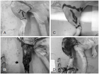
Figure 1 - In A and B, flap with pedicle of proximal flow (G1). In C and D, flap with pedicle of distal flow (G2).
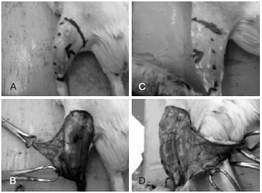
Figure 2 - In A and B, flap with pedicle of lateral base (G3). In C and D, flap with pedicle of medial base (G4).
G1 - 22 animals with a proximal flow pedicled flap; G2 - 24 animals with a distal flow pedicled flap; G3 - 22 animals with a lateral based flap; and G4 - 22 animals with a medial based flap.
After the skin incision, the base of the pedicle was maintained according to the operating group. Flap avulsion was performed using moderate progressive traction through 4 hooks attached to the flap edges, with sufficient force to ensure complete detachment of the bed right at the flap base. After 5 minutes, the flap was repositioned and sutured to the bed of origin.
The rats were observed daily for 7 days for signs of flap necrosis (Figure 3); thereafter, they were euthanized with a lethal dose of thiopental.
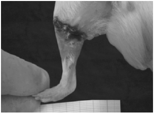
Figure 3 - Partial flap necrosis.
The necrotic area and the total flap area were measured after complete resection of the avulsed flap. The flap was positioned on the operating table and photographed (Figure 4). The pictures were analyzed with the aid of ImageJ software (National Institute of Mental Health, USA)4 to calculate the area. The total flap area (cm2), necrotic area (cm2), and relationship among the areas (percentage) were then determined.
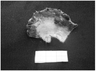
Figure 4 - Flap positioned on the surgical table for posterior quantification of the necrotic and total flap areas with the aid of the ImageJ program.
Statistical analysis was performed using the Kruskal-Wallis non-parametric test for independent samples among the 4 groups. The Dunn test of multiple comparisons was used to assess differences between paired groups. The significance level was 95% (P < 0.05). Statistical analysis was performed using the program Prism 4b for Macintosh, Version 4.0 (GraphPad Software, Inc, San Diego, CA, USA). Exclusion criteria included postoperative death and partial flap autophagy.
RESULTS
After animals that experienced flap autophagy or postoperative death were excluded, 17 rats remained in G1 and G2, 15 remained in G3, and 16 remained in G4. G1 (n = 17) had a mean total flap area of 12.41 cm2, an average necrotic area of 0.51 cm2, and a mean ratio between the necrotic area and the total area of 0.041 (4.1%). In G2 (n = 17), the mean total flap area was 5.63 cm2, the average necrotic area was 3.64 cm2, and the mean ratio between the necrotic area and the total area was 0.39 (39%). G3 (n = 15) had a mean total flap area of 3.88 cm2, a mean necrotic area of 0.39 cm2, and a mean ratio between the necrotic and total area of 0.09 (9%). In G4 (n = 16), the mean total flap area was 4.25 cm2, the average necrotic area was 0.75 cm2, and the mean ratio between the necrotic area and the total area was 0.08 (8%).
Comparisons of means showed statistically significant differences among the 4 groups (P < 0.0001) (Figure 5). Dunn's test showed significant differences between pairs G1 and G2, G2 and G3, and G2 and G4 (P < 0.05). There was no significant difference detected between G1 and G3, G1 and G4, and G3 and G4 (P > 0.05).
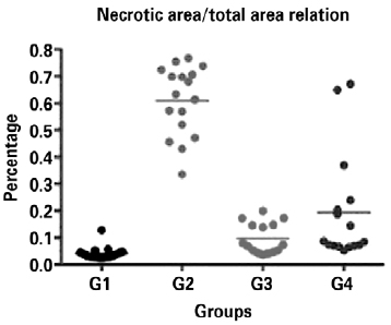
Figure 5 - Relationships between the necrotic area and the total flap area in the 4 groups.
DISCUSSION
Simple repositioning of an avulsed flap in clinical practice usually results in total or partial necrosis of the repositioned tissue5. It is important to develop an experimental model of flap avulsion that can be used to test different therapeutic modalities to improve the outcome of the repositioned flap.
Three models of flap avulsion in the rat tail have been described6-8; however, none of these models have been established in the hind limb. We believe that it is important to develop a flap avulsion model on the hind limb of rats since lower limb injuries are most commonly seen in clinical practice in trauma centers. Furthermore, with the use of 4 different flap orientations, we observed that distal flow flaps are the most severely affected and have worse outcomes.
The distal flow avulsion model (G2) represents a more serious injury thantheproximal,medial, or lateralflow models. This type of injury involves a greater degree of ischemia and congestion that results in a more extensive necrotic area. Thus, we suggest that the distal flow pedicle (G2) model should be used to test drugs that have the potential to improve flap viability since it allows easier observation of drug action.
CONCLUSIONS
The group with the distal flow pedicle (G2) showed a higher necrotic area relative to the total flap area, making it the most suitable group for testing therapeutic agents for avulsed flaps.
REFERENCES
1. Mandel M. The management of lower extremity degloving injuries. Ann Plast Surg. 1981;6(1):1-5.
2. Milcheski DA, Nakamoto HA, Tuma Jr. P, Gemperli R, Ferreira MC. Tratamento cirúrgico de ferimentos descolantes nos membros inferiores: proposta de protocolo de atendimento. Rev Col Bras Cir. 2010;37(3):199-203.
3. Milcheski DA, Ferreira MC, Tuma Jr P, Nakamoto HA. Modelo experimental para estudo de desenluvamento cutâneo no membro inferior. Rev Bras Cir Plást. 2011;26(2):328-31.
4. Image J 1.42q for Macintosh. Versão 10.2. Wayne Resband National Institutes of Health, USA. Disponível em: http://rsbweb.nih.gov/ij/download.html.
5. Jeng SF, Hsieh CH, Kuo YR, Wei FC. Technical refinement in the management of circumferentially avulsed skin of the leg. Plast Reconstr Surg. 2004;114(5):1225-7.
6. Oztuna V, Eskandari MM, Unal S, Colak M, Karabacak T. The effect of pentoxifylline in treatment of skin degloving injuries: an experimental study. Injury. 2006;37(7):638-41.
7. Kurata T, O'Brien BM, Black MJ. Microvascular surgery in degloving injuries: an experimental study. Br J Plast Surg. 1978;31(2):117-20.
8. Wang ZT, Guo SZ, Xiu ZF, Lu KH, Li QS. A new model of skin avulsion injuries in rats. Zhonghua Zheng Xing Wai Ke Za Zhi. 2008;24(3):212-5.
1. PhD, Faculty of Medicine, University of São Paulo (USP), assistant physician, Division of Plastic Surgery, Hospital das Clínicas (HC-FMUSP), full member of the Sociedade Brasileira de Cirurgia Plástica (Brazilian Society of Plastic Surgery) - SBCP, São Paulo, SP, Brazil.
2. Full professor of Plastic Surgery, FMUSP, full member of the SBCP, São Paulo, SP, Brazil.
Correspondence to:
Dimas Andre Milcheski
Rua Alves Guimarães, 855 - ap. 54 - Pinheiros
São Paulo, SP, Brazil - CEP 05410-001
E-mail: drdimasandre@gmail.com
Submitted to SGP (Sistema de Gestão de Publicações/Manager Publications System) of RBCP (Revista Brasileira de Cirurgia Plástica/Brazilian Journal of Plastic Surgery).
Article received: October 1, 2012
Article accepted: November 5, 2012
This study was performed at the Laboratory of Microsurgery (LIM/04), Department of Plastic Surgery, Faculdade de Medicina da Universidade de São Paulo (Faculty of Medicine, University of São Paulo), São Paulo, SP, Brazil.
 Read in Portuguese
 Read in English
 PDF PT
 Print
Print
 Send this article by email
 How to Cite
 Mendeley
 Pocket