


Case Report - Year 2013 - Volume 28 -
Delayed reconstruction of the vulvar and lower abdominal regions as a complementary treatment for postsurgical sequelae of bladder exstrophy correction
Reconstrução tardia da região abdominal inferior e vulvar como tratamento complementar para sequelas pós-correção de extrofia vesical
ABSTRACT
Patients with malformations of the exstrophy-epispadias complex, including bladder exstrophy, may present for correction of deformities and sequelae in abdominal area, after primary treatment of urogenital malformations, performed early in life and in multiple stages. The secondary correction with aesthetic and functional goals is usually performed after growth and definition of hairy and fat distribution as well as after completion of urological treatment. Psychological aspects should also be considered. We report three female patients, with a history of bladder exstrophy correction in the neonatal period, presenting multiple deformities in the abdominal and vulvar areas, treated at our institution.
Keywords: Bladder exstrophy, Urogenital surgery, Abdominoplasty, Scar, Diastasis.
RESUMO
Os portadores de malformações do complexo extrofia-epispádia, incluindo a extrofia vesical, podem se apresentar para correções de deformidades e sequelas abdominais após o tratamento primário das malformações urogenitais, realizado nos primeiros anos de vida e em múltiplos estágios. A correção secundária, com objetivos estéticos e funcionais, é normalmente realizada após o crescimento e definição da distribuição pilosa e adiposa, bem como após a finalização do tratamento urológico. Os aspectos psicológicos também devem ser considerados. Relatamos uma série de três casos de pacientes do sexo feminino, com antecedente de correção de extrofia vesical no período neonatal, apresentando múltiplas deformidades na região abdominal e vulvar, submetidas à reconstrução em nosso serviço.
Palavras-chave: Extrofia vesical, Cirurgia Urogenital, Abdominoplastia, Cicatriz, Diástase.
Bladder exstrophy is one of the most severe malformations of the genitourinary tract; it is considered the most common form of the so-called exstrophy-epispadias complex (EEC), representing nearly 50-60% of cases. Its incidence is approximately 1 in 50,000 live births, and the male/female sex ratio is 2.3:1 1-7. Bladder exstrophy is very complex from the perspective of reconstruction by urologists and pediatric surgeons; multiple treatment stages are usually necessary depending on the protocol used in each patient group 1,2,3,6,7,8.
Multiple surgeries requiring skin and muscle mobilization are needed to cover the defect and primary deformity due to the separation of pubic bones and musculature; these result in local alterations involving the flattening or depression of the vulvar and lower abdominal regions 1,2,3,5-9.
Functional correction and improved physical development are the main requirements of definitive treatment; they usually determine the eventual secondary reconstructions aiming at aesthetic and functional improvements in older patients in order to enhance body image. However, scars, infra-umbilical depression, and deformities of the vulvar region may cause dissatisfaction, ultimately affecting quality of life 2,5-8,10.
Herein, we report a case series of 3 female patients with a history of neonatal bladder exstrophy correction who were submitted to reconstruction of the abdominal and vulvar region at the Plastic Surgery Service of Santa Casa de Misericórdia, São Paulo, Brazil.
CASE 1
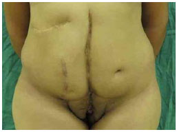
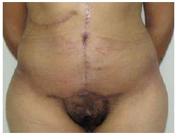
The patient was a 20-year-old nulliparous female who underwent neonatal bladder exstrophy correction involving 6 surgeries at another service. From a functional perspective, the patient reported urinary and fecal continence, no difficulty maintaining sexual relations, and regular menstrual cycles. She presented with deformities in the lower abdominal region, including multiple transverse and oblique scars, pubic region depression, and lateral/inferior displacement of the umbilical scar. The vulvar region exhibited anteriorization of the introitus and displacement of the anterior vulvar commissure, resulting in a hairless central area. Pre-operative imaging examination did not reveal significant alterations.
The deformities were corrected in a single stage by using modified abdominoplasty aiming to correct the vulvar deformities. A standard arciform incision was created, and the detachment was performed conventionally in the supra-aponeurotic plane. As a consequence of the more lateral position of the pubic bones, which exhibited a diastasis of approximately 7 cm, the patient exhibited diastasis of the rectus abdominis muscles approximately 4 and 6 cm in the supra- and infra-umbilical regions, respectively. Plicature was performed conventionally. The umbilical scar was maintained and repositioned. The scar tissue in the mons veneris region was excised, and the medial superior mobilization of the hairy area was performed along with repositioning of the anterior vulvar commissure. Up-on the patient's request, we also reduced the labia minora. No postoperative complications were observed.
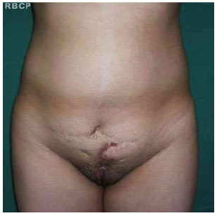
Pre operative
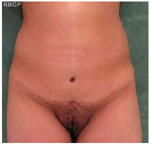
Postoperative 6 months
CASO 2
The patient was an 18-year-old nulliparous female with a history of neonatal bladder exstrophy correction and 4 surgeries reported. From a functional perspective, the patient reported urinary and fecal continence as well as regular menstrual cycles. She presented with deformities in the lower abdominal region, including multiple scars, pubic region depression, and the absence of an umbilical scar. The patient also presented with areas of lipodystrophy in the anterior region and flanks.
A modified abdominoplasty was performed with redundant tissue resection in the hypogastric region and the creation of a final transverse scar. In addition to diastasis of the pubic bones (approximately 5 cm), diastasis of the rectus abdominis muscles of approximately 3 and 5 cm in the supra- and infra-umbilical regions, respectively, was observed. Plicature was conducted conventionally. In addition, to neo-omphaloplasty with local flaps and dermal fixation to the musculoaponeurotic plane, liposuction and lipectomy were performed. The mons veneris was reconstructed by using the Poullard technique. No postoperative complications were observed.
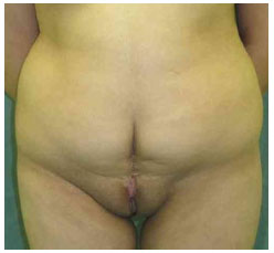
Pre operative
Postoperative 6 months
CASO 3
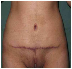
The patient was a 16-year-old nulliparous female with a history of neonatal bladder exstrophy correction; she had undergone 15 surgeries including colostomy (in the right hypochondrium) and bladder augmentation with external urinary diversion (Mitrofanoff) in the right vulvar region. The patient presented with deformities in the lower abdominal region, including multiple widened and depressed scars in the median, paramedian, oblique in the right hypochondrium, and transverse supra-pubic regions. Depression and widening of the mons veneris region and absence of an umbilical scar were also evident.
Scar revision in the right hypochondrium and vertical abdominoplasty were performed, creating a final median scar. Besides diastasis of the pubic bones (approximately 6 cm), we observed diastasis of the rectus abdominis muscles approximately 4 and 6 cm in the supra- and infra-umbilical regions, respectively. Plicature was conducted conventionally. Neo-omphaloplasty was performed with local rectangular flaps. The standard treatment in the mons veneris region was modified because of the presence of urinary diversion; it was performed with revision of the median scar (Poullard technique) and small complementary skin resection in the bilateral inguinocrural regions to avoid distorting the urinary diversion trajectory. No postoperative complications were observed.
Pre operative
Postoperative 6 months
DISCUSSION
The etiology of EEC malformations involves exaggerated development of the cloacal membrane, preventing the medial migration and development of the mesodermal tissues that would form the musculoaponeurotic structures of the abdominal wall. Without adequate protection, the cloacal membrane eventually ruptures; depending on the embryonic development stage, this rupture may generate complications ranging from isolated balanic epispadias to cloacal exstrophy, which is the most severe form 2-6,8,11.
A series of malformations including external rotation of the hip bones, shortening/inferiorization of the pubic bones, diastasis of the rectus abdominis muscles, inferiorization of the umbilical scar, and predisposition to inguinal hernia formation consequently occur. The perineal structures present anteriorly with alterations in the pelvic muscles that may predispose the patient to incontinence and prolapse. Cases of bladder exstrophy exhibit vesicoureteral reflux in addition to mucosal exposure and reduced capacity. Male patients exhibit alterations on the cavernous bodies, including shortening and dorsal curvature of the penis (i.e., chordee). Meanwhile, female patients usually exhibit a bifid clitoris and alteration of the anterior vulvar commissure as well as a higher occurrence of bicornuate uterus. 1,2,4-8,11
The objectives of the initial reconstruction in patients with EEC are as follows: (1) protection and preservation of the lower and upper urinary tract, (2) socially acceptable urinary continence, and (3) adequate reconstruction of the abdominal and external genital regions from aesthetic and functional perspectives.1,3,5-8
Medical teams responsible for these patients should focus on the genitourinary tract, prioritizing functional reconstruction 3,5,6,7,8. In older children, particularly adolescents and young adults, dissatis-faction with the appearance of the abdominal and external genital regions can compromise psychosocial state and consequently quality of life. 2,3,5,6,8,11,12
There are various techniques for corrections focusing on aesthetic and functional improvements. Some authors report the use of expanders, which may be positioned in both the subcutaneous and intermuscular planes, and subsequently advancing and/or overlapping the flaps 3,8,10. Joniau et al. 7 emphasize the importance of maintaining and repositioning the umbilical scar whenever possible. Vanderbrink et al. 6 and Cook et al. 5 describe the correction of the vulvar region in the mons veneris using inferolaterally based local flaps as well as the importance of waiting until the pubic hair is established; they also describe the correction of bifid clitoris and repositioning of the anterior vulvar commissure by using local flaps. Cederna 9 describes the use of the Poullard technique for median scars. Meanwhile, Cocke 2 reports the correction of abdominal alterations by using a modified abdominoplasty adapted to correct vulvar alterations in association with the infra-umbilical median scar. Finally, Moura et al. 12 describe the use of a conventional abdominoplasty in a single case with the need to reposition a preexisting urinary diversion (using the Mitrofanoff technique) below the original umbilical scar.
Smaller repairs may eventually be performed in older children; however, this should be delayed in the majority of the cases to allow greater physical development, particularly the establishment of pubic hair and adequate adipose distribution, and thus more definitive treatment 1,2,6.
CONCLUSSION
Secondary alterations of the vulvar and lower abdominal regions for the correction of bladder exstrophy may require functional/aesthetic reconstruction aiming to improve quality of life. The techniques used should be adequate for the correction of case-specific deformities, including the presentation of EEC alteration as well as sequelae and secondary deformities.
REFERENCES
1- Anderl H, Jakse G, Marberger H. Reconstruction of abdominal wall and mons pubis in females with exstrophy. Urology (1983);22(3):247-250.
2- Cocke WM. Secondary reconstruction of abdominal wall defects associated with exstrophy of the bladder. Ann Plast Surg (1993);31:459-461.
3- Marconi F, Messina P, Pavanello P, De Castro R. Cosmetic reconstruction of the mons veneris and lower abdminal wall by skin expansion as the last stage of the surgical treatment of bladder exstrophy: A reporto f three cases. Plast Reconst Surg (1993);91(3):551-555.
4- Barroso Jr U, Macedo Jr A, Ortiz V. Extrofia de Bexiga. In: Melega JM, Ed. Cirurgia Plástica - Fundamentos e Arte: Cirurgia reparadora de tronco e membros. Rio de Janeiro: Medsi; 2003. P.291-306.
5- Cook AJ, Farhat WA, Cartwright LM, Khoury AE, Salle JLP. Simplified mons plasty: A new technique to improve cosmesis in females with the exstrophy-epispadias complex. J Urol (2005);173:2117-2120.
6- Vanderbrink BA, Stock JA, Hanna MK. Aesthetic aspects of bladder exstrophy: results of puboplasty. J Urol (2006);176:1810-1815.
7- Joniau S, Stoel AM, Van-Poppel H, Hierner R. Salvage reconstructive surgery in an adult patient with failed previous repair of an exstrophy-epispadias complex. An operation with a functional and aesthetic purpose. Int Braz J Urol (2007);33(6):810-814.
8- Eid JF, Rosenberg P, Rothaus K, Mininberg D, Hoffman L, Vaughan ED. Use of tissue expanders in final reconstruction of infrapubic midline scar, mons pubis and vulva after bladder exstrophy repair. Urology (1993);41(5):426-430.
9- Cederna JP. Re: Secondary reconstruction of abdominal wall defects associated with exstrophy of the bladder - letters. Ann Plast Surg (1994);33(5):571.
10- Byrd HS, Hobar PG. Abdominal wall expansion in congenital defects. Plast Reconst Surg (1989);84(2):347-352.
11- Sripathi V, Sen S, Ahmed S. Normal umbilicus and infraumbilical abdominal wall in bladder exstrophy. Br J Urol (1997);80:672.
12- Moura T, Lima Jr JE, Sakae E, Aki F, Giron AM, Ferreira MC. Classic abdominoplasty: A new approach to the correction of the abdominal wall deformity in patients with bladder exstrophy. Clinics (2009);64(9):929-31.
1. Master in Surgery. - Attending Physician in the Department of Plastic Surgery ISCMSP
2. Doctor of Surgery. - Chief of Plastic Surgery ISCMSP.
Daniel Francisco Mello
Rua Nanuque, 335, ap 84. Vila Leopoldina
05302-031 São Paulo/SP
mello.plastica@gmail.com
Article received:30/09/2013
Article acceped:30/102013
Work was performed at the Pontifícia Universidade Católica (PUC-SP), Campus Sorocaba.
 Read in Portuguese
 Read in English
 PDF PT
 Print
Print
 Send this article by email
 How to Cite
 Mendeley
 Pocket