


Case Report - Year 2014 - Volume 29 -
Subtotal calcanectomy for the treatment of calcaneal pressure sores with associated osteomyelitis: a report of 2 cases
Calcanectomia subtotal para tratamento de úlcera de pressão com osteomielite associada: relato de 2 casos
ABSTRACT
INTRODUCTION: Feet wounds are very common and require multidisciplinary approach for prevention, treatment and rehabilitation. When involving the calcaneus, they offer even greater difficulty and may complicate with osteomyelitis. Debridement of devitalized tissue and antibiotics are important steps for treatment. For the reconstruction, local or free flaps are needed. However, not all patients, due to systemic conditions or local blood supply, are not candidates for this type of reconstruction and some times are submitted to amputations.
CASES REPORT: The authors report two cases in which subtotals calcanectomies were used for the treatment of wounds in the calcaneus. In both cases, amputations were avoided.
Keywords: Calcaneus; Osteomyelitis; Pressure Ulcer.
RESUMO
INTRODUÇÃO: Feridas em pés são muito frequentes e requerem abordagem multidisciplinar para a sua prevenção, tratamento e reabilitação. Quando acometem o calcâneo, oferecem dificuldade ainda maior e podem apresentar complicação com a ocorrência de osteomielite. Debridamento de tecido desvitalizado e antibioticoterapia são etapas obrigatórias para o tratamento. Na reconstrução, retalhos locais ou livres são necessários. Porém, nem todos os pacientes, devido a condições sistêmicas ou de vascularização local, são candidatos a esse tipo de reconstrução e acabam sendo submetidos a amputações.
RELATO DE CASO: Os autores relatam dois casos nos quais foram utilizadas calcanectomias subtotais para o tratamento de feridas em calcâneo. Em ambos os casos, foram evitadas as amputações.
Palavras-chave: Calcâneo; Osteomielite; Úlcera por Pressão.
Feet wounds occur very frequently and require a multidisciplinary approach for their prevention, treatment, and rehabilitation1. Ulcers normally originate from pressure or trauma, and occur in patients with decreased sensibility due to diabetes, paraplegia, poliomyelitis, mielomeningocele, fracture, or rheumatoid arthritis2,3.
When the calcaneus bone is affected, the wounds may present complications and evolve into osteomyelitis. Debridement of dead tissue and antibiotics are mandatory in the treatment. Local or free flaps are required for reconstruction4,5. However, because of systemic or local vascularization conditions, not all patients are suitable candidates for this type of reconstruction and amputations eventually become unavoidable.
Total or subtotal calcanectomy is an alternative procedure to more radical amputations2,6,7,8-11.
OBJECTIVE
The objective of the present study was to report two cases of subtotal calcanectomy performed for the treatment of calcaneal wounds.
METHOD
Case 1
A female patient with type 2 diabetes underwent a hemicolectomy because of a malignant neoplasm. In the postoperative period, she presented complications of anastomotic dehiscence and septic shock. She was underwent a new abdominal surgery and remained hospitalized in the intensive care unit (ICU) for 23 days. During this time, because of hemodynamic instability and mobilization difficulty, she developed a pressure ulcer on the right calcaneus (Figure 1), with radiological symptoms of osteomyelitis and an instep wound. Several attempts of debridement and partial skin flaps were made, with success only on the dorsal wound. As the next option would have been a transtibial amputation, we opted for debridement of dead tissue (Figure 2) and to allow primary wound closure, partial calcanectomy (Figure 3). Staphylococcus aureus and Pseudomonas aeruginosa were isolated from a culture of the material obtained from the debridement.
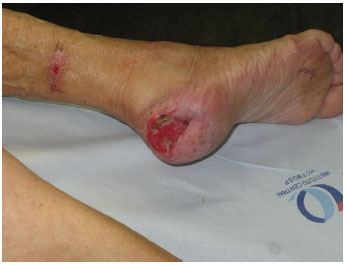
Figure 1. Chronic wound on the calcaneus.
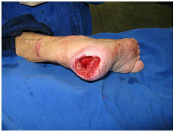
Figure 2. Appearance of the wound after debridement and calcanectomy.
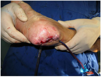
Figure 3. Immediate postoperative appearance.
Case 2
The patient was a 31-year-old man with type 1 diabetes, diabetic retinopathy (with very low bilateral visual accuracy), diabetic nephropathy (requiring hemodialysis at home), and peripheral neuropathy. In February 2011, he experienced a fall from his own height that resulted in a serious cranio-encephalic trauma; he underwent an orotracheal intubation and remained hospitalized in the ICU. During this time, he developed pressure ulcers in both calcaneus bones. He presented fever from an unidentifiable origin, except for a radiological indication of calcaneal osteomyelitis. He underwent debridement of dead tissue and partial calcanectomy to allow primary wound closure. Enterococcus faecium and Acinetobacter baumannii were isolated from a culture of the material obtained from the debridement.
RESULTS
Case 1
The patient had a good prognosis in the postoperative period. She was released from the hospital on the 37th day after the surgery. After 5 months, she could walk with crutches (Figures 4 and 5).
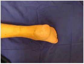
Figure 4. Late postoperative appearance.
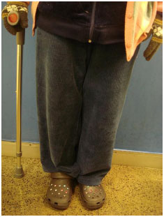
Figure 5. Patient in the orthostatic position.
Case 2
The patient remained hospitalized until the 25th day postoperatively with intravenous antibiotics administration. He was released without any signs of complications. During the ambulatory follow-up, he showed a minimal postoperative wound dehiscence, which was treated with bandages. Presently, 2 months after the surgery, the wound has cicatrized and the patient has started rehabilitation.
DISCUSSION
Wounds on the plantar surface of the foot and on the calcaneus represent a challenge for the plastic surgeon because of the low amount of neighboring tissue available for reconstruction. In the presence of osteomyelitis, the treatment is even more difficult, as it involves, besides debridement of dead tissue, the use of vascularized tissue to cover the wound and requires prolonged intravenous administration of antibiotics. To cover the wound after debridement, free or reticulated flaps can be used4,5. However, owing to comorbidities, previous clinical conditions, or local vascularization conditions, a transtibial amputation may eventually be necessary. A total or subtotal calcanectomy can be performed as an alternative procedure to more radical amputations6-11.
In the cases presented here, the wound could be accessed after debridement of skin necrosis, although other incisions may help expose the calcaneus12. The bone tissue affected by osteomyelitis is, obviously, debrided. Nevertheless, even if it is not affected by the disease, part of the calcaneus is removed to allow primary wound closure.
The expected local complication is the dehiscence of the postoperative wound. Despite closure without tension, the region has low vascularization. Such complication was observed in Case 2. Jeremy Cook and colleagues13 analyzed several calcanectomies and observed a 71.4% rate of wound healing in the first year. Vasculopathy and wound depth were the factors that mostly affected the wound healing process. There was wound recurrence after complete closure in 40% of the cases, and eight patients required a transtibial amputation of the same limbed for which calcanectomy was performed. Therefore, these patients must be observed for a prolonged time, with preventive care to avoid the development of new wounds; furthermore, intravenous administration of antibiotics is recommended, based on the bone tissue culture4,5. In both cases reported in this work, bacteria were isolated and the patients remained hospitalized for treatment with antibiotics for approximately 1 month. The presence of bacteria in the lesions, as in all open wounds, is very common. In the analyses of Crandall and colleagues2, involving 31 cases, the most frequently isolated bacteria were Staphylococcus aureus and Proteus spp.
The selection of patients probably affects the results. For this selection, Smith and colleagues6 used the ankle/brachial index, albumin levels, and lymphocyte count. Thereby, they intended to evaluate the vascularization of the limb and the nutritional state of the patients. Our patients underwent this procedure without such evaluation, as the treatment was performed to preserve the limb. An unsuccessful therapy would have resulted in a transtibial amputation.
An important concern is rehabilitation. After a transtibial amputation, the use of prosthesis can, in most cases, restore the patient's ability to walk to the same level as before the amputation14. In the cases reported by Smith and colleagues6, after a subtotal calcanectomy, a hard foot orthosis was designed especially for their patients who retained their ability to walk without any functional impairment. Yildirim5, on the other hand, in their series of nine cases, used distally based neurocutaneous flaps, and all patients returned to their preoperative state, eight of whom without using any type of orthosis. In this work, the patient in case 1 can walk with crutches and the patient in case 2 has recently started rehabilitation. A longer follow-up time will be necessary to assess the existence of any functional sequela.
CONCLUSION
The authors described two cases in which subtotal calcanectomy was successfully performed for the treatment of calcaneal wounds.
REFERENCES
1. Singh N, Armstrong DG, Lipsky BA. Preventing foot ulcers in patients with diabetes. JAMA. 2005;293(2):217-28.
2. Crandall RC, Wagner FW Jr. Partial and total calcanectomy: a review of thirty-one consecutive cases over a ten-year period. J Bone Joint Surg Am. 1981;63(1):152-5.
3. Alcaraz P, Aubran C, Jaoua S, Roudier C, Mattei JP, Announ N, et al. Calcaneal osteomyelitis due to fistulization of an ulcerated rheumatoid nodule. Joint Bone Spine. 2006;73(1):102-4.
4. Anderson RB, Foster MD, Gould JS, Hanel DP. Free tissue transfer and calcanectomy as treatment of chronic osteomyelitis of the os calcis: a case report. Foot Ankle. 1990;11(3):168-71.
5. Yildirim S, Gideroglu K, Aköz T. The simple and effective choice for treatment of chronic calcaneal osteomyelitis: neurocutaneous flaps. Plast Reconstr Surg. 2003;111(2):753-60.
6. Smith DG, Stuck RM, Ketner L, Sage RM, Pinzur MS. Partial calcanectomy for the treatment of large ulcerations of the heel and calcaneal osteomyelitis. An amputation of the back of the foot. J Bone Joint Surg Am. 1992;74(4):571-6.
7. Perez ML, Wagner SS, Yun J. Subtotal calcanectomy for chronic heel ulceration. J Foot Ankle Surg. 1994;33(6):572-9.
8. Isenberg JS, Costigan WM, Thordarson DB. Subtotal calcanectomy for osteomyelitis of the os calcis: a reasonable alternative to free tissue transfer. Ann Plast Surg. 1995;35(6):660-3.
9. Baumhauer JF, Fraga CJ, Gould JS, Johnson JE. Total calcanectomy for the treatment of chronic calcaneal osteomyelitis. Foot Ankle Int. 1998;19(12):849-55.
10. Bollinger M, Thordarson DB. Partial calcanectomy: an alternative to below knee amputation. Foot Ankle Int. 2002;23(10):927-32.
11. Randall DB, Phillips J, Ianiro G. Partial calcanectomy for the treatment of recalcitrant heel ulcerations. J Am Podiatr Med Assoc. 2005;95(4):335-41.
12. Fisher TK, Armstrong DG. Partial calcanectomy in high-risk patients with diabetes: use and utility of a "hurricane" incisional approach. Eplasty. 2010;10:e17.
13. Cook J, Cook E, Landsman AS, Basile P, Dinh T, Lyons T, Rosenblum B, Giurini J. A retrospective assessment of partial calcanectomies and factors influencing postoperative course. J Foot Ankle Surg. 2007;46(4):248-55.
14. Pinzur MS, Gottschalk F, Smith D, Shanfield S, de Andrade R, Osterman H, et al. Functional outcome of below-knee amputation in peripheral vascular insufficiency. A multicenter review. Clin Orthop Relat Res. 1993;(286):247-9.
1 - Physician - Resident Physician, Clinical Hospital of the Faculty of Medicine of the University of São Paulo
2 - Plastic Surgeon - Plastic Surgeon, Clinical Hospital of the Faculty of Medicine of the University of São Paulo
3 - Plastic Surgeon - Preceptor Physician in the Plastic Surgery Course at the Faculty of Medicine of the University of São Paulo
4 - Physician - Resident Physician, Clinical Hospital of the Faculty of Medicine of the University of São Paulo
5 - Plastic Surgeon - Assistant Physician in the Plastic Surgery Course at the Faculty of Medicine of the University of São Paulo
6 - Plastic Surgeon - Full Professor in the Plastic Surgery Course at the Faculty of Medicine of the University of São Paulo
Institution: Clinical Hospital of the Faculty of Medicine of the University of São Paulo.
Corresponding author:
Lincoln Saito Millan
Plastic Surgery Service of the University of São Paulo
Street Doutor Enéas de Carvalho Aguiar, 255, 8º andar, sala 8128
E-mail: lincolnsaito@gmail.com
Article received: April 19, 2011
Article accepted: May 23, 2011
 Read in Portuguese
 Read in English
 PDF PT
 Print
Print
 Send this article by email
 How to Cite
 Mendeley
 Pocket