


Case Report - Year 2015 - Volume 30 -
Gestational gigantomastia: how to address this clinical situation
Gigantomastia gestacional: como abordar essa situação clínica
ABSTRACT
Gestational gigantomastia is a rare disorder characterized by an excessive and rapid enlargement of the breasts, resulting in edema and venous congestion of breast tissue. It is a painful condition that causes skin ulceration and local infection. In some cases, these complications lead to an emergency mastectomy or induced abortion. The most probable etiology is an abnormal stimulation of breast tissue that is probably triggered by an abnormally elevated level of hormones or by the hypersensitivity of breast tissue to normal hormone levels. Although the breast volume decreases after pregnancy, it rarely returns to its original state; therefore, breast reduction through mastectomy or mammaplasty is usually necessary. Moreover, there is a high likelihood of recurrence in subsequent pregnancies. The authors report a case of gigantomastia in a primigravida that required pregnancy termination, because of the risk of maternal death, and a subsequent breast reduction surgery.
Keywords: Pregnancy; Breast; Gigantomastia; Mammaplasty; Mastectomy.
RESUMO
A gigantomastia gestacional é uma desordem rara, na qual ocorre o crescimento excessivo e rápido das mamas, culminando com edema e congestão venosa das mesmas, além de ocasionar dor, ulceração da pele e infecção local. Estas complicações, em alguns casos, levam à necessidade de mastectomia de emergência ou abortamento induzido. A hipótese etiológica mais aceita é a de que exista uma estimulação anormal do tecido mamário, que pode ser desencadeada por níveis excessivos de hormônios ou por uma hipersensibilidade deste tecido a níveis hormonais normais. Apesar de a mama regredir após a gestação, raramente volta ao estado original; portanto, a redução da mama através de mastectomia ou mamoplastia geralmente é necessária. Além disso, é grande o risco de recorrências em gestações futuras. Os autores relatam o caso de gigantomastia em uma primigesta, com necessidade de interrupção da gravidez, devido ao risco de morte materna, e posterior intervenção cirúrgica com mamoplastia redutora.
Palavras-chave: Gravidez; Mama; Gigantomastia; Mamoplastia; Mastectomia.
Gigantomastia is defined as an excessive breast hypertrophy of unknown etiology. Cases have been described as juvenile, pregnancy-induced, drug-induced, and idiopathic gigantomastia; some of these cases have been associated with autoimmune disease and a family history1,2.
Gestational gigantomastia is a complication that occurs in approximately 1 in every 28,000-118,000 pregnancies3,4. It occurs in the first trimester of pregnancy in approximately 64% of massive breast hypertrophy cases, and breast tissue can enlarge as much as 20 times. Some factors have been implicated in its etiology, such as changes in the corpus luteum and placental hormones, human chorionic gonadotropin, and hyperprolactinemia2. It is generally agreed that the etiology is an abnormal stimulation of the breast tissue, probably triggered by an abnormally elevated level of hormones or the hypersensitivity of this tissue to normal hormone levels3-5.
Treatment modalities include hormone therapy and surgery, or a combination of both. Because there is a high likelihood of recurrence in subsequent pregnancies, mastectomy is often preferred over breast reduction surgery. The differential diagnosis of gigantomastia includes a thorough investigation to exclude malignant breast neoplasm and fibroepithelial tumors, such as fibroadenoma and phyllodes tumor5.
In addition to being a rare condition, gestational gigantomastia is unpredictable because it can occur in multigravidas without a history of this disorder, as was described by Silva Filho et al.4, Antevski et al.6, and Bloom and Nahabedian2. In some cases, the pregnancy is carried to full term with the use of medication alone, and breast reduction is performed during puerperium. Another case report described a patient with a twin pregnancy and myasthenia gravis with comorbidity who underwent a C-section at 38 weeks of gestation with simultaneous breast reduction to avoid subsequent intervention and the risk of myasthenia exacerbation. In the case reported by Bloom and Nahabedian2, the patient underwent breast reduction after a spontaneous abortion. She became pregnant again 1 month after the surgery with no recurrence of gigantomastia. Finally, Swelstad et al.3 reported a case of a primigravida whose general health status was seriously compromised as a result of complications from gigantomastia, and who underwent total mastectomy at 28 weeks of gestation and breast reconstruction after the delivery.
CASE REPORT
A 22-year-old primigravida with gestational gigantomastia was referred to the Mastology and Plastic and Reconstructive Surgery Departments of the University Hospital of the Federal University of Paraná.
The patient, who had been reduction until then and had no family history of gigantomastia, reported the gradual enlargement of both breasts since the beginning of her pregnancy. The increasing size of her breasts was causing necrosis and local infection that represented a threat to her life; therefore, the pregnancy, which was being monitored at a prenatal clinic, had to be terminated in March 2011, at 17 weeks of gestation. During hospitalization, she exhibited breast hypertrophy with lymphatic and venous stasis, as well as ulcerations on the skin with areas of necrosis and cellulite (Figures 1 and 2).
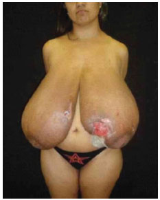
Figure 1. The 22-year-old patient with gestational gigantomastia. She had ulcers on both breasts.
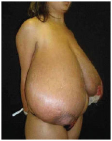
Figure 2. Lateral view.
In June 2011, the patient underwent breast reduction with Thorek's technique; 4690 g and 4435 g of tissue were removed from the right and left breasts, respectively (Figure 3).
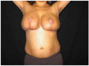
Figure 3. Photograph at 2 months postoperation.
The patient's condition evolved favorably, with no breast hematomas. The Brown compression dressing was removed on the 7th day postoperatively and the graft integration was 100%.
DISCUSSION
Gestational gigantomastia is a complication that usually requires surgical intervention. In these cases, resection of the breast tissue is hampered by venous congestion and increased vascular plexuses, which increase bleeding during surgery; in addition, the tissue is denser and less fatty. The transposition of the nipple-areolar complex (NAC) is hampered by the inadequate vascularization of the local pedicle, and grafting of the NAC is therefore required2.
Gigantomastia does not necessarily occur during the first pregnancy1. However, the occurrence of gigantomastia during a previous pregnancy almost always complicates subsequent pregnancies6. It is very important to note that the breast tissue that remains after breast reduction can regrow during subsequent pregnancies6.
Although the condition may not occur during the first, second, or third pregnancy, once massive breast hypertrophy has occurred, all subsequent pregnancies are complicated if the patient is not submitted to breast reduction or mastectomy7. This recurrence is observed even in pregnancies that progress to spontaneous abortion. When there are no complications, such as necrosis and/or infection, volume reduction through mammaplasty should be postponed to after the fourth month postpartum7,8.
The histopathology report revealed lobular hypertrophy and hyperplasia consisting of secretory acini. Some authors currently propose the use of less aggressive procedures, such as the use of dopaminergic receptor stimulators. Fetal growth restriction occurs in some cases, although no placental changes were observed in the reported cases. This is probably caused by nutrients that are essential to the fetus being used in the process of excessive breast enlargement. The proposed treatment for massive breast hyperplasia during pregnancy focuses on improving the actual status of the patient, especially addressing the pain caused by gigantomastia. Bromocriptine and/or progesterone, which reduce pain and gland swelling, have been proposed as medical therapies4,8.
Bromocriptine has been used by many authors, at doses of 5.0-7.5 mg/day, with good results, even when initiated after the 7th month of gestation. However, the drug does not prevent or minimize the occurrence of necrosis and subsequent ulceration of the breast skin; in addition, some patients have complained of nausea and emesis. Progesterone is not currently used because of its poor results when compared with bromocriptine4,7.
Reduction mammaplasty should be indicated, although spontaneous involution has been reported.
When there are no complications, such as necrosis and/or infection, reduction mammaplasty should be postponed to after the 4th month postpartum4.
In the reported case, mammaplasty was chosen because the patient had infection and local ulcers. Therefore, a smaller surgical treatment that carries lower morbidity was the treatment option. The patient is still young and wishes to become pregnant in the future; therefore, the definitive adenomastectomy procedure will be performed at a subsequent stage.
REFERENCES
1. Amini P, Stasch T, Theodorou P, Altintas AA, Phan V, Spilker G. Vertical reduction mammaplasty combined with a superomedial pedicle in gigantomastia. Ann Plast Surg. 2010;64(3):279-85. http://dx.doi.org/10.1097/SAP.0b013e3181b0a5d8. PMid:20179473.
2. Bloom SA, Nahabedian MY. Gestational macromastia: a medical and surgical challenge. Breast J. 2008;14(5):492-95. http://dx.doi.org/10.1111/j.1524-4741.2008.00628.x. PMid:18657144.
3. Swelstad MR, Swelstad BB, Rao VK, Gutowski KA. Management of gestational gigantomastia. Plast Reconstr Surg. 2006;118(4):840-48. http://dx.doi.org/10.1097/01.prs.0000232364.40958.47. PMid:16980844.
4. Silva AR FO, Burlá JM, Jesus NR, Gomes ND, Gonzales AB. Relato de Caso: hipertrofia maciça das mamas na gravidez. Rev. Bras. Ginecol. Obstet. 2002;24(6):413-17. http://dx.doi.org/10.1590/S0100-72032002000600009
5. Dancey A, Khan M, Dawson J, Peart F. Gigantomastia-a classification and review of the literature. J Plast Reconstr Aesthet Surg. 2008;61(5):493-502. http://dx.doi.org/10.1016/j.bjps.2007.10.041. PMid:18054304.
6. Antevski B, Jovkovski O, Filipovski V, Banev S. Extreme gigantomastia in pregnancy: case report-my experience with two cases in last 5 years. Arch Gynecol Obstet. 2010,284(3): 575-78. http://dx.doi.org/10.1007/s00404-010-1714-8. PMid:20978777.
7. Mojallal A, Moutran M, Shipkov C, Saint-Cyr M, Rohrich RJ, Braye F. Breast reduction in gigantomastia using the posterosuperior pedicle: an alternative technique, based on preservation of the anterior intercostal artery perforators. Plast Reconstr Surg. 2010;125(1):32-43. http://dx.doi.org/10.1097/PRS.0b013e3181c49561. PMid:20048594.
8. Foustanos A, Panagiotopoulos K, Skouras G. Intraoperative modification of Pitanguy technique of reduction mammaplasty for elevation of the nipple-areola complex in case of severe breast ptosis. Aesthetic Plast Surg. 2011;35(1):55-60. http://dx.doi.org/10.1007/s00266-010-9556-0. PMid:20725725.
Universidade Federal do Paraná (UFPR), Curitiba, PR, Brasil
Institution: Study performed at the Serviço de Cirurgia Plástica, Hospital de Clínicas, Universidade Federal do Paraná, Curitiba, PR, Brazil.
Corresponding author:
Renato da Silva Freitas
Serviço de Cirurgia Plástica e Reparadora, Hospital das Clínicas, Universidade Federal do Paraná
Rua General Carneiro, 181 - Curitiba, PR, Brazil
Zip Code 82060-900
E-mail: dr.renato.freitas@gmail.com
Article received: September 5, 2011.
Article accepted: December 1, 2011.
 Read in Portuguese
 Read in English
 PDF PT
 Print
Print
 Send this article by email
 How to Cite
 Mendeley
 Pocket