


Case Reports - Year 2016 - Volume 31 -
Macrodactyly: a retrospective study of four cases
Macrodactilia: estudo retrospectivo de quatro casos
ABSTRACT
INTRODUCTION: Macrodactyly is a rare anomaly of unknown etiology. The first cases were described in 1821 by Klein. It represents approximately 1% of all congenital anomalies. It appears at birth and is characterized by excessive growth of the fingers, toes, or of the entire limb; however, its appearance may be delayed, with symptoms of nerve compression, and may present with carpal tunnel syndrome.
METHODS: Retrospective study of four cases of macrodactyly treated at the Hospital da Santa Casa Misericórdia in Campo Grande, MS, in the last 10 years
RESULTS: We describe four cases of macrodactyly: three in the fingers and one affecting the first toe. All patients were treated with surgical procedures, one with amputation of phalanges and metacarpals.
CONCLUSIONS: Amputation is a surgical option recommended in some cases, as is the early treatment of carpal tunnel syndrome.
Keywords: Fingers/abnormalities; Toes/abnormalities; congenital deformities of the limbs; amputation.
RESUMO
INTRODUÇÃO: A macrodactilia é uma anomalia rara e de etiologia desconhecida. Os primeiros casos foram descritos, em 1821, por Klein. Representa aproximadamente 1% de todas as anomalias congênitas. Surge no nascimento e caracteriza-se pelo crescimento dos dedos das mãos, dos pés ou de todo o membro; entretanto, pode se apresentar mais tardiamente, com os sintomas de compressão de nervo, podendo associar-se à síndrome do túnel do carpo.
MÉTODOS: Estudo retrospectivo de quatro casos de macrodactilia atendidos no Hospital da Santa Casa Misericórdia de Campo Grande, MS, nos últimos 10 anos.
RESULTADOS: Descrevemos quatro casos de macrodactilia, sendo três em quirodáctilos e um acometendo primeiro pododáctilo. Todos os pacientes tratados com procedimentos cirúrgicos, um dos casos com amputação de falanges e metacarpo.
CONCLUSÕES: É recomendada a amputação como opção cirúrgica em alguns casos e o tratamento precoce da síndrome do túnel do carpo quando presente.
Palavras-chave: Dedos/anormalidades; Dedos do pé/anormalidades; Deformidades congênitas dos membros; amputação.
Macrodactyly is a rare anomaly of unknown etiology. The first cases were described in 1821 by Klein. It represents approximately 1% of all congenital anomalies, and is characterized by excessive growth of the fingers, toes, or the entire limb. However, its appearance may be delayed, with symptoms of nerve compression1-3, and may present with carpal tunnel syndrome. The growth of all digital structures is observed. The nerves thicken and are tortuous. The symptoms include joint stiffness, ulceration of the fingertips, triggering, pain, and paresthesia. The growth of the fingers can be part of the Klippel-Trenaunay-Weber and Proteus syndromes2.
This disease is bilateral in 6% of cases1. Two forms of macrodactyly have been described, static and progressive. The static form appears at birth; the growth rate of the digit is proportional to the other fingers. The progressive form, the most common type, may or may not appear at birth, but appears in childhood, with a disproportionate growth of the finger or toe compared to the other digits, and leads to angular deviation. It is associated with syndactyly in 10% of patients2,4.
This anomaly is extremely difficult to treat. Several techniques have been described to reduce the size of the finger or toe: dermolipectomy, reductional osteotomy of phalanges, and epiphysiodesis. The surgical results are unsatisfactory, and often result in amputation of digits or entire digital rays of the hand. In the compression seen in carpal tunnel syndrome, the opening of the carpal tunnel is required, and in some cases, the restoration of thumb opponency3.
This report describes a series of four cases of macrodactyly through a review of medical records.
CASE REPORTS
Case 1
A male child was admitted at the Hospital da Santa Casa Misericórdia de Campo Grande, MS at 4 years of age, with digital gigantism of the second and third fingers of the right hand (Figure 1), without syndromic association. Physical examination revealed an increased palmar volume, gigantism of the second and third fingers, axis deviation, finger hyperextension, ankylosis of the joints, and thenar muscle atrophy. Radiological examination of the left hand showed growth of bone, soft tissues, and deviation of the fingers adjacent to the axis of each finger.
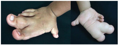
Figure 1. Macrodactyly - 2° and 3rd fingers of the right hand.
The patient underwent dermolipectomy of the palmar region, and disarticulation of phalanges, second and third fingers, and the second metacarpal. After 1 year, an additional intervention was performed, with disarticulation of the third metacarpal and dermolipectomy of the palmar region. The histopathological examination revealed lipofibromatosis. For the assessment of the result, the opponency sensitivity test was performed. Recovery of opponency was observed with pincer movements, apprehension of cylindrical objects, and positioning of the abducted thumb. After 5 years of outpatient postoperative follow-up, he presented without complications and healed with good appearance. There was no bone growth in the postoperative period.
Case 2
A male teenager was admitted at the Hospital da Santa Casa Misericórdia de Campo Grande, MS at 15 years of age, with gigantism of the first right toe (Figure 2), without syndromic association. Physical examination revealed an increase in the volume of the first toe, difficulty in wearing closed shoes, and recurrent trauma. The radiological examination showed an increase of volume, axis deviation, ankylosis of joints, and increased bone growth of the phalanges and first metatarsals.
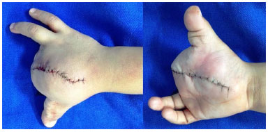
Figure 2. 7th postoperative day - disarticulation 3rd metacarpal and palmar dermolipectomy.
He underwent soft tissue resection with closure using a rotational flap. He did well postoperatively, and felt more comfortable wearing closed shoes. After 3 years of postoperative outpatient follow-up, he presented no complications. There was no bone growth in the postoperative period. Healing appeared good.
Case 3
A female child was admitted at the Hospital of the Santa Casa Mercy of Campo Grande, MS at 11 years of age, with gigantism in the fourth right finger (Figure 3), without syndromic association. Physical examination revealed an increase in the volume of the fourth finger and slight bone growth in the phalanges on radiography.
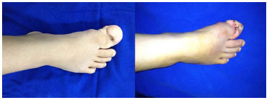
Figure 3. Macrodactyly in 1st toe of the right foot.
The patient underwent soft tissue debulking and reductional osteotomy of the phalanges. She showed good functional recovery. After 7 years of outpatient follow-up, she presented without complications, and healed with good appearance. There was no bone growth in the postoperative period.
Case 4
A male child was admitted at the Hospital da Santa Casa Misericórdia de Campo Grande, MS at 7 years of age, with gigantism in the second and third left fingers, without syndromic association. Physical examination revealed an increase in the volume of the left fingers with axis deviation (Figure 4), difficulty in pinching, holding of objects, and writing. Radiography revealed increased bone growth in the phalanges.
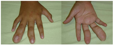
Figure 4. Macrodactyly of 4th finger right hand.
The patient underwent dermolipectomy of the affected fingers, reductional osteotomy of phalanges, and epiphysiodesis, with repositioning of the axis. He had a good postoperative course, and improvement in pinching and manipulation of objects. After 6 years of outpatient follow-up, he presented without complications and healed with good appearance. There was no bone growth in the postoperative period.
DISCUSSION
Macrodactyly is a rare congenital anomaly, with syndromic association in some cases3,5,6. It is essential to perform a complete preoperative assessment, to rule out syndromic associations. A complete physical examination is required, evaluating motor function, sensitivity, pincer movements, and day-to-day movement difficulties. Additional tests, such as computed tomography, nuclear magnetic resonance imaging, and arteriography, may be used.
Abnormal blood or humoral supply to the nerve is a possible cause of this malformation, and might stimulate overgrowth of tissues7. However, nerve overgrowth induces compression and further functional loss. The reduction of digits can lead to an unsatisfactory result and the resection of digital rays may be indicated. In addition, decompression of the median nerve and restoration of thumb opponency may be required1.
In this series of cases, an individualized approach was always used, to ensure both an aesthetic and functional result.
COLLABORATIONS
ETAF Analysis and/or data interpretation; statistical analysis; writing of the manuscript; conception and design of the study.
KGA Critical review of its contents; completion of operations and/or experiments; final approval of the manuscript.
GRC Statistical analysis.
GSMO Statistical analysis.
LHFT Statistical analysis.
PYMGA Completion of operations and/or experiments.
REFERENCES
1. Monteiro VA, Chiconelli JR, Almeida SF. Macrodactilia: estudo retrospectivo de sete casos. Rev Bras Ortoped. 1998;33(1):54-8.
2. Ezaki M, Kay SPJ, Light TR, Tonkin MA, Wood VE, Dobyns JH. Congenital hand deformities. In: Green DP, Hotchikiss RN, Pederson WC, eds. Green's operative hand surgery. New York: Churchill Livingstone; 1999. p.533-44.
3. Mirza MA, King ET, Reinhart MK. Carpal tunnel syndrome associated with macrodactyly. J Hand Surg Br. 1998;23(5):609-10. PMID: 9821604
4. Al-Qattan MM. Lipofibromatous hamartoma of the median nerve and its associated conditions. J Hand Surg Br. 2001;26(4):368-72.
5. Tan O, Atik B, Dogan A, Alpaslan S, Uslu M. Middle phalangectomy: a functional and aesthetic cure for macrodactyly. Scand J Plast Reconstr Surg Hand Surg. 2006;40(6):362-5.
6. Syed A, Sherwani R, Azam Q, Haque F, Akhter K. Congenital macrodactyly: a clinical study. Acta Orthop Belg. 2005;71(4):399-404. PMID: 16184993
7. Lagoutaris ED, DiDomenico LA, Haber LL. Early surgical repair of macrodactyly. J Am Podiatr Med Assoc. 2004;94(5):499-501
1. Santa Casa de Campo Grande, Campo Grande, MS, Brazil
2. Sociedade Brasileira de Cirurgia Plástica, São Paulo, SP, Brazil
3. Sociedade Brasileira de Medicina Hiperbárica, Campo Grande, MS, Brazil
Institution: Santa Casa de Campo Grande, Campo Grande, MS, Brazil.
Corresponding author:
Elson Taveira Adorno Filho
Rua São Paulo, 661 - Monte Castelo
Campo Grande, MS, Brazil Zip Code 79010-050
E-mail: elsonadorno@hotmail.com.br
Article received: June 27, 2013.
Article accepted: September 1, 2013.
Conflicts of interest: none.
 Read in Portuguese
 Read in English
 PDF PT
 Print
Print
 Send this article by email
 How to Cite
 Mendeley
 Pocket