


Case Reports - Year 1998 - Volume 13 -
Microsurgical Reimplantation of Total Scalp Avulsion - Case Report
Reimplante Microcirúrgico de uma Avulsão Total de Couro Cabeludo - Relato de Caso
ABSTRACT
A 25 year-old woman, victim of a total avulsion of the scalp including a large portion of the forehead, when the rotative machine caught her hair. Successfu1 reimplantation was carried out using microsurgery. Reimplantation should be attempted in all scalp avulsions.
Keywords: microsurgery; total scalp; microsurgical reimplantation.
RESUMO
Paciente de 25 anos de idade, sexo feminino, vítima de avulsão total de couro cabeludo, incluindo grande parte da pele da região frontal, decorrente de trauma provocado por uma máquina rotativa. Conseguiu-se, com sucesso, o reimplante através de microcirurgia.
Palavras-chave: microcirurgia; escalpo total; reimplante microcirúrgico
In our society, where agricultural activities are predominant, the mechanization process came up with new kinds of accidents, moreover those caused by the rotative axes of plows.
When the axes used in agricultural equipment catch plowmen clothes or long hair they cut off certain body segments.
The mechanism of avulsion of the scalp in accidents caused by plows generally occurs in the subgalea region. In the total avulsion, many other structures may be evolved like the cutaneous portion of the face, eyelids and ears. In these regions, the frontal and occipital muscles present less resistance than the galea.
The resulting sequelae from the total avulsion of the scalp with exposure of the skull arc verv difficult to reconstruct and generally cause irreparable aesthetic deformities. It is known that in such case a microsurgical reimplantation is the best choice of treatment(1, 2, 3, 4, 5).
The first reimplantation of the scalp was performed in 1974 and published in 1976 bv Miller et cols(2).
CASE REPORT
Patient CGQ, 25 years old, female, white, single, plowman, evangelical, entered the emergency room of Hospital de Base de Bauru (Bauru, SP, Brazil) two hours after the trauma, on October 28, 1990.
The rotative axis of a farm tractor caught the patient's hair, which led to the total avulsion of the scalp.
It was possible to observe a portion of the frontal region of the face, the eyebrows and part of the upper eyelids in the amputated segment, which was washed with saline solution, warped in gauze and placed then in a plastic bag to be cooled in an icebox.
Laboratorial and radiological tests (skull X-ray) were normal.
The patient was submitted to surgery under general anesthesia about one hour after the first aid. Cleaning with water and neutral soap and debridement were performed.
The scalp was trichotomized and washed once more in saline solution. Next, a wide artery and vein were identified for a surgical anastomosis.
The left superficial temporal artery was dissected as it presented better condition than all other vessels, which were very damaged.
A terminoterminal microsurgical anastomosis of the superficial temporal artery was performed using 10-0 nylon. After the release of the arterial flow, a little was waited for bleeding, in order to facilitate the identification of vessels. The distal stump of the superficial temporal vein was anastomosed with its proximal stump.
The scalp was sutured with 5-0 nylon and the skin of the face with nylon 6-0. Draining with Penrouse drains, cotton plaster and smooth binding were done. Antibiotics (cephalosporin, 3mg/day), platelet antiaggregative (acetylsalicylic acid, 1g/day) and anticoagulator (Liquemine) were used.
The surgery lasted about four hours and the postoperative did not have any intercurrellces. Hair growth was normal.
DISCUSSION
Due to religious matters, the patient had very long hair. Besides d1at, she neglected warping her hair while working near the plow. These factors led to this kind of accident.
The avulsion mechanism in these cases is generated by a continuous rotative movement causing a very intensive traction that results in the avulsion of the affected structure.
Using a hairnet would be an effective way to avoid this kind of accident and such procedure should be obligatory in agriculture.
Microsurgery has led to an adequate restoration (figs. 1, 2, 3, 4, 5) as it immediately restablished the blood circulation. Generally, the anastomosis of a vein and an artery is enough to restore the circulation in scalp (fig. 3), bur it is advisable to restore as many vessels as possible.
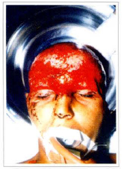
Fig. 1 - Preoperative frontal view 2 hours after the trauma.
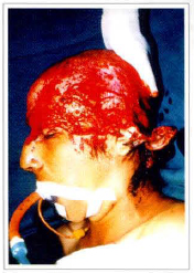
Fig. 2 - Preoperative profile view showing laceration of the left ear.
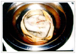
Fig. 3 - Preoperative view of the avulsioned segment in saline solution after trichotomy and cleaning.
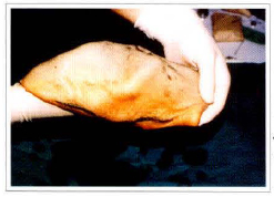
Fig. 4 - Preoperative frontal view of the avulsioned segment where the eyebrows can be visualized.
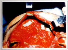
Fig. 5 - Transoperative view of the microanastomosis performed in the arteria and left superficial temporal vein.
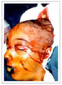
Fig. 6 - Transoperative view of the sutures after vascular reimplantation.
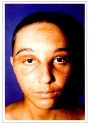
Fig. 7 - 30 days postoperative frontal view. Painless complete integration of the avulsioned segment.
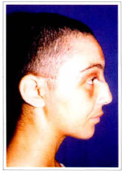
Fig. 8 - 30 days postoperative right profile view.
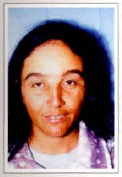
Fig. 9 - Postoperative view after 6 years and 10 months. Frontal view showing hair growth.
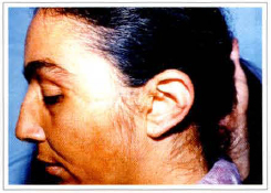
Fig. 10 - Postoperative view after 6 years and 10 months. Left profile view, showing face and ear scar sequelae.
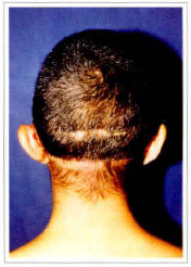
Fig. 11 - 30 days postoperative rear view.
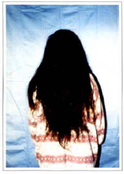
Fig. 12 - Postoperative view after 6 years and 10 months showing hair length, due ro religious beliefs.
The great avulsions look more catastrophic, but they present better results due to the presence of wide remaining vessels in the amputated segments.
REFERENCES
1. GATTI, JE and LAROSSA, D. Scalp Avulsions and Review of Successful Reimplantation. Ann. Plast. Surg. 1981; 6:127.
2. MILLER, GDH, AMSTEE, EJ, SNELL, JA. Sucessful Reimplantation of an Avulsed Scalp by Microvascular Anastomosis. Plast. Reconst. Surg. 1976; 58:133.
3. NAHAI, F, HURTEAU, J, VASCONES, LO. Reimplantation of an Entire Scalp and Ear by Microvascular Anastomosis of only one Artery and one Vein. Br. J. Plast. Surg. 1978; 31:339.
4. SAKAI, S, SOEDAS, S, ISHII, Y. Avulsion of the Scalp wich one is the best Artery for Anastomosis? Ann. Plast. Surg. 1990; 24:350.
5. STRATOUDAKIS, AC and SAVITSKY, LB. Microsurgical Reimplantation of Avulsed Scalp. Ann. Plast. Surg. 1981; 7:312.
I - Associate Member of the SBCP. Chief of the Plastic Surgery Department of Associação Hospitalar de Bauru - Brazil.
II - Titular Member of the SBCP. Tirular Member of the Brazilian Society of Reconstructive Microsurgery. Medical chief of Centro Brasileiro de Cirurgia Plástica and Clínica Dandreia e Miura (Plastic Surgery Residency Program).
Address for Correspondence:
Clínica de Cinrrgia Plástica Dr. Carlos Augusto Cameschi
R. Capitão Gomes Duarte 10-5 - Altos da Cidade
Bauru - SP - 17040-020
Phone: (55 14) 234-5550
 Read in Portuguese
 Read in English
 PDF PT
 Print
Print
 Send this article by email
 How to Cite
 Mendeley
 Pocket