


Case Report - Year 2019 - Volume 34 -
Boerhaave syndrome: A rare complication of cosmetic surgery
Síndrome de Boerhaave: uma complicação rara em pós-operatório de cirurgia estética
Laurinda Castellani1,*
ABSTRACT
Introduction: The objective of this case report is to alert surgeons to a possible postoperative complication of long cosmetic surgery under general anesthesia. Boerhaave syndrome is a serious life-threatening disease that requires diagnosis within 12 hours and proper treatment. Case report: A 58-year-old female patient presented with vomiting and nausea after abdominoplasty and mastopexy under general anesthesia. Results: The patient underwent dermolipectomy and mastopexy using the inferior pedicle technique under spinal anesthesia. Four hours after the operation, she presented several episodes of vomiting. Ten hours after the operation, she reported painful swallowing followed by generalized severe pain and presented severe dyspnea, sweating, pallor, and a 90/50 mmHg blood pressure. As the condition worsened, the patient was referred to the intensive care unit where she was intubated and underwent laboratory tests, thoracentesis, and radiological examinations. The patient currently uses an esophageal prosthesis. Conclusions: The literature suggests avoiding prolonged surgery, especially under general anesthesia, because of the risk of carbon dioxide retention, which may lead to postoperative emetic crisis in patients with a history of esophageal disease. It also suggests paying attention to symptoms, not excluding the possibility of Boerhaave syndrome.
Keywords: Spontaneous rupture; Plastic surgery; Esophageal motility disorders; Esophagus; General anwesthesia.
RESUMO
Introdução: Este estudo se trata de um relato de caso que tem por objetivo alertar os cirurgiões para uma possível complicação em pós-operatório de cirurgias estéticas longas sob anestesia geral. A Síndrome de Boerhaave é uma doença grave que ameaça a vida do paciente e merece um diagnóstico precoce até 12hs e um tratamento adequado.
Relato de caso: A paciente no pós-operatório de cirurgia plástica abdominal e mastopexia apresentou, após anestesia geral, crises de vômito e náuseas.
Resultados: Paciente com 58 anos do sexo feminino submetida à dermolipectomia abdominal e mastopexia pela a técnica de pedículo inferior sob raquianestesia, onde após um período de quatro horas do término da cirurgia apresentou vários episódios de vômitos. Após 10 horas do ato cirúrgico apresentou queixa de algia ao deglutir, seguida de algia intensa generalizada, dispneia intensa, sudorese, palidez, PA 90x50mmhg. Com a piora do quadro a paciente foi encaminhada para a unidade de terapia intensiva onde foi entubada. Foram realizados exames laboratoriais, toracocentese e exames radiológicos. Atualmente, a paciente encontra-se com prótese esofágica.
Conclusões: Fazendo a correlação com a bibliografia, no caso em tela sugere-se evitar cirurgias prolongadas, principalmente sob anestesia geral onde pode ocorrer a retenção de gás carbônico, que pode levar a crise emética no pós-operatório em pacientes com antecedentes de doença esofagiana e estar atentos aos sintomas, não descartando a possibilidade da ocorrência da Síndrome Boerhaave.
Palavras-chave: Ruptura espontânea; Cirurgia plástica; Transtornos da motilidade esofágica; Esôfago; Anestesia geral
INTRODUCTION
This is a case report study on the rare occurrence of Boerhaave syndrome in a patient who underwent abdominoplasty with general anesthesia.
The patient presented vomiting and nausea after abdominoplasty and mastopexy under general anesthesia, which, together with the use of an abdominal binder, resulted in the syndrome. It is important to emphasize that the location and sudden onset of pain may be confused with myocardial infarction or pulmonary embolism1.
Boerhaave syndrome or spontaneous rupture of the esophagus was first described in 1724 by Hermann Boerhaave2. It is a severe life-threatening disease that requires diagnosis within 12 hours and proper treatment. It is a relatively rare syndrome, but has a high mortality rate (35%). In fact, it is considered the most lethal type of rupture in the digestive tract4.
Boerhaave syndrome should be considered in the postoperative period of abdominal dermolipectomy under general anesthesia, which in this study was associated with mastopexy and postoperative vomiting and nausea.
Given the information provided here, the subject of this study is extremely important, especially considering the lack of studies on the association of Boerhaave syndrome with plastic surgery.
The objective of this study was to alert plastic surgery professionals about Boerhaave syndrome, which may occur after abdominoplasty under general anesthesia.
CASE REPORT
Patient A.M.R.L.P., a 58-year-old woman, visited our clinic to undergo abdominal dermolipectomy and mastopexy.
At physical examination:
Weight: 92 kg; Gynecological history: 2PN2CP, gestational diabetes; Cardiorespiratory system: denied dyspnea on exertion or in decubitus position, walked
30 to 40 minutes a day;
Genitourinary system: no complaints; Gastrointestinal system: denied epigastric pain; Motor system: rotator cuff tendon rupture in the right shoulder, with a prosthesis
placed under general anesthesia;
Family history: maternal breast cancer; Habits: denied smoking or alcoholism; Others: not using any medications; Physical examination: abdominal apron with stretch marks in the hypogastric region,
Pfannenstiel scar, and grade II breast ptosis (Regnault classification).
Laboratory and radiological examinations, and preanesthetic consultation showed no abnormalities, and the patient was considered eligible for surgical dermolipectomy and mastopexy.
On May 25, 2007, she underwent abdominal dermolipectomy and mastopexy using the inferior pedicle technique under spinal anesthesia. The surgery lasted 4 hours with no complications.
Four hours after the end of surgery, the patient presented several episodes of vomiting alternating with periods of improvement after antiemetic medication.
Ten hours after surgery, she reported painful swallowing, which improved with xylocaine spray, and the patient passed the night without other complications.
At 6:45 am on May 26, 2007, she reported generalized severe pain and dyspnea, which improved with administration of corticosteroids and intravenous bronchodilator. The patient was discharged at 11:30 am with no complaints.
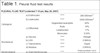
On the same date, at 2:30 pm, the patient returned with severe dyspnea, sweating, pallor, and a 90/50 mmHg blood pressure. She received supportive care, but the condition worsened, and she was referred to the intensive care unit, where she was intubated and underwent thoracentesis. The aspirated fluid had characteristics of gastric juice, and the amylase values were increased in the biochemical test (Table 1).
| PLEURAL FLUID TEST (collected 7:15 pm, May 26, 2007) | |||
|---|---|---|---|
| Cytological | a) Erythrocytes | ***** | 280.000/mm3 |
| b) Leukocytes | ***** | 5000.000/mm3 | |
| c) Differential | Segmented neutrophils | 60% | |
| Rods | 18% | ||
| Lymphocytes | 17% | ||
| Monocytes | 0,50% | ||
| Biochemical | a) Glucose | ***** | 60mgdl |
| b) LDH | ***** | 2.980U/L | |
| c) Total proteins | ***** | 55g/dl | |
| d) Amylase | ***** | 2.800U/L | |
| Bacteriological | a) Gram bacterioscopy |
***** | Gram-positive cocci isolated and in pairs |
Source: ICU, Santa Casa de Itapeva.
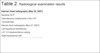
The mediastinal shift worsened, but the diagnosis was not conclusive. The patient’s general condition was poor, with sepsis and severe changes in laboratory parameters (Table 2).
| Internal chest radiography (May 26, 2007) |
|---|
| Baseline RCP |
| Subcutaneous emphysema in the LHT |
| Tracheal tube |
| Internal chest radiography (May 27, 2007) |
| Left hypolucent hemothorax |
| LHT 1/3 medium/inferior veiling |
| Right mediastinal shift |
Source: UTI Santa Casa de Itapeva.
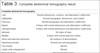
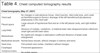
After these examinations, the general surgery team drained the left hemothorax (LHT) and requested computed tomography of the abdomen (Table 3) and chest (Table 4). After several examinations, the patient’s condition continued to deteriorate, with LHT opacity 10 days later.
| Complete abdominal tomography | |
|---|---|
| Liver | Normal dimensions, contours, and attenuation coefficients |
| Gallbladder | Normal topography, dimensions, and content |
| Pancreas | Normal topography and attenuation coefficients |
| Spleen | Entopic with normal dimensions and attenuation coefficient. |
| Left/right kidney | Entopic with normal dimensions and attenuation coefficients |
| Adrenals | No signs of injury |
| Bladder | Entopic with normal wall and content |
| Uterus and attached structures | No evidence of abnormalities |
| Abdominal vessels | Normal size and topography |
| Free liquids | Absent |
| Lymph node enlargement | Absent |
| Bone structures | Preserved appearance |
| ***** | Small amount of air in the anterior abdominal wall |
Source: ICU, Santa Casa de Itapeva.
| Chest tomography (May 27, 2007) | |
|---|---|
| Chest wall | No changes in subcutaneous tissue, chest muscles, intercostal area, and costal arches Anterosuperior thoracic drainage of the left hemothorax Subcutaneous emphysema in the chest wall |
| Pleural space | Large amount of hypotensive fluid and air in the left pleural cavity, and right midline shift |
| Lung fields | Absence of nodules, cavitations, or bilateral lower lobe intraparenchymal collections: small right and left lower lobes |
| Mediastinum | No significant changes in attenuation coefficient Absence of lymph node enlargement |
| Mediastinal vessels | Normal base vessels, without changes in arteries and veins |
| Heart area | Pericardium and cardiac chambers without changes |
| Esophagus | Preserved appearance |
| Vertebral bodies and diaphragm | No visible injuries |
| Adrenals | No signs of injury |
| ID | Large left hydropneumothorax with mediastinal shift Atelectasis/pulmonary condensation of small bilateral right lower lobe and left lower lobe Subcutaneous emphysema |
Source: ICU, Santa Casa de Itapeva
After several radiological examinations and laboratory tests, including upper digestive endoscopy, the patient was transferred to a hospital with a thoracic surgery service, where she underwent a thoracotomy for LHT decortication and was diagnosed intraoperatively at approximately 30 days after her plastic surgery. Currently, the patient is well recovered and uses an esophageal prosthesis.
DISCUSSION
Boerhaave syndrome is characterized by secondary esophageal perforation and a sudden increase in esophageal intraluminal pressure. It is common to find an etiological factor, which is often an internal pressure trauma. Although the classic definition of spontaneous rupture is incorrect, it is universally accepted. This syndrome results from increased intra-abdominal pressure due to vomiting, vomiting efforts, or other efforts. Pressure is rapidly transmitted to the esophagus, and a perforation may occur owing to distension of the mucosa and esophageal muscles, which cannot resist high pressure5.
In 80% of cases, the most common cause is vomiting or effort to vomit due to excessive food or alcohol ingestion5.
This perforation is often longitudinal and usually occurs on the left lateral wall5 of the distal esophagus, above the esophagogastric transition and diaphragm. Thus, it may produce an esophageal-pleural fistula with subsequent pleural effusion and/or pneumothorax.
It is more frequent in men between 40 and 60 years of age6. The main symptom is severe, abrupt, progressive retrosternal pain. The classic Mackler’s triad7, including vomiting, chest pain, and cervical emphysema, is uncommon. Hematemesis occurs in 22% of cases. Dyspnea occurs in 61% of cases, and cyanosis is also common. When an esophageal-pleural fistula is present, the typical pleural fluid presents nonspecific findings such as low pH and increased amylase8, and the diagnosis can be confirmed by finding food in the fluid. Other signs and symptoms include dysphagia, tachycardia, and hypotension, which may progress to sepsis and death.
Many diagnostic tools are available, including clinical diagnosis with history, physical examination, and radiological findings. Physical examination shows decreased vesicular murmur and fever in 30% of cases, with possible cervical crepitus and decreased airborne noises. Laboratory tests may show leukocytosis in some cases, without further changes9.
Chest radiography is useful for initial diagnosis, and the most common finding is pleural effusion, usually on the left side. Other findings include pneumothorax, often on the left side5, hydropneumothorax, pneumomediastinum, and subcutaneous emphysema.
Esophagography would reveal typical contrast extravasation into the pleural cavity and provide information on the size and location of the perforation, which are useful in choosing the most adequate surgical approach9.
Computed tomography can confirm the diagnosis or replace esophagography in intolerant patients. Early diagnosis and proper treatment are essential to prevent mediastinitis, sepsis, and shock, often associated with the second phase of the disease
Choosing the surgical technique to treat the spontaneous esophageal perforation can be controversial, and time from onset is a decisive factor. When the diagnosis is established within the first 24 hours, cardiorespiratory stabilization, clinical support, and antibiotic therapy are recommended, as well as primary closure of the perforation through thoracotomy associated with mediastinal drainage.
After this period, the presence of edema, tissue necrosis, and mediastinal infection can make surgery extremely difficult. The most important complication of primary closure is the occurrence of fistulas.
Conservative treatment can be a choice in cases of small perforations, where the patient has minor symptoms and limited mediastinal lesions.
In addition to surgical correction, treatment with parenteral nutrition support, nasogastric tube, broad-spectrum antibiotics, and gastric acid secretion inhibitors is essential for these patients.
Late-diagnosed cases often require some form of esophageal exclusion, usually cervical esophagostomy and gastrostomy associated with jejunostomy for nutritional support.
Spontaneous esophageal rupture is reported in the literature to occur in 10% of patients with reflux esophagitis, duodenal diverticulum, and carcinoma, but can also occur in patients without previous esophageal disease1.
CONCLUSION
Postemetic rupture of the esophagus is a serious condition that is often overlooked in the initial evaluation. Vomiting crisis, left pleural effusion, thoracentesis with fluid aspiration, and suspected digestive secretion with increased amylase levels are diagnostic factors that indicate surgical treatment. Postoperative evolution is usually complicated with vital organ failure and requires intensive support. Mortality and morbidity rates are high10.
The present case leads us to conclude that prolonged surgeries should be avoided, especially under general anesthesia, because of the risk of carbon dioxide retention, which may lead to postoperative emetic crisis, mostly in patients with a history of esophageal disease. Above all, it is important to pay attention to symptoms, not excluding the possibility of Boerhaave syndrome.
No cases of Boerhaave syndrome in the postoperative period of abdominal plastic surgery were found in the literature.
REFERENCES
1. Rodrigues JJG, Machado MCC, Rasslan S. Clínica Cirúrgica. Barueri (SP): Manole; 2008.
2. Derbes VJ, Mitchell Júnior RE. Hermann Boerhaave’s Atrocis, nec descripti prius, morbi historia, the first translation of the classic case report of rupture of the esophagus, with annotations. Bull Med Libr Assoc. 1955 Apr;43(2):217-40.
3. Passos Filho O, Cangussu HC, Lopes RH, Oliveira ATT, et al. Síndrome de Boerhaave: Relato de caso. Rev Col Bras Cir. 2013 Fev;40(1):83-84. DOI: https://doi.org/10.1590/S0100-69912013000100016
4. Soto RL, Hernandez JLM, Garcia FA, Sanchez NA, et al. A Ruptura Espontânea do Esôfago: Um problema no diagnóstico de emergência. UM Med Interna. 2003 Fev;20(2).
5. Correia Neto A. Clínica Cirúrgica. São Paulo (SP): Sarvier;1988. v.4
6. Atallah FN, Riu BM, Nguyen LB, Seguin PO, Fourcade OA. Boerhaave’s syndrome after postoperative vomiting. Anesth Analg. 2004 Apr;98(4):1164-6. DOI: https://doi.org/10.1213/01.ANE.0000101981.85523.82
7. Godinho M, Wiezel EHB, Marchi E, Módena SF, Paula RA. Ruptura espontânea do esôfago - síndrome de Boerhaave. Rev Col Bras Cir. 2012;39(1):83-84. DOI: https://doi.org/10.1590/S0100-69912012000100017
8. Sabiston JR, David C. Tratado de Cirurgia. 14ª ed. Rio de Janeiro (RJ): Guanabara Koogan; 1993.
9. Coelho JCU. Aparelho digestivo: clínica e cirurgia. 3ª ed. São Paulo (SP): Atheneu; 2006.
10. Mota HJ, Ximenes Netto M, Medeiros AC. Ruptura pós-emética do esôfago: a síndrome de Boerhaave. J Bras Pneumol. 2008 Ago;33(4):480-483. DOI: https://doi.org/10.1590/S1806-37132007000400019
1. Clínica Castellani, Itapeva, SP, Brazil.
Corresponding author: Laurinda Castellani Praça Dr Esperidiao Lúcio Martins, 93, Centro, Itapeva, SP, Brazil. Zip Code: 18400-020. E-mail: l.castellani@hotmail.com
Article received: October 17, 2018.
Article accepted: June 22, 2019.
Conflicts of interest: none.
 Read in Portuguese
 Read in English
 PDF PT
 Print
Print
 Send this article by email
 How to Cite
 Mendeley
 Pocket