


Case Report - Year 2019 - Volume 34 -
Static lower lip reanimation as a filler complication of facial paralysis
Reanimação estática do lábio inferior na paralisia facial como complicação de preenchimento

ABSTRACT
Facial rejuvenation procedures to circumvent traditional surgery have become increasingly popular to promote a youthful appearance with minimally invasive procedures such as injectable botulinum toxin, soft-tissue fillers, and chemical peels. Nevertheless, complications can occur even with an astute and experienced injector. Here we present the case of a patient who underwent static lip reanimation using a dermoadiposal flap for right facial nerve damage following nodule removal as a filler complication. A "modified bulls horn approach" to the upper lip lift was performed around the nasal wings and columella and along the right nasolabial fold. The flap was de-epithelized and harvested. Using the open tip of a small liposuction cannula, the distal portion of the flap was tunneled and fixed directly in a C-loop fashion using U stitches, transfixing the flap to the periosteum of the zygomatic arch. At 3 years follow-up, no significant complications were observed, and the patient reported no functional limitations or dissatisfaction with the scars in the nasolabial fold or around the nasal wings and columella.
Keywords: Surgery plastic; Dermal fillers; Facial paralysis; Facial Injuries; Reconstruction.
RESUMO
Procedimentos de rejuvenescimento facial substitutos da cirurgia tradicional tornaram-se cada vez mais populares para promover uma aparência jovial com procedimentos minimamente invasivos, como toxina botulínica injetável, preenchimento de tecidos moles e peelings químicos. No entanto, complicações podem ocorrer mesmo na presença de um injetor habilidoso e experiente. Apresentamos o caso de uma paciente submetida a reanimação labial estática usando retalho dermoadiposo para lesão do nervo facial direito após remoção de nódulos como complicação de preenchimento. A "abordagem modificada de bull horn" foi realizada para elevação do lábio superior em torno das asas nasais e columela e ao longo do sulco nasolabial direito. O retalho foi desepitelizado e obtido. Usando a ponta aberta de uma pequena cânula de lipoaspiração, a porção distal do retalho foi encapsulada e fixada diretamente em C-loop e foram utilizados pontos U, transfixando o retalho para o periósteo do arco zigomático. Nos três anos de seguimento não foram observadas complicações significativas e a paciente não relatou nenhuma limitação funcional ou insatisfação com o aspecto das cicatrizes no sulco nasolabial e ao redor das asas nasais e da columela.
Palavras-chave: Cirurgia Plástica; Preenchedores Dérmicos; Paralisia Facial; Traumatismos Faciais; Reconstrução
Introduction
Highly effective procedures used for facial rejuvenation include injectable neurotoxins, soft-tissue fillers, and chemical peels. Soft-tissue fillers have an impressive ability to volumize and reshape aging, sagging skin with a small time commitment and minimal side effects. A variety of filling agents have been approved by the United States Food and Drug Administration (FDA) to correct moderate to severe facial wrinkles such as hyaluronic acid, poly-l-lactic acid, calcium hydroxyapatite, and polymethylmethacrylate. Although not FDA approved and forbidden in many countries for the treatment of facial aesthetic contours, silicone is used “off-label” for cosmetic purposes for treating acne scars or performing facial augmentation.
Reported complications after soft-tissue augmentation may be related to poor injection technique or the filler material itself. Complications such as visible product, hypertrophic scarring, nodule formation, and the Tyndall effect (bluish discoloration from hydroxyapatite fillers) can occur if the soft-tissue synthetic materials are placed too superficially. Persistent and painful erythematous nodules have been associated with every type of injectable filler as granulomatous reactions. Their treatment ranges from antibiotics to surgical removal.
Here we present the case of a patient who underwent static lip reanimation for facial nerve damage following nodule removal as a filler complication.
Case report
A 50-year-old woman presented at our clinic with a lower lip deformity. She had a history of facial rejuvenation using silicone to restore the volume of the nasolabial folds and cheeks. Persistent erythematous nodules granulomatous reactions occurred and were removed surgically.
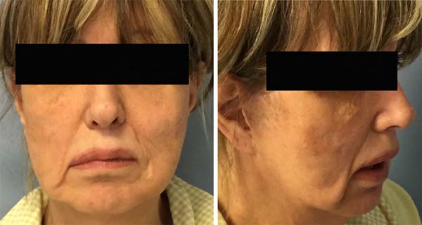
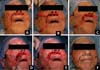
On examination 2 years after primary surgery, she had a bilaterally elongated upper lip, a right lower lip drop with evident lip dynamic distortion when she smiled, and bilateral cheek deformity probably due to marginal mandibular nerve damage (Figure 1). Using a flexible ruler, the distances of the modiolus to the cupid’s and commissure to the cupid’s bow were 4.5 and 3.5 cm on the paralyzed side and 4 and 3 cm on the non-paralyzed side.
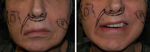
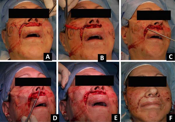
Preoperative markings in unilateral facial palsy are essential. With the patient seated upright, the nasolabial fold and cheek deformities were marked. A “modified bull’s horn approach” was taken to the upper lip lift1. The flap markings included the “bull’s horn” drawn around the nasal wings and columella and its pedicle along the right nasolabial fold (Figure 2). A line was drawn from the columellar-labial fold and extended laterally around the nasal wings to define the superior limit of the bull’s horn flap. Another line was drawn parallel to the first one in a lower position to define the inferior border of the flap, meeting the upper one at the lateral edges. Under local anesthesia and sedation, the flap was de-epithelized, and a dermoadiposal flap was harvested from the lateral left margin to the lateral right margin, and its pedicle was left attached to the upper ptotic right lip. Using the open tip of a small liposuction cannula, the flap was tunneled and fixed directly in a C-loop fashion using U stitches, and the flap was transfixed to the periosteum of the zygomatic arch. The traction tension of the flap was adjusted to balance the paralyzed and nonparalyzed sides of the upper and lower lips. During surgery, a first fat-graft injection was also made to correct cheek contour deformity, while a second fat-graft session was planned 3 months later to improve the aesthetic results (Figure 3).
Fat tissue was harvested using dry technique with a 3-mm multi-hole barbed cannula and a 10-mL syringe under manually generated negative pressure. The fat was centrifuged at 1000 rpm for 3 minutes and finally injected with a 19G needle. The high-density fraction of the concentrated adipose tissue was used to restore the cheek deformities, while the low-density fraction was used for aesthetic refinements of the face2.
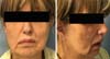
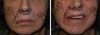
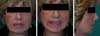
No significant complications were observed, and the patient reported no functional limitations or dissatisfaction with the aspect of the scars in the nasolabial fold or around the nasal wings and columella. The upper and lower lips appeared to be well positioned with a slight deviation of the cupid’s bow toward the reconstructed side because of the overcorrection. An adequately shaped, intense smile was observed despite some incoordination with the non-paralyzed side. Postoperatively, the distances of the modiolus to the cupid’s bow and commissure to the cupid’s bow were 4 and 3 cm, respectively, on both sides. Balance and adequate positioning of philtrum on the median line were evaluated, dividing the distance from the paralyzed side with the sum of the measurements of the paralyzed and non-paralyzed sides. The pre- and postoperative values of 53.3% and 50%, respectively, for the modiolus and commissure distances suggested a static good outcome. The results at 3 years of follow-up are shown in Figure 4.

Discussion
Facial paralysis is a profoundly disfiguring condition with significant psychological and functional consequences. Prior to the introduction of surgical reanimation, treatment was based on medical therapies (botulinum toxin injection) and prostheses3. Over time, static surgical procedures were introduced; thereafter, the use of temporalis and masseteric muscle transfers became popular.
Recently, nerve grafts and free muscle flap transfers have been considered by many surgeons the ideal procedures for long-standing facial paralysis to restore symmetry at rest and during smiling. However, facial movements are generally unidirectional and localized and cannot precisely reproduce the function of multiple muscles responsible for oral expressions4,5.
Differences in patients, facial perceptions, and various treatment strategies preclude a direct comparison of reconstructive techniques.
Tissue transfer is correlated to major trauma and donor site morbidity; it can require a two-stage operation, prolonged re-innervation time, and multiple revision procedures over time6. Even if it is performed at specialized centers, a patient’s baseline health status requires evaluation. Thus, elderly or frail patients may not choose microneurovascular tissue transfer.
Static restoration of the lower face is generally used to achieve symmetry at rest. Slings such as harvested tensor fascia lata are commonly used7. Even if static procedures cannot achieve mimic movements, they generate limited trauma and can be completed in a single surgical stage.
Here we presented an easy way to correct a lip deformity using a modified bull’s horn approach. The original technique was first described by Ramirez et al1 for an upper lip lift. It consisted of an excision of the white part of the upper lip directly beneath the nose in the shape of a bull’s horn, with advancement of the inferior border of the incision to the area directly beneath the nose. We used the same approach to harvest a dermoadiposal flap by using commonly discarded tissues and adding the pedicle flap markings in the nasolabial fold with a final concealed scar.
The patient was informed of the different techniques available to correct her defect as a consequence of facial nerve damage. She did not suffer from any particular disease and was considered a good candidate for a temporalis muscle transposition with a facelift. The patient refused this option because she did not want to undergo an invasive procedure. Because she complained of lower lip elongation, another option was offered to correct her defect as described.
A fat transfer procedure was carried out simultaneously with the flap transfer to correct a volume defect. At 3 months after the first surgery, a second fat transfer session was planned to improve the residual cheek deformity bilaterally as a consequence of the nodule removal. At 3 years of follow-up, the asymmetry correction appeared stable and the dermoadiposal flap was a good autologous suspension material for static correction of the lip deformity.
REFERENCES
1. Ramirez OM, Khan AS, Robertson KM. The upper lip lift using the ‘bull’s horn’ approach. J Drugs Dermatol. 2003 Jun;2(3):303-6.
2. Caggiati A, Germani A, Di Carlo A, Borsellino G, Capogrossi MC, Picozza M. Naturally adipose stromal cell-enriched fat graft: comparative polychromatic flow cytometry study of fat harvested by barbed or blunt multihole cannula. Aesthet Surg J. 2017 May;37(5):591-602. PMID: 28052909 DOI: https://doi.org/10.1093/asj/sjw211
3. Biglioli F, Frigerio A, Colombo V, Colletti G, Rabbiosi D, Mortini P, Dalla ET, Lozza A, Brusati R. Masseteric-facial nerve anastomosis for early facial reanimation. J Craniomaxillofac Surg. 2012 Feb;40(2):149-55. DOI: https://doi.org/10.1016/j.jcms.2011.03.005
4. Biglioli F, Frigerio A, Autelitano L, Colletti G, Rabbiosi D, Brusati R. Deep-planes lift associated with free flap surgery for facial reanimation. J Craniomaxillofac Surg. 2001 Oct;39(7):475-81. DOI: https://doi.org/10.1016/j.jcms.2010.09.003
5. Chuang DC. Technique evolution for facial paralysis reconstruction using functioning free muscle transplantation - experience of Chang Gung Memorial Hospital. Clin Plast Surg. 2002 Oct;29(4):449-59. DOI: https://doi.org/10.1016/S0094-1298(02)00021-4
6. Harrison DH, Grobbelaar AO. Pectoralis minor muscle transfer for unilateral facial palsy reanimation: an experience of 35 years and 637 cases. J Plast Reconstr Aesthet Surg. 2012;65(7):845-50. DOI: https://doi.org/10.1016/j.bjps.2012.01.024
7. Rose EH. Autogenous fascia lata grafts: clinical applications in reanimation of the totally or partially paralyzed face. Plast Reconstr Surg. 2005 Jul;116(1):20-32;discussion:33-5. DOI: https://doi.org/10.1097/01.PRS.0000169685.54862.18
1. Istituto Dermopatico dell’Immaticolata IDI, Rome, Italy.
Corresponding author: Rosaria Laporta Via dei Monti di Creta, 104, Rome, RM, Italy. Zipe Code: 00167 E-mail: r.laporta@idi.it
Article received: November 11, 2018.
Article accepted: April 16, 2019.
Institution: Istituto Dermopatico Dell’immacolata, Idi, Irccs, Rome, Italy.
Conflicts of interest: none.
 Read in Portuguese
 Read in English
 PDF PT
 Print
Print
 Send this article by email
 How to Cite
 Mendeley
 Pocket