


Case Report - Year 2019 - Volume 34 -
Use of pectoralis major fascia in dorsal nasal augmentation: case report
Uso de fáscia peitoral maior em preenchimento de dorso nasal: relato de caso
Camila Matos Versiani1,* ; Lucas Silva Couto2; Andreia Souto da Motta1; Marcus Vinícius Capanema Gonçalves1; David Santiago Ordonez Arizaga1; Klaus Rodrigues de Oliveira1; Waldemar Chaves Nascimento Brandão Penna1; Sérgio Moreira da Costa1
ABSTRACT
Increasing the nasal dorsum in rhinoplasty is the focus of several studies that seek the best graft sources and surgical techniques. The use of cartilage from the nasal septum, ear shell, or costal arches is already established for this purpose. In recent years, methods have been sought to reduce the palpability and dispersibility of cartilaginous grafts. Thus, synthetic materials such as SURGICEL® and autologous materials such as fascia have been explored. Temporal fascia are more widely used but require a new surgical incision, increasing surgical time and morbidity. Also described is the use of fascia lata and rectus abdominis fascia, which are comparatively thicker and less flexible. In many rhinoplasty procedures, it is necessary to remove the costal cartilage, which allows the collection of fascia from the major chest muscles through the same surgical incision. Thus, we describe the use of major chest muscle fascia and chopped costal cartilage in structured rhinoplasty to increase the dorsum.
Keywords: Rhinoplasty; Autologous transplantation; Fascia; Costal cartilage; Graft survival
RESUMO
O aumento do dorso nasal nas rinoplastias é foco de estudo de diversos trabalhos que buscam as melhores fontes de enxerto e técnicas cirúrgicas. A utilização de cartilagem já é consagrada para este fim, a partir do septo nasal, da concha auricular ou dos arcos costais. Nos últimos anos, têm-se buscado meios para reduzir a palpabilidade e dispersibilidade dos enxertos cartilaginosos. Assim, são descritos materiais sintéticos, como o SURGICEL®; e, autólogos, representados pelas fáscias. A fáscia temporal é mais amplamente utilizada, porém requer uma nova incisão cirúrgica, aumentando o tempo e a morbidade da cirurgia. É também descrito o uso de fáscia lata e fáscia reto abdominal, comparativamente mais espessas e menos flexíveis. Em muitos casos de rinoplastia fazse necessária a retirada da cartilagem costal, o que permite a coleta de fáscia do músculo peitoral maior pela mesma incisão cirúrgica. Dessa forma, descrevemos a utilização da fáscia do músculo peitoral maior envolvendo cartilagem costal picada, em uma rinoplastia estruturada com aumento do dorso.
Palavras-chave: Rinoplastia; Transplante autólogo; Fáscia; Cartilagem costal; Sobrevivência de enxerto
INTRODUCTION
Increasing the nasal dorsum in rhinoplasty demands good preoperative planning, intraoperative execution, and postoperative care. Synthetic, autologous, and homologous materials, mainly cartilage, have been used for this purpose1,2.
The use of cartilage has been widely studied in recent decades. The graft is obtained from septal, conchal, or costal cartilage and can be used and shaped in various ways, surviving as a living tissue with a low occurrence of absorption, extrusion, and infection. In addition, it provides almost no stimulus to the immune response1,3.
Minced cartilage is a versatile option for filling and camouflaging cartilaginous grafting. The process is performed manually to obtain cubes smaller than 1 mm2. The use of fascia to integrate the chopped cartilage aims to improve graft contour and reduce palpability and dispersibility; the use of temporal fascia for this purpose is most frequently described1,3.
Here we report the case of a patient who underwent bilateral rhinoplasty and mastopexy with prostheses. In this case, we used fragments of major chest fascia to integrate the chopped costal cartilage using a technique similar to that described by Erol in 20004.
CASE REPORT
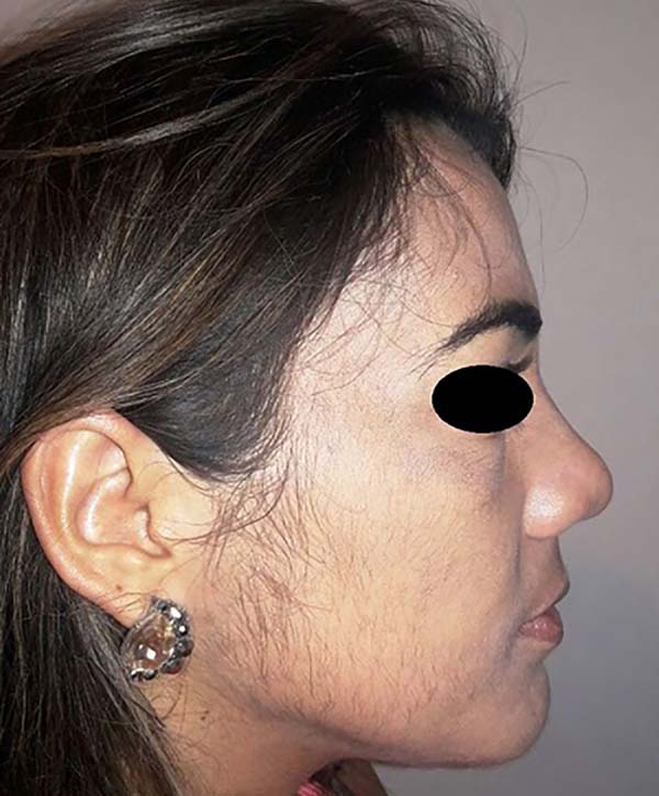
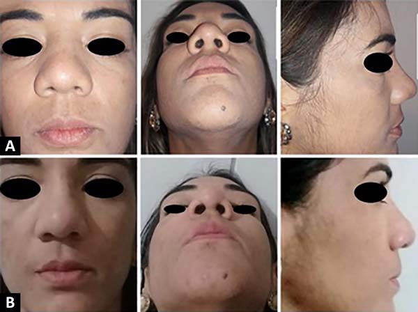
R.K.S.M., a 35-year-old patient, underwent structured rhinoplasty with increased dorsum and mastopexy with prostheses on January 26, 2018 (Figure 1).

A bilateral incision was made in the mammary sulcus with dissection to the right up to the pectoralis major muscle and the collection of a 4 cm × 4 cm portion of its fascia (Figures 2-5).

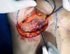
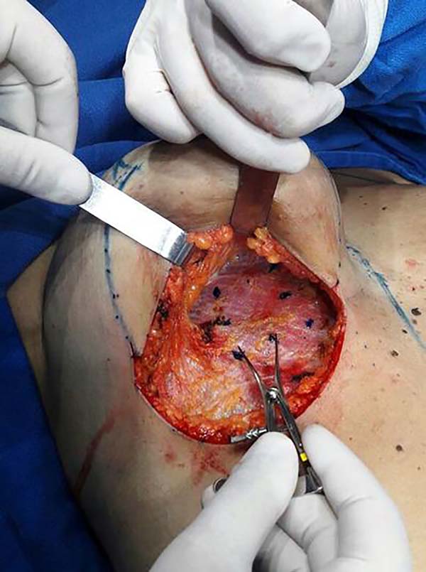


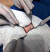
As described by Robotti and Penna5, the fibers of the pectoralis major muscle are divulgated but not separated. The sixth costal arch is accessed and bone and cartilage are differentiated through color discrimination and needle palpation. Thus, an entire fragment of cartilage was collected and the perichondrium was left intact. The wound is filled with saline solution, the integrity of the pleura is confirmed by ventilatory pressure by the anesthesiologist, and the planes are closed.
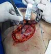
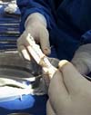
The collected fascia was wrapped in a 1-mL syringe and one of its extremities was sutured, thus creating an open bag in one of its extremities as in the Turkish delight technique4. Through the open end, the small cylindrical bag was filled with the costal cartilage manually minced into small cubes. The open end was then sutured (Figure 6).

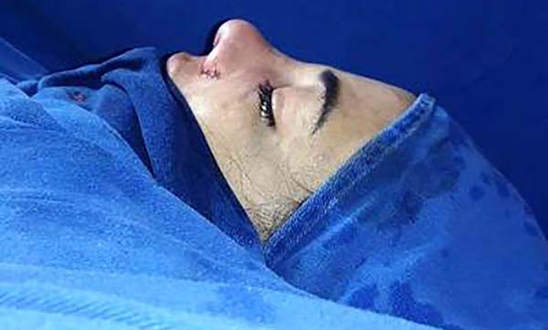
The cylinder of the chopped cartilage involved by the fascia was positioned on the patient’s nasal dorsum, promoting its increase in size (Figure 7). Mastopexy was performed with the inclusion of prostheses. The surgery proceeded uneventfully, and the patient was discharged on the 1st postoperative day in excellent general condition. In outpatient follow-up, we observed the stability of the graft on the nasal dorsum 17 months after surgery (Figure 8).


DISCUSSION
Deep temporal fascia is frequent in primary and secondary rhinoplasty because it has good flexibility and thickness, a low absorption rate, and is resistant to infection. Thus, the fascia can be used to cover the nasal osteocartilaginous framework and prevent irregularities from becoming apparent, especially in patients with thin skin. It is also used for nasal back augmentation and to better define the tip5.
The use of fascia involving chopped cartilage also brings other benefits. It has already been demonstrated that the chopped parts coated by fascia coalesce into a mass with an organized architecture and chondrocytes that present normal metabolic activity. Thus, the graft has adequate durability and stability2.
Collecting temporal fascia is a simple procedure, but it requires a second incision in a separate surgical field, which adds time and morbidity to the surgery as well as a new scar. There are reports of the use of fascia lata for the same purpose, but this has relative disadvantages, such as less flexibility. The occurrence of alopecia close to a temporal surgical incision was also mentioned6.
The fascia of the rectus abdominis muscle is comparatively thicker and less compliant in addition to being important to abdominal wall integrity and contractility. Studies by As’adi et al. in 20149 and Cerkes and Basaran in 201610, which used fascia of the rectus abdominis muscle and chopped cartilage for increased nasal dorsum, demonstrated good long-term graft viability. However, further studies are needed on rectus abdominis muscle stability and contraction7,8,9,10.
The major chest fascia, a thin layer of connective tissue that covers the major chest muscle, is histologically and macroscopically similar to the temporal fascia. It is routinely dissected and removed in radical oncological mastectomies. No specific surgical maneuvers are necessary to avoid dehiscence of the pectoralis major muscle nor are there any other complications. Furthermore, removal of the major chest fascia in mastectomies does not influence postoperative bleeding or seroma formation7.
In patients in need of nasal dorsal augmentation or revision rhinoplasty, autologous costal cartilage is usually collected, as good quality cartilage is required for nasal augmentation or structuring. Costal cartilage has great advantages over auricular cartilage with respect to quantity and quality7,8.
To remove the cartilage when the fifth, sixth, or seventh costal arch is chosen, the major pectoral fascia is accessed through the same incision. In this way, one part of the fascia can be collected for use in rhinoplasty, thus avoiding another surgical incision and its inherent morbidity7. In the present case, the patient underwent bilateral mastopexy and rhinoplasty. Through the mastopexy incision, it is possible to collect the pectoral fascia and costal cartilage.
CONCLUSION
The collection and use of chest fascia in cases requiring costal cartilage eliminates the need for another incision and scar in addition to reducing surgical morbidity and time. The fascia of the pectoralis major muscle is a good alternative for use in rhinoplasty.
COLLABORATIONS
|
CMV |
Conceptualization, Methodology, Visualization, Writing - Original Draft Preparation, Writing - Review & Editing |
|
LSC |
Analysis and/or data interpretation, Writing - Original Draft Preparation |
|
ASM |
Writing - Original Draft Preparation |
|
MV |
Analysis and/or data interpretation, Writing - Original Draft Preparation |
|
DSOA |
Writing - Original Draft Preparation |
|
KRO |
Writing - Original Draft Preparation |
|
WCNBP |
Final manuscript approval, Realization of operations and/or trials, Supervision |
|
SMC |
Supervision |
REFERENCES
1. Baser B, Kothari S, Thakur M. Diced cartilage: na effective graft for posttraumatic and revision rhinoplasty. Indian J Otolaryngol Head Neck Surg. 2013 Aug;65(Suppl 2):356-359. DOI: https://doi.org/10.1007/s12070-012-0525-6
2. Park P, Jin HR. Diced cartilage in fascia for major nasal dorsal augmentation in Asians: a review of 15 consecutive cases. Aesth Plast Surg. 2016 Dec;40(6):832-839. DOI: https://doi.org/10.1007/s00266-016-0698-6
3. Bussi M, Palonta F, Toma S. Grafting in revision rhinoplasty. Acta Otorhinolaryngol PMID: 23853414Ital. 2013;33(3):183-189.
4. Erol OO. The Turkish delight: a pliable graft for rhinoplasty. Plast Reconstr Surg. 2000 May;105(6):2229-41. DOI: https://doi.org/10.1097/00006534-200005000-00051
5. Park SW, Kim JH, Choi CY, Jung KH, Song JW. Various applications of deep temporal fascia in rhinoplasty. Yonsei Med J. 2015 Jan;56(1):167-174. DOI: https://doi.org/10.3349/ymj.2015.56.1.167
6. Hodgkinson DJ, Valente V. The versatile posterior auricular fascia in secondary rhinoplasty procedures. Aesth Plast Surg. 2017 Aug;41(4):893-897. DOI: https://doi.org/10.1007/s00266-017-0824-0
7. Xavier R. Pectoralis major fascia in rhinoplasty. Aesth Plast Surg. 2015 Mar;39(3):300-305. DOI: https://doi.org/10.1007/s00266-015-0461-4
8. Daniel RK, Sajadian A. Secondary rhinoplasty: management of the overresected dorsum. Facial Plast Surg. 2012 Aug;28(4):417-26. DOI: https://doi.org/10.1055/s-0032-1319840
9. As’adi K, Salehi SH, Shoar S. Rib diced cartilage-fascia grafting in dorsal nasal reconstruction: a randomized clinical trial of wrapping with rectus muscle fascia vs deep temporal fascia. Aesthet Surg J. 2014 Aug;34(6):NP21-31. PMID: 24879882 DOI: https://doi.org/10.1177/1090820X14535078
10. Cerkes N, Basaran K. Diced Cartilage Grafts Wrapped in Rectus Abdominis Fascia for Nasal Dorsum Augmentation. Plast Reconstr Surg. 2016 Jan;137(1):43-51. PMID: 26368329 DOI: https://doi.org/10.1097/PRS.0000000000001876
1. Hospital Felício Rocho, Belo Horizonte, MG, Brazil.
2. Santa Casa de Misericórdia, Belo Horizonte, MG, Brazil.
Corresponding author: Camila Matos Versiani Rua Platina, 56, Apto 201, Belo Horizonte, MG, Brazil. Zip Code: 30411-092 E-mail: camilamversiani@hotmail.com
Article received: October 25, 2018.
Article accepted: January 22, 2019.
Institution: Hospital Felício Rocho, Belo Horizonte, MG, Brazil.
Conflicts of interest: none.
 Read in Portuguese
 Read in English
 PDF PT
 Print
Print
 Send this article by email
 How to Cite
 Mendeley
 Pocket