


Reviw Article - Year 2019 - Volume 34 -
Treatment of keloids: a literature review
Tratamento para queloides: revisão de literatura
Pedro Martins Corrêa1,* ; Camila Eugênia Fonseca Passos1; Eduardo Mesquita de Souza1; Guilherme Augusto Sousa Batista1; Jéssica De Oliveira Jacintho1; Luiza Barbosa de Oliveira1; Oscar Campos Lisboa1; Joyce de Sousa Fiorini Lima1
ABSTRACT
Introduction: Keloids are characterized by an abnormal
response to dermal trauma, resulting in fibroblast proliferation,
excessive collagen production, and impairment of adjacent
healthy tissue. The diagnosis is clinical, and many conservative
and surgical methods can be used as treatments. However,
data on the efficacy of these treatments are limited, and
there is no consensus regarding the best treatment option.
This gap needs to be filled by developing comprehensive
evidence-based therapies.
Methods: A non-systematic
literature review of keloid scars was carried out using
PubMed, Scielo, MEDLINE, UptoDate, and dermatology
and dermatological surgery textbooks.
Literature review: The search retrieved relevant information on surgical and
adjuvant therapies used for keloids, including excision,
intralesional injections, cryotherapy, laser therapy, silicone
gel sheeting, radiation therapy, and pressure therapy. These
data are crucial because, in addition to complaints of pain,
itching, and restriction of movement, the main reason for
seeking treatment for keloids is for cosmetic and aesthetic
improvement, and the rates of recurrence and treatment
failure are high, emphasizing the importance of creating
awareness regarding the available procedures and their
effectiveness.
Conclusion: Many surgical and adjuvant
therapies for keloids are available. Nonetheless, there is no
consensus on a universally accepted treatment. Therefore,
additional high-quality studies are needed to identify the most
effective therapeutic approaches to achieve better results.
Keywords: Keloid; Plastic surgery; Scar; Therapy; Relapse.
RESUMO
Introdução: Queloides surgem de resposta excessiva à lesão da derme, resultando em proliferação de fibroblastos, produção exagerada de colágeno e comprometimento da pele sadia adjacente. O diagnóstico é clínico e muitos métodos conservadores e cirúrgicos já foram utilizados para tratamento. Porém, dados da eficácia desses tratamentos são limitados e não há consenso na literatura quanto a melhor técnica a ser empregada, permanecendo uma lacuna que necessita ser preenchida, a fim de que seus usos sejam indicados com maior confiabilidade, em um modelo de medicina baseada em evidências.
Métodos: Revisão não sistemática da literatura sobre "queloides" nas bases de dados PubMed, Scielo, MEDLINE, UptoDate e livros-texto das áreas de Dermatologia e Cirurgia Dermatológica.
Revisão de Literatura: Foram enumeradas e abordadas as principais informações sobre técnicas cirúrgicas e adjuvantes empregadas para essas lesões, que são: excisão, injeções intralesionais, crioterapia, laserterapia, revestimento com gel de silicone, radioterapia e pressoterapia. Torna-se relevante o levantamento dessas informações, tendo em vista que, além de poder causar dor, prurido e restrição de movimento, o principal motivo da procura de assistência médica para queloide é devido ao aspecto cosmético/estético, e as taxas de reincidência e falha terapêutica ainda são altas, sendo necessário conscientizar o paciente sobre o procedimento e seus efeitos.
Conclusão: São muitos os tratamentos disponíveis para o queloide, sejam cirúrgicos ou não, todavia não há consenso sobre uma abordagem universalmente aceita. São necessários mais estudos, com a finalidade de definir a melhor conduta e atingir melhores resultados, visto a qualidade mediana das evidências apresentadas nos estudos.
Palavras-chave: Queloide; Cirurgia plástica; Cicatriz; Terapêutica; Recidiva
INTRODUCTION
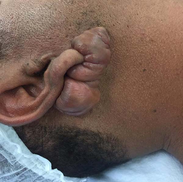
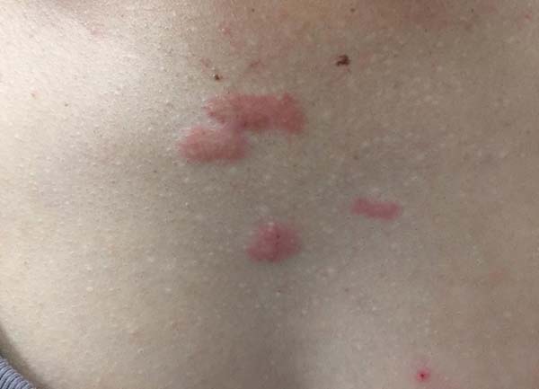
Keloids are abnormal, benign, erythematous, fibrous proliferations formed on the skin and usually develop after dermal trauma caused by burns, surgeries, acne, piercings, and tattoos.1 Healing extends beyond the wound margins surrounding adjacent normal skin2 (Figures 1 and 2).
The pathogenesis involves excessive fibroblast proliferation and abnormal collagen production. Wound tension and infectious processes promote hypoxia and increased deposition of extracellular matrix components leading to the formation of thick collagen bundles with a hyaline aspect. Lesions usually occur on the face, ear lobes, trunk, and neck potentially causing functional and cosmetic impairments, which may lower self-esteem and quality of life1.
The incidence and prevalence of keloids are unknown but affect predominantly young adults. Moreover, the prevalence varies according to race and is higher in Black and Asian populations, but similar between sexes. Keloids have a genetic basis, with an autosomal dominant inheritance of incomplete penetrance and manifest in genetic syndromes3.
The diagnosis is usually clinical and based on medical histories as well as the shape, size, and growth pattern of lesions. Biopsies may be performed in cases in which diagnoses are uncertain. The most common symptoms are pain and itching. After burn injuries, temperature dysregulation, dry skin, neuropathic pain, and impairment of mechanical function may occur due to destruction of hair follicles, sweat glands, capillaries, and nerve endings4.
The objective of treatment is to reduce complaints and scar volume and promote functional and cosmetic improvements. Furthermore, the chosen surgical approach needs to include the possible postoperative risk of overhealing.
It is known that the delay in epithelialization is related to an increased occurrence of keloids. Therefore, it is necessary to stimulate rapid re-epithelization with respect to physiological mechanisms and skin anatomy5.
For this reason, the risk of recurrence is a significant limiting factor in therapeutic success. Recurrence is more common in body regions subject to mechanical stress, and increasing patient awareness regarding this fact is fundamental. Current treatment options fail to achieve satisfactory scar reduction. Moreover, despite the large number of treatment options, their quality of evidence is low and intervention outcomes have poor consistency and predictability. This limitation has caused confusion about which treatments and techniques should be considered primary, secondary, or tertiary.
OBJECTIVE
The objectives of this review were to outline the main treatment options for keloids and address their advantages and disadvantages.
METHODS
A non-systematic literature review of keloids was conducted using PubMed, Scielo, DynaMed, MEDLINE, UptoDate, and textbooks in dermatology and dermatological surgery to identify the main surgical and adjuvant therapies. The descriptors used were keloids, treatment, surgery, and healing.
LITERATURE REVIEW
Keloids can become lesions that are healed in a timely manner preventing the development of the pathophysiological features of the original lesion. Current surgical approaches should include adjuvant therapies to avoid the risk of recurrence potentially leading to the formation of larger keloids. The most commonly used therapies are:
3.1 - Excision
Excision consists of the surgical removal of the keloid and is considered a secondary or tertiary treatment option for mature keloids6. The fusiform excision of lesions at an angle of 30° to skin tension lines is recommended. Recurrence can be avoided by reducing the tension at wound closure, leaving the edges everted, and using resorbable thread in the fascia or subcutaneous tissue7.
Avoiding excessive manipulation and trauma, removing foreign bodies, and preventing bruises and infections help improve the quality of scars. Surgical correction by Z-plasty or W-plasty is possible8.
Surgical excision alone should be avoided because of the risk of recurrence in 45-100% of cases, and therefore should be associated with adjuvant therapies. The use of pressure dressings after surgery is also recommended9.
Surgical excision combined with radiotherapy is an effective treatment for large keloids according to international clinical recommendations; however, this approach should be used as a last resort. The combination of surgery with radiotherapy in patients who failed a first treatment increased the recurrence rate from 8% to 28%10. A previous study demonstrated that surgical excision of foot keloids associated with intralesional corticosteroid injections and silicone gel sheeting decreased the 1-year recurrence rate by 78.5%11.
3.2 - Intralesional injections
Intralesional injections involve the intralesional application of medications such as corticosteroids. The most commonly used medication is triamcinolone acetonide which is considered a primary therapy and is usually combined with silicone gel sheeting for keloid scars smaller than 0.5 cm. It is also the first-choice treatment in isolation or combination with intralesional fluorouracil or cryotherapy for keloids larger than 0.5 cm6.
Corticosteroids act by decreasing the synthesis of collagen and glycosaminoglycan and inhibiting fibroblast proliferation. These drugs are effective in reducing pain and itching because of their anti-inflammatory and vasoconstrictive properties. The rate of response to treatment is 50-100%, and the 5-year recurrence rate is ≤50%. Injections are painful and have adverse effects in 63% of cases, including dermal atrophy, hypopigmentation, and telangectasis7.
Cryotherapy facilitates corticosteroid injection but causes residual vitiligo. The use of thermal bags before cryotherapy applications and slow administration can reduce pain1. Injectable corticosteroids are also used in adjuvant therapy after surgical excision7, whereas the use of triamcinolone after surgical excision is effective for keloids larger than 4 cm12.
Intralesional verapamil may be as effective as intralesional triamcinolone in treating keloids but has fewer adverse effects13. However, a randomized study found that the risk of recurrence using verapamil was higher14. Another randomized trial demonstrated that the effectiveness of the two treatments was similar, but the delay in treatment outcomes was longer with verapamil15.
Intralesional 5-fluorouracil (5-FU) is used for lesions refractory to corticosteroid treatment. This antimetabolite chemotherapeutic agent interferes with DNA and RNA synthesis and induces fibroblastic apoptosis; it also inhibits type I collagen production by affecting transforming growth factor-beta (TGF-β) signaling16.
The adverse effects of 5-FU include pain and hyperpigmentation7. This medication can be used together with intralesional corticosteroids and as an adjuvant after surgical excision8. The rate of success of treatment in isolation and in combination with corticosteroids is 45-78% and 50-96%, respectively. However, the quality of evidence of 5-FU treatments is low17.
Botulinum toxin type A injections have been used as a new treatment option. It acts by decreasing tension in the tissue near the keloid scar during the healing process and affecting apoptosis and cell proliferation. A study found that the rate of recurrence in 80 patients with keloid lesions who underwent excisional surgery followed by a single application of 5-FU and botulinum toxin on day 9 after surgery was 4%. Therefore, the authors recommended the routine application of this toxin after resection; however, further studies are needed to assess the effectiveness of this approach18.
3.3 - Dermal regeneration matrix
The dermal regeneration matrix (DRM) (Integra®) consists of a bilaminar structure formed by a dermal component (bovine collagen and chondroitin-6-phosphate) and an epidermal component made with a 100 µm thick synthetic silicone sheet19.
It has a three-dimensional structure with 50 ± 20 µm pores and resembles normal skin. The matrix is completely degraded after 30 days. The outer silicone layer acts as a protective barrier and prevents the loss of liquids. After neovascularization, the silicone sheet is replaced with a thin autologous epidermal graft, and the epidermal cells in the grafts proliferate and attach to the neodermis19.
DRMs promote wound healing without wound tension, do not trigger immune responses, contain a thin epidermal graft, do not require adjuvant therapies, and the results resemble healthy skin. DRMs are sterile and their implantation is relatively simple. The disadvantages are its high cost, risk of infection between the two layers of the matrix, and the need for two surgical procedures19,20.
A study evaluated the use of DRMs for keloids and found that the aesthetic results were acceptable and that the skin remained flat and infection free. Moreover, there were no long-term complaints or evidence of scarring at donor sites. Limitations included a small sample size and a follow-up period of <24 months20.
3.4 - Cryotherapy
Cryotherapy involves freezing keloids with liquid nitrogen. Studies recommend the monthly administration of intralesional corticosteroids with or without adjuvant cryotherapy as the primary treatment for large keloids6.
The mechanism of action involves cellular and microvascular damage, leading to necrosis and consequent involution of the lesions without affecting the connective tissue framework. This approach induces immune modulation and tumor cell apoptosis. The mechanism of action depends on the freezing and thawing rate, tissue type, and temperature9.
This therapy causes rapid freezing of the scar tissue from the lesion center to the outer superficial area. Cryotherapy differs from cryogenic contact therapy, which partially freezes the lesion. Intralesional cryotherapy can be performed using a probe, which is inserted in the scar and allows dermal targeting without affecting the epidermis21.
Side effects include blisters, pain, and hypo- or hyper-pigmentation. A study reported a 67.4% reduction in keloid volume in a 6-month period using a single session of intralesional cryotherapy, as well as improvement in redness and absence of hypopigmentation and recurrence9.
Evidence suggests that intralesional cryotherapy reduces scar volume and symptoms and has few adverse effects. However, more unbiased, robust, and comparative studies are needed to confirm this evidence21. A randomized trial demonstrated that intralesional cryotherapy and brachytherapy were less effective than surgical excision in patients with resistant keloids. Intralesional cryotherapy also reduces the volume of primary keloids22.
3.5 - Laser therapy
Targeted treatments include pulsed dye laser (PDL), carbon dioxide ablative fractional resurfacing (AFR), and intense pulsed light (IPL)23. PDL or AFR are indicated for smaller keloids refractory to silicone gel sheeting, intralesional corticosteroids, and 5-FU24.
Combined treatments, such as PDL and AFR, are recommended over single treatments by improving the aesthetic results and reducing symptoms25.
The most common complications of PDLs are transient purpura, mild or moderate erythema, and edema. Blistering or scabbing of the skin may occur in the early post-treatment stages. Hypo- or hyper-pigmentation may also occur, especially in patients with darker skin. AFRs may cause delayed healing, ulcers, and post-inflammatory hyperpigmentation23,26.
3.6 - Silicone gel sheeting
Silicone gel sheeting is used as a primary therapy to prevent and treat keloid scars smaller than 0.5 cm in moderate to high-risk patients6,26. The anti-scarring mechanism is unclear, but may be related to occlusion and hydration of the stratum corneum, generation of static electricity, and decreases in the number of mast cells. Silicone has been shown to increase collagenase activity and TGF β-2 levels and to regulate both cell proliferation and differentiation27.
This treatment should be performed after complete scar healing and for at least 12 hours a day for 2 to 3 months while maintaining a hygienic field6. A possible adverse reaction is folliculitis7. Silicone can be applied as sheets or gels, and both formulations are safe, effective, and produce good aesthetic results28.
3.7 - Radiation therapy
Radiation therapy consists of applying ionizing radiation to keloids using different therapeutic sessions and on different days. This therapy inhibits fibroblast proliferation and stimulates fibroblast differentiation27. Several studies have demonstrated the high efficacy of this method in reducing keloid recurrence when administered immediately after surgical excision29.
Radiotherapy as an adjuvant for operated keloids can be performed using different techniques, including TGF-β therapy, conventional X-ray, single-dose radiotherapy, and electron-beam radiotherapy. Electron irradiation is superior to conventional irradiation because of its better dose distribution in the target tissue and less penetration into deeper adjacent tissues30.
The dose used for treating keloids should be adapted according to the location of the lesion, and the highest doses should be used at high-risk sites. The effectiveness of this approach in keloid prevention depends on the biologically effective dose, and no consensus has been reached on the best dosage. A minimum follow-up of 2.5 years with postoperative electron irradiation is necessary to achieve satisfactory results regarding scar quality and reduction of recurrence30.
A study analyzed the treatment of 174 ear lobe keloids using a total dose of 10 Gy in 2 sessions or 15 Gy in 3 sessions. After 18 months of follow-up, the recurrence rate was 4% with no significant difference between doses31. Another study found that the overall relapse rate was 5.6% after a mean follow-up period of 40 months, and this rate was lower in patients who received doses higher than or equal to 20 Gy32.
Electron irradiation is well-tolerated by patients. Nonetheless, potential adverse effects include transient hyperpigmentation of the treated area and persistent peeling for approximately 3 months. In addition, it should be used with caution in children and young adults since a few body regions are sensitive to radiation in these populations. A study reported the potential risk of malignancy of cells subjected to radiotherapy27.
3.8 - Pressure therapy
Pressure therapy is a prophylactic approach involving the application of continuous pressure to keloids for several weeks to inhibit the development of new scars or to reduce the size of pre-formed scars27,33. This therapy can be performed using compression bandages or elastic compression stockings27. Pressure earrings, metal clips, and magnets, with or without silicone sheeting, have been used for treating ear lobe keloids34.
The mechanism of action is not well established but seems to be related to the occlusion of small blood vessels by pressure, leading to oxygen and nutrient deprivation and consequently decreasing fibroblast proliferation and collagen synthesis. In addition, decreased capillary blood flow reduces the level of alpha-2-macroglobulins resulting in the inhibition of collagenases27,34.
The optimal applied pressure is difficult to determine. Notwithstanding, this value should exceed capillary blood pressure, but should not decrease peripheral blood pressure (20-30 mmHg). A prospective study evaluated 11 patients with keloids initially treated with surgical resections, followed by pressure therapy after 15 to 21 days. Magnets with pressures between 33 and 47 mmHg were used, and the recurrence-free rate was 90.91% at the end of a 4-6-month follow-up period34.
Nonetheless, evidence supporting the use of this technique is limited. A meta-analysis that evaluated 6 high-quality studies showed no significant difference in the results between burn patients treated with or without pressure therapy35. The therapy has no significant side effects, except for possible pressure discomfort27. Moreover, treatments are simple and affordable, and therefore, essential in regions with limited technological and pharmacological resources27,34.
3.9 - Tissue adhesives (Prineo®)
Prineo® is a double polyester mesh system containing the glue octyl-2-cyanoacrylate and belongs to the group of tissue adhesives. Molecular reactions begin when the adhesives come into contact with the wound and the assembly adheres to keratinocytes36.
Prineo® should be applied to the margins of the incision as a thin layer in a single movement. After application, the product dries in approximately 5 min and patients may feel a warm sensation at the treatment site during the application37.
Tissue adhesives are applied quickly and easily, promote good tensile strength throughout the wound, form an antimicrobial barrier, and cause less pain during removal. In addition, scar enlargement using this method is smaller than that using suture closures. Prineo® is recommended for extensive wounds with higher tensions36.
A study compared Prineo® and surgical sutures and found that there were no significant differences (p > 0.05) in scar quality including aesthetics. In the study sample (n = 101), there was only one reported case of a keloid associated with surgical sutures and no cases associated with Prineo® (p = 0.042). The most relevant adverse effects are contact dermatitis in response to the formaldehyde released during the polymerization reaction between cyanoacrylate and the skin36.
A literature review has shown that this method is not widely used for treating keloids but is an additional option. Therefore, further studies on the use of Prineo® for keloid prevention and treatment are needed.
CONCLUSION
Several surgical and adjuvant therapies are available for treating keloids. However, the number of high-quality studies on this topic is small and there is no consensus on a universally accepted approach. Decisions regarding the best therapies are individualized and vary according to the size of the lesion, lesion recurrence, and available medical resources. Intralesional corticosteroid injections associated with other methods such as silicone gel sheeting or pressure therapy produce good aesthetic results in smaller lesions, whereas triamcinolone injections combined with cryotherapy yields good results in larger lesions.
COLLABORATIONS
|
PMC |
Analysis and/or data interpretation, Conception and design study, Conceptualization, Investigation, Methodology, Writing - Original Draft Preparation |
|
CEFP |
Analysis and/or data interpretation, Conception and design study, Conceptualization, Investigation, Methodology, Writing - Original Draft Preparation |
|
EMS |
Analysis and/or data interpretation, Conception and design study, Conceptualization, Investigation, Methodology, Writing - Original Draft Preparation |
|
GASB |
Analysis and/or data interpretation, Conception and design study, Conceptualization, Investigation, Methodology, Writing - Original Draft Preparation |
|
JOJ |
Analysis and/or data interpretation, Conception and design study, Conceptualization, Investigation, Methodology, Writing - Original Draft Preparation |
|
LBO |
Analysis and/or data interpretation, Conception and design study, Conceptualization, Investigation, Methodology, Writing - Original Draft Preparation |
|
OCL |
Analysis and/or data interpretation, Conception and design study, Conceptualization, Investigation, Methodology, Writing - Original Draft Preparation |
|
JSFL |
Final manuscript approval, Supervision |
REFERENCES
1. Pereira ALC, Leal F, Azulay DR, Azulay RD. Dermatologia. 6ª ed. Rio de Janeiro (RJ): Guanabara Koogan; 2013.
2. Shin JY, Lee JW, Roh SG, Lee NH, Yang KM. A comparison of the effectiveness of triamcinolone and radiation therapy for ear keloids after surgical excision: a systematic review and meta-analysis. Plast Reconstr Surg. 2016 Jun;137(6):1718-25. PMID: 27219228 DOI: https://doi.org/10.1097/PRS.0000000000002165
3. Goldstein BG, Goldstein AO. Keloids and hypertrophic scars. Dellavalle RP, Levy ML, Corona R, editors. Post TW. Waltham, MA: UpToDate; 2019; [acesso em 2019 fev 16]. Disponível em: https://www.uptodate.com/contents/keloids-and-hypertrophic-scars
4. Khansa I, Harrison B, Janis JE. Evidence-based scar management. Plast Reconstr Surg. 2016 Sep;138(Suppl 3):165S-78S. DOI: https://doi.org/10.1097/PRS.0000000000002647
5. Gauglitz GG. Management of keloids and hypertrophic scars: current and emerging options. Clin Cosmet Investig Dermatol. 2013;6:103-114. DOI: https://doi.org/10.2147/CCID.S35252
6. Gold MH, McGuire M, Mustoe TA, Pusic A, Sachdev M, Waibe J, et al. Updated international clinical recommendations on scar management: part 2 - algorithms for scar prevention and treatment. Dermatol Surg. 2014 Aug;40(8):825-31.
7. Arno AI, Gauglitz GG, Barret JP, Jeschke MG. Up-to-date approach to manage keloids and hypertrophic scars: a useful guide. Burns. 2014 Nov;40(7):1255-66. PMID: 24767715 DOI: https://doi.org/10.1016/j.burns.2014.02.011
8. Metsavaht LD. Abordagem cirúrgica de cicatrizes. Surg Cosmet Dermatol. 2016;8(1):11-20.
9. Berman B, Maderal A, Raphael B. Keloids and hypertrophic scars: pathophysiology, classification, and treatment. Dermatol Surg. 2017 Jun;43(Suppl 1):S3-S18. DOI: https://doi.org/10.1097/DSS.0000000000000819
10. Keeling BH, Whitsitt J, Liu A, Dunnick CA. Keloid removal by shave excision with adjuvant external beam radiation therapy. Dermatol Surg. 2015 Aug;41(8):989-92. DOI: https://doi.org/10.1097/DSS.0000000000000417
11. Park TH, Park JH, Chang CH. Clinical features and outcomes of foot keloids treated using complete surgical excision and full thickness skin grafting followed by corticosteroid injections. J Foot Ankle Res. 2013;6:26. DOI: https://doi.org/10.1186/1757-1146-6-26
12. Rasheed I, Malachy A. The management of helical rim keloids with excision, split thickness skin graft and intralesional triamcinolone acetonide. J Cutan Aesthet Surg. 2014 Jan/Mar;7(1):51-53. DOI: https://doi.org/10.4103/0974-2077.129981
13. Verhiel S, Grzymala AP, van der Hulst R. Mechanism of action, efficacy, and adverse events of calcium antagonists in hypertrophic scars and keloids: a systematic review. Dermatol Surg. 2015 Dec;41(12):1343-50. DOI: https://doi.org/10.1097/DSS.0000000000000506
14. Danielsen PL, Rea SM, Wood FM, Fear MW, Viola HM, Hool LC, et al. Verapamil is less effective than triamcinolone for prevention of keloid scar recurrence after excision in a randomized controlled trial. Acta Derm Venereol. 2016 Aug;96(6):774-8.
15. Ahuja RB, Chatterjee P. Comparative efficacy of intralesional verapamil hydrochloride and triamcinolone acetonide in hypertrophic scars and keloids. Burns. 2014 Jun;40(4):583-8. PMID: 24182692 DOI: https://doi.org/10.1016/j.burns.2013.09.029
16. Ledon JA, Savas J, Franca K, Chacon A, Nouri K. Intralesional treatment for keloids and hypertrophic scars: a review. Dermatol Surg. 2013 Dec;39(12):1745-57. PMID: 24299571 DOI: https://doi.org/10.1111/dsu.12346
17. Bijlard E, Steltenpool S, Niessen FB. Intralesional 5-fluorouracil in keloid treatment: a systematic review. Acta Derm Venereol. 2015;95:778-782.
18. Wilson AM. Eradication of keloids: surgical excision followed by a single injection of intralesional 5-fluorouracil and botulinum toxin. Can J Plast Surg. 2013;21(2):87-91. DOI: https://doi.org/10.1177/229255031302100208
19. Pereima MJL, Goulart BC, Pereima RR, Feijó R, Freitas JL. Diminuição do tempo de maturação de matrizes de regeneração dérmica quando associados a uso de curativos de pressão negativa. Rev Bras Queimaduras. 2013;12(3):145-152.
20. Nguyen KT, Shikowitz L, Kasabian AK, Bastidas N. A novel approach to keloid reconstruction with bilaminar dermal substitute and epidermal skin grafting. Plast Reconstr Surg. 2016 Jul;138(1):235-9. PMID: 26986991 DOI: https://doi.org/10.1097/PRS.0000000000002242
21. O’Boyle CP, Shayan-Arani H, Hamada MW. Intralesional cryotherapy for hypertrophic scars and keloids: a review. Scars Burn Heal. 2017 Jan/Dec;3. DOI: https://doi.org/10.1177/2059513117702162
22. Bijlard E, Timman R, Verduijn GM, Niessen FB, Hovius SER, Mureau MAM. Intralesional cryotherapy versus excision with corticosteroid injections or brachytherapy for keloid treatment: Randomised controlled trials. J Plast Reconstr Aesthet Surg. 2018 Jun;71(6):847-856. PMID: 29426811 DOI: https://doi.org/10.1016/j.bjps.2018.01.033
23. Brewin MP, Lister TS. Prevention or treatment of hypertrophic burn scarring: a review of when and how to treat with the pulsed dye laser. Burns. 2014 Aug;40(5):797-804. PMID: 24439925 DOI: https://doi.org/10.1016/j.burns.2013.12.017
24. Jin R, Huang X, Li H, Yuan Y, Li B, Cheng C, et al. Laser therapy for prevention and treatment of pathologic excessive scars. Plast Reconstr Surg. 2013 Dec;132(6):1747-58. PMID: 24281600 DOI: https://doi.org/10.1097/PRS.0b013e3182a97e43
25. Hultman CS, Friedstat JS, Edkins RE, Cairns BA, Meyer AA. Laser resurfacing and remodeling of hypertrophic burn scars: the results of a large, prospective, before-after cohort study, with long-term follow-up. Ann Surg. 2014 Sep; 260(3):519-29.
26. O’Brien L, Jones DJ. Silicone gel sheeting for preventing and treating hypertrophic and keloid scars. Cochrane Database Syst Rev. 2013 Sep;1(9):CD003826. DOI: https://doi.org/10.1002/14651858.CD003826.pub3
27. Fernandes WS, Ferreira RCA. Queloide: uma revisão dos tratamentos atualmente disponíveis. R Bras Ci Saúde. 2014;18(2):181-186. DOI: https://doi.org/10.4034/RBCS.2014.18.02.14
28. Puri N, Talwar A. The efficacy of silicone gel for the treatment of hypertrophic scars and keloids. Journal of Cutaneous and Aesthetic Surgery. 2009 Jul/Dec;2(2):104-6. DOI: https://doi.org/10.4103/0974-2077.58527
29. Lee SY, Park J. Postoperative electron beam radiotherapy for keloids: treatment outcome and factors associated with occurrence and recurrence. Ann Dermatol. 2015 Feb;27(1):53-58. DOI: https://doi.org/10.5021/ad.2015.27.1.53
30. Oliveira Júnior B, Schellini SA, Lastória JC, Carvalho LR, Stolf HO, Oliveira ALPD. Tratamento de queloides usando radioterapia pósoperatória com elétrons: estudo comparativo e randomizado com dois esquemas. Surg Cosmet Dermatol. 2013;5(1):16-26.
31. Ogawa R, Huang C, Akaishi S, Dohi T, Sugimoto A, Kuribayashi S, et al. Analysis of surgical treatments for earlobe keloids: analysis of 174 lesions in 145 patients. Plast Reconstr Surg. 2013 Nov;132(5):818e-825e. PMID: 24165633 DOI: https://doi.org/10.1097/PRS.0b013e3182a4c35e
32. Renz P, Hasan S, Gresswell S, Hajjar RT, Trombetta M, Fontanesi J. dose effect in adjuvant radiotherapy for the treatment of resected keloids. Int J Radiat Oncol Biol Phys. 2018 Sep;102(1):149-154. PMID: 29970316 DOI: https://doi.org/10.1016/j.ijrobp.2018.05.027
33. Monstrey S, Middelkoop E, Vranckx JJ, Bassetto F, Ziegler UE, Meaume S, et al. Updated scar management practical guidelines: non-invasive and invasive measures. J Plast Reconstr Aesthet Surg. 2014 Aug;67(8):1017-25. DOI: https://doi.org/10.1016/j.bjps.2014.04.011
34. Quintero-Larrovere M, Soto-Montenegro AE, Lozada-Urbani JA. Uso de imanes en el tratamiento de queloides auriculares. Cir Plást Iberolatinoam. Madrid. 2017 Abr/Jun;43(2):163-174.
35. Anzarut A, Olson J, Singh P, Rowe BH, Tredget EE. The effectiveness of pressure garment therapy for the prevention of abnormal scarring after burn injury: a meta-analysis. J Plast Reconstr Aesthet Surg. 2009 Jan;62(1):77-84. PMID: 18249046 DOI: https://doi.org/10.1016/j.bjps.2007.10.052
36. Cammarota MC, Moura LG, Ribeiro Junior ISMAR, Lima RQD, Almeida CMD, Daher L, Daher JC. Comparação de resultado estético em cicatrizes com uso de fios cirúrgicos e Prineo®. Rev Bras Cir Plást. 2017;32(1):101-108.
37. Lipsett S. Minor wound repair with tissue adhesives (cyanoacrylates). Wolfson AB, Wiley JF, editors. Post TW. Waltham, MA: UpToDate; 2019; [acesso em 2019 fev 16]. Disponível em: https://www.uptodate.com/contents/minor-wound-repair-with-tissue-adhesives-cyanoacrylates
1. Universidade Federal de Ouro Preto, Escola de Medicina, Ouro Preto, MG, Brazil.
Corresponding author: Pedro Martins Corrêa Rua Darcy Vargas, 40, Apto 102, Nova Suiça, Belo Horizonte, MG, Brazil. Zipe Code: 30421-093. E-mail: pedromc1987@gmail.com
Article received: December 19, 2018.
Article accepted: April 21, 2019.
Conflicts of interest: none.
 Read in Portuguese
 Read in English
 PDF PT
 Print
Print
 Send this article by email
 How to Cite
 Mendeley
 Pocket