


Case Report - Year 2020 - Volume 35 -
Peculiarities of cutaneous angiosarcoma
Angiossarcoma cutâneo e suas peculiaridades
Sinval Soares Cruvinel1 ; Victor Parreira Bizinoto1,*; Nádia Cristine Nunes Côrtes1; Alexandre Motta Macedo1; Daniel Miranda Alves Pereira1; Antônio Ricardo Duarte1
ABSTRACT
Cutaneous angiosarcoma is a rare soft tissue sarcoma with a poor prognosis and an incidence of approximately 2.0%. This entity manifests as bruises, violaceous nodules and plaques, and diffuse hemorrhagic lesions with infiltrative growth. Here we report a case of an 80-year-old Caucasian man who presented with a nodular, hard, and dark lesion present in the upper third of the right ear for more than 1 year. The treatment of cutaneous angiosarcoma is multidisciplinary, including surgery alone or combined with radiotherapy for early lesions and chemotherapy for disseminated lesions. Cutaneous sarcomas are rare, and their appropriate treatment and follow-up are critical.
Keywords: Hemangiosarcoma; Sarcoma; Skin neoplasms; Skin; Prognosis.
RESUMO
O angiossarcoma cutâneo é um sarcoma raro de tecido mole com prognóstico ruim, tendo a incidência em torno de 2,0% entre os sarcomas. Esta entidade pode se apresentar de várias formas clínicas, quais sejam, como lesão com aspecto de local contundido, nódulo, placa violácea e áreas hemorrágicas infiltrativas planas. Relatamos um caso de um homem leucoderma de 80 anos, cuja história se iniciou há mais de um ano com o surgimento de lesão nodular, rugosa e escura em terço superior da orelha direita. O tratamento do angiossarcoma cutâneo é multidisciplinar, sendo a cirurgia isolada ou associada à radioterapia (RT) usada para lesões iniciais e quimioterapia (QT) recomendada em lesões disseminadas. Os sarcomas cutâneos são tumores raros na rotina do cirurgião plástico, sendo crucial que, mediante suspeita, seja realizado tratamento e seguimento de maneira adequada.
Palavras-chave: Hemangiossarcoma; Sarcoma; Neoplasias cutâneas; Pele; Prognóstico
INTRODUCTION
The term “sarcoma” is derived from the Greek “sarkos” (flesh) and “oma” (tumor). Sarcomas comprise a heterogeneous group of mesenchymal neoplasms and are classified into primary bone sarcomas and soft tissue (cutaneous) sarcomas1.
Among soft tissue sarcomas, cutaneous angiosarcomas deserve special attention and can be divided into:
Idiopathic angiosarcoma: the most common type, especially in the elderly, characterized by ecchymotic plaques and/or friable violaceous nodules, ulcerated or not, usually on the head and neck;
Angiosarcoma secondary to chronic lymphedema: a violaceous nodule or infiltrated plaque more common in mastectomized patients who underwent axillary clearance (Stewart-Treves syndrome);
Post-radiation angiosarcoma: a rare sarcoma associated with conservative treatment of breast cancer usually presenting as infiltrated plaques or nodules adjacent to the irradiated area; and
Low-grade angiosarcomas1. Cutaneous angiosarcoma is a rare tissue soft tissue sarcoma with an incidence of approximately 2% and a poor prognosis2.
This disease is most common in the age group >60 years and in men (2:1 ratio)3. Its clinical forms include bruises, violaceous nodules and plaques, and diffuse hemorrhagic lesions with infiltrative growth3. These lesions are histologically indistinguishable and comprise a network of dermal vascular channels varying in size from small capillaries to sinusoidal spaces interspersed with normal endothelium4. One study showed that clinical outcomes were not favorable even after complete tumor resection, as for other sarcomas5.
CASE REPORT
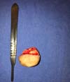
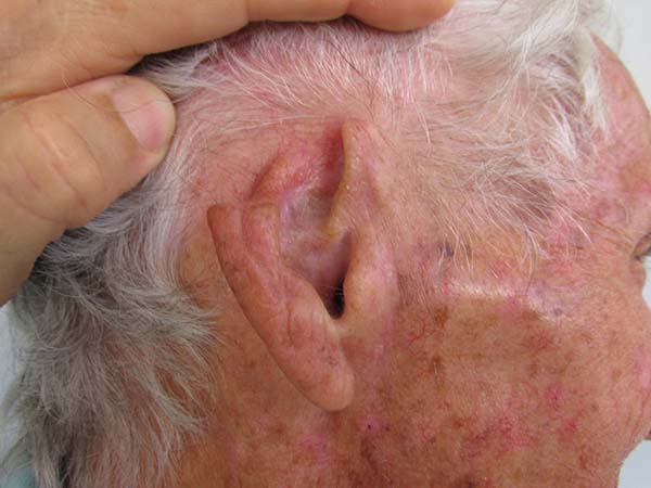
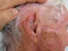
An 80-year-old man presented with a lesion present in the upper third of the ear for more than 1 year (Figures 1, 2, and 3). The patient reported no itching or pain. He reported that the lesion growth was slow and progressive, which motivated him to seek a specialized evaluation.

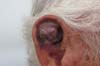
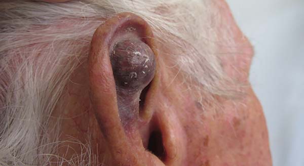
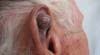
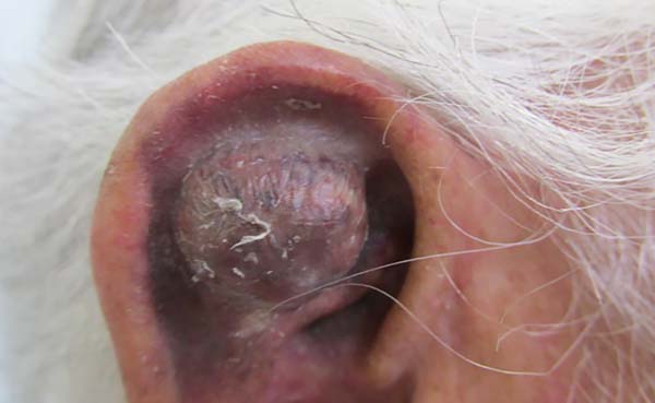
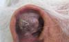
The examination showed a hyperchromic, nodular, and hard lesion in the upper third of the right ear. The lesion had regular borders but no inflammatory signs or secretions. There was no evidence of enlarged cervical lymph nodes.
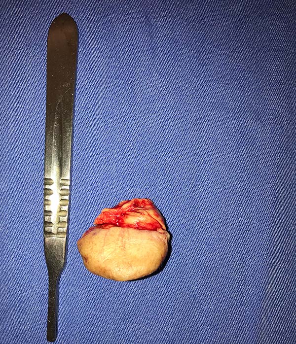
Surgical resection of the upper third of the right ear was performed under local anesthesia (Figures 4 and 5) and the wound edges were sutured. Surgical reconstruction was scheduled for a later date (Figure 6) because of the uncertain nature of the lesion and a high suspicion of malignancy.
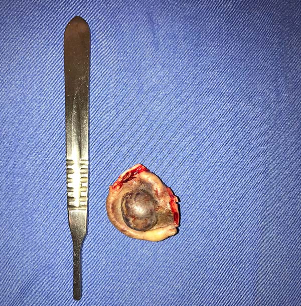
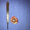
A histopathological examination showed the presence of tumors with poorly differentiated mesenchymal cells compatible with cutaneous angiosarcoma. An immunohistochemical examination showed positive human anti-CD34.
DISCUSSION
Cutaneous angiosarcoma should be treated by a multidisciplinary team according to the lesion extent, anatomical location, and patient preferences. Surgical resection alone or in combination with radiotherapy is used for early lesions, although wide-margin resection is not always feasible.
Chemotherapy is indicated for disseminated tumors and can be combined with radiotherapy for the locoregional treatment of extensive lesions or as neoadjuvant therapy. The most commonly used chemotherapeutic agents are doxorubicin, cyclophosphamide, methotrexate, and vincristine. The concomitant use of alpha-interferon and 13-cis-retinoic acid in advanced disease is reportedly effective.
Treatment should be aggressive in previously irradiated areas. Despite this wide range of treatment options, recurrence is common and the prognosis is poor.
Angiosarcoma originating from chronic lymphedema is aggressive, and complete amputation of the affected limb increases patient survival.
Among soft tissue sarcomas of the head and neck, angiosarcoma has a high rate of lymph node and distant metastases, corresponding to 50% of cases; the most commonly affected organ is the lung. The prognosis is poor, and the 5-year survival rate is less than 10–30%.
CONCLUSION
Cutaneous sarcomas are rare; however, aggressive treatment and adequate follow-up are crucial in suspected cases. Complete tumor resection, chemotherapy, and radiotherapy are the primary treatments for angiosarcoma depending on patient preference.
COLLABORATIONS
|
SSC |
Analysis and/or data interpretation, Data Curation, Final manuscript approval, Realization of operations and/or trials, Supervision, Visualization, Writing - Review & Editing |
|
VPB |
Analysis and/or data interpretation, Realization of operations and/or trials, Writing - Original Draft Preparation, Writing - Review & Editing |
|
NCNC |
Writing - Original Draft Preparation, Writing - Review & Editing |
|
AMM |
Writing - Original Draft Preparation |
|
DMAP |
Writing - Original Draft Preparation, Writing - Review & Editing |
|
ARD |
Writing - Original Draft Preparation, Writing - Review & Editing |
REFERENCES
1. Fernandes S, Pinto GM, Moura C, Afonso A, Cardoso J. Sarcomas cutâneos – do diagnóstico ao tratamento. J Port Soc Dermatol Venereol. 2012;70(3):319.
2. Ishida Y, Otsuka A, Kabashima K. Cutaneous angiosarcoma: update on biology and latest treatment. 2018;30(2):107-12. DOI: https://doi.org/10.1097/CCO.0000000000000427
3. Kim JDU, Santos ABO, Kulcsar MAV, Cernea CR, Brandão LG. Tumores cutâneos raros em cabeça e pescoço: experiência de 4 anos em uma instituição terciária. Rev Bras Cir Cabeça Pescoço. 2014;43(2):63-71.
4. Fleury Junior LFF, Sanches JA. Sarcomas cutâneos primários. An Bras Dermatol. 2006;81(3):207-21.
5. Fujisawa Y, Yoshino K, Fujimura T, Nakamura Y, Okiyama N, Ishitsuka Y, et al. Cutaneous angiosarcoma: the possibility of new treatment options especially for patients with large primary tumor. Front Oncol. 8:46. DOI: https://doi.org/10.3389/fonc.2018.00046
1. Universidade Federal de Uberlândia, Uberlândia, MG, Brazil.
Corresponding author: Victor Parreira Bizinoto Avenida Mato Grosso, 3395, Apto 202, Umuarama, Uberlândia, MG, Brazil. Zip Code: 38402-043. E-mail: carlosgoye.m@gmail.com
Article received: February 13, 2019.
Article accepted: July 8, 2019.
Conflicts of interest: none.
 Read in Portuguese
 Read in English
 PDF PT
 Print
Print
 Send this article by email
 How to Cite
 Mendeley
 Pocket