


Review Article - Year 2020 - Volume 35 -
Biomechanical phenomena involved in facial trauma: an integrative review
Fenômenos biomecânicos envolvidos nos traumas faciais: uma revisão integrativa
THAIS MACIEL VALENTE1 ; THIAGO MACIEL VALENTE1,*; FRANCISCO DE ASSIS CRESCENCIO VERGETTI2; MURILO ALVES TEIXEIRA3
ABSTRACT
Introduction: Trauma is defined as an injury that leads to changes in an individual's structure due to the energy
exchange between tissues and the environment. Because of
its location, the maxillofacial skeleton is commonly affected
by trauma. Besides, existing studies that seek to address the
theme commonly do so in a fragmented way, focused only on
a bone structure. Therefore, the present study was proposed
as an attempt to bridge this gap in today's literature.
Methods: The search was performed on the platforms PubMed, LILACS, and Cochrane Library using the descriptors: "biomechanical
phenomena," "facial injuries" and "fractures, bone," finding
321 articles. The inclusion criteria were: studies published in
the last five years, available in full, in English or Portuguese.
After using these filters, 50 studies were found, and after analytical reading of the title and available summary, 44 studies were excluded.
Discussion: The mandible is more vulnerable
to lateral than frontal impacts; it was shown that in lateral
impacts, the most significant stress force was exerted on structures ipsilateral to the impact. It was also demonstrated
that dentition's partial or total absence presented greater stress forces on the condyle. In the orbit, there are mainly edge fractures and globe/floor fractures. The first are fractures that tend to be smaller and anteriorly arranged, whereas those
on the floor would be the opposite.
Conclusion: In short, several factors can influence the occurrence of facial trauma;
among them are the biomechanical phenomena involved.
Keywords: Biomechanical phenomena; Facial bones; Facial injuries; Bone fractures; Oral surgery.
RESUMO
Introdução: O trauma é definido como um agravo que leva a alterações na estrutura do indivíduo por causa da troca de energia entre os tecidos e o meio. Por causa da sua localização, o esqueleto maxilofacial é comumente acometido por traumas. Além disso, os estudos existentes que buscam abordar a temática comumente a abordam de maneira fragmentada, focada apenas em uma estrutura óssea. Portanto, o presente estudo foi proposto como tentativa de minorar essa lacuna existente na literatura hodierna.
Métodos: A busca foi realizada nas plataformas PubMed, LILACS e Cochrane Library utilizando os descritores: "biomechanical phenomena", "facial injuries" e "fractures, bone", encontrando 321 artigos. Os critérios de inclusão foram: estudos publicados nos últimos 5 anos, disponíveis integralmente, nos idiomas inglês ou português. Após a utilização desses filtros foram encontrados 50 estudos, e após leitura analítica do título e do resumo disponível, foram excluídos 44 estudos.
Discussão: A mandíbula é mais vulnerável aos impactos laterais do que frontais, evidenciou-se que nos impactos laterais a maior força de estresse era exercida em estruturas ipsilaterais ao impacto. Também se demonstrou que a ausência parcial ou total de dentição apresentavam maiores forças de estresse ao côndilo. Na órbita há principalmente fraturas de borda e fraturas de globo/assoalho. A primeira são fraturas que tendem a ser menores e dispostas anteriormente, já as de assoalho, seria o inverso.
Conclusão: Em suma, existem vários fatores que podem influenciar na ocorrência do trauma de face, dentre elas estão os fenômenos biomecânicos envolvidos.
Palavras-chave: Fenômenos biomecânicos; Ossos faciais; Traumatismos faciais; Fraturas ósseas; Cirurgia bucal
INTRODUCTION
Trauma is defined as an injury that leads to changes in an individual’s structure due to the energy exchange between tissues and the environment1. Because of its location, the maxillofacial skeleton is commonly affected by trauma2. Epidemiology varies according to the demography, geography, and economics of the place where the study is carried out3. Automobile accidents have been one of the leading causes of facial fractures, in addition to aggressions or falls3.
These types of fractures can often be associated with severe concomitant injuries, such as traumatic brain injuries4. This relationship emphasizes the need to perform a careful physical examination, complemented by imaging and hematological exams, and to research the history of trauma to ensure that no injuries are neglected and the need for the evaluation of a multidisciplinary team for the proper treatment of the patient4.
However, some studies report the reduction of traumas related to these factors in some locations; it is assumed that the improvement of traffic safety laws and safer roads are why this change3.
Given the complexity of the trauma, the average hospital stay is seven days, with the most affected site being the middle third of the face5 and ratifying that the importance of knowing the epidemiology of maxillofacial trauma is essential to improve the quality of care and promote, mainly, strategies for its prevention2.
Studying the biomechanics of trauma is also essential at the time of diagnosis for proper treatment. However, it is challenging to generate a practical and ethically acceptable study to provide valid information6.
An adequate understanding of the injured region’s anatomy, along with the trauma history, also knowing its biomechanics, helps when planning treatment7.
Thus, there is little data in the literature on the subject due to ethical limitations. Besides, studies that seek to address the theme commonly do so in a fragmented way, focused only on a bone structure. Therefore, the present study was proposed as an attempt to bridge this gap in today’s literature.
OBJECTIVE
This work aims to review the current literature about the biomechanical phenomena involving facial trauma.
METHODS
It is an integrative review, whose guiding question elaborated for the beginning of the search was: “What are the main factors that influence the facial trauma kinematics?”.
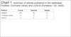
The search was carried out in July 2019 on the PubMed, LILACS, and Cochrane Library platform using the descriptors present on the “health science descriptors (DECS)” platform: “biomechanical phenomena,” “facial injuries” and “fractures, bone,” thus finding 321 articles.
The inclusion criteria were: studies published in the last five years, available entirely on the web, in English or Portuguese. After using these filters, 50 studies were found, of which, after analytical reading of the title and abstract available on the platform, 44 studies were excluded.
Most of them were excluded because they only addressed surgical treatment, some were excluded because they were literature reviews, since studies of the type of reviews and editorials were not considered to be included in the sample, and one study presented Mandarin as a language, being taken from the sample. Thus, six articles were selected to compose the review sample (Chart 1).
| Platform | Found | Selected | Sample |
|---|---|---|---|
| PubMed | 321 | 50 | 6 |
| Cochrane Library | 0 | 0 | 0 |
| LILACS | 5 | 0 | 0 |
RESULTS
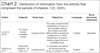
The selected articles were organized in a table so that the topics considered relevant for each study were exposed, such as author and date of publication, type of study, study subjects, objectives, language, and bone structures affected, as set out in Chart 2.
| Author | Subjects | Study type | Objectives | Language | Bone structures addressed |
|---|---|---|---|---|---|
| Liu et al., 201811 | 3D virtual master mandible model | Original article | This study examined the distribution of stress to the mandible without third molars and with different IM3 orientations resulting from a 2000-Newton test of anterior midline impact force or mandible body. | English | Mandible |
| Patel et al., 201716 | Study with cadavers. 10 orbits of 5 heads. | Original article | To elucidate and define the biomechanical factors involved in orbital floor fractures. | English | Orbit |
| Kang e Chung, 201519 | Male, 52 years old | Case report | Description of a case report with literature review. | English | Orbit |
| Tuchtan et al., 201515 | Postmortem corpses and 3D models | Original article | Evaluate the dispersion of force not only in the mandible, but also in the brain. | English | Mandible |
| Santos et al., 201513 | 3D Models | Original article | Analyze stress distributions from traumatic loads applied to the symphyseal, parasympathetic areas and regions of the mandibular body, in the edentulous mandible of the elderly using finite element analysis (FEA). | English | Mandible |
| Gayathri et al., 201621 | Female, 36 years old. | Case report | Clarify the consultations mentioned above. The article also aims to explore the biomechanics involved in such combined fractures and analyze treatment probabilities. | English | Styloid process |
Regarding the dates, there were three articles from 2015, the most recent was from 2018 and the others from 2016 and 2017, thus obeying the inclusion criteria, which allow the inclusion of articles published in the last five years, and they were not found in 2019 articles of the year.
In the studies that comprised the sample, the majority (3/5) used 3D models for the analysis, while the other studies used human and human cadavers while still alive - when they were case reports. All studies were published in English.
Therefore, based on the articles’ reading, they were divided into three categories to be discussed with more outstanding care:location of the impact as determinants of the fracture, the influence of the molars for the injury, importance of the clinic for the proper management of trauma face
DISCUSSION
Impact site as determinant for fracture
Several factors can predispose to facial trauma, such as male gender, advanced age8,9; and sports practice, for example, sports such as basketball, football, and baseball10. Besides these, there is no doubt about the influence of the place where the impact occurred for the predisposition to certain fractures, as evidenced by the studies that composed the sample addressing fractures in the mandible, orbit, and styloid process.
Mandible
Regarding the mandible, in the study by Liu et al., 201811, two regions of this bone structure were mainly addressed: the mandible and the angle’s condyle. Observing that the chin region’s impacts exerted more significant stress under the condyle, while lateral impacts exerted greater stress on the condyle and, subsequently, on the mandible angle11.
The mandible is more vulnerable to lateral than frontal impacts, with greater impact resistance only the nasal and zygomatic bones, whose areas are more sensitive12.
Besides, it was evidenced that in the lateral impacts, the most significant stress force was exerted in structures ipsilateral to the impact, being, therefore, in the lateral impact, the condyle, followed by the ipsilateral angle, more susceptible to fractures11.
One explanation for this finding is the ability to dissipate stress and absorb it by the bone structures closest to the impact, being the structures contralateral to the impact less susceptible to fractures11.
Another study, by Santos et al., in 201413, agreed with the one mentioned above, concerning condyle fractures, whose impact exerted on the symphysial and parasymphysial region presented a more significant burden agreeing about greater stress in regions ipsilateral.
Another type of condyle fracture is the guardsman fracture, a bilateral fracture of these structures concomitant with the fracture of the mandible’s symphysis, being the injury mechanism often a fall without the attempt to cushion the impact with the hands, as in the elderly or individuals after a syncope14.
Among the directions of the impact, as evidenced by Tuchtan et al., in 201515, the uppercut simulated blow, popularly called “hook,” generates greater forces than the frontal and lateral impacts, even affecting the occipital bone, with more significant damage to the chin.
Orbit
Another area addressed in the articles that comprised the sample was the orbit, with mainly two presentations, edge fractures and globe/floor fractures. The first refers to fractures that tend to be smaller and anteriorly arranged; on the floor, the opposite occurs1616. the floor
It is assumed that the relationship between the fracture size dimension and its disposition on the anteroposterior axis is due to the decrease in the thickness of the orbital bones, which tend to decrease as they turn posteromedially, as demonstrated by Patel et al. in 201716.
Besides, orbital floor fractures can be divided into blow-out and blow-in. The first, when there is an invagination of bone fragments into the maxillary sinus, usually occurring in major trauma to the zygoma or orbit. Blow-in, on the other hand, occurs when fragments turn into the eye socket, occurring when there is an increase in pressure in the maxillary sinus, as in a situation where the tire bursts close to the patient’s face17.
There are two main theories for blow-out orbit fractures, the hydraulic and buckling theory. The first one states that the eyeball’s hydraulic pressure is transmitted to the orbit wall, generating fracture of the orbit18. The buckling theory states that the direct impact on the lower edge of the orbit can cause a temporary deformation of it without fracturing it; however, the impact is transmitted to the floor of the orbit18; it may be accompanied by clinical signs, such as hematoma, lower eyelid edema and irregularities in the lower edge of the orbit19.
Styloid process
Another structure is added that was addressed by the articles that composed the sample: the styloid process. Lesions can be divided into intrinsic and extrinsic. The intrinsic originates from the muscles inserted in this structure may occur due to spasms, convulsions, laughter, and excessive coughing. On the other hand, the extrinsic is an impact on the structure in situations where it is already prone to fracture, or even in the anterior region of the mandible20,21.
Influence of molars for injury
Another study reports that during a lateral impact, the presence of the impacted third molar can decrease the risk of fracture in the condyle; however, it can increase the risk of fracture of the ipsilateral mandibular angle11.
According to a meta-analysis published in 2017, the risk of fractures of the mandible angle in individuals with third molars is almost three times higher than in individuals who do not have this dentition22. This information has been ratified by Tuchtan et al. (2015) 15, a study in which it showed that it presented greater stress forces in the condyle in the partial or total absence of dentition. Furthermore, in the study by Brucoli et al., In 201923, it was also seen that the complete eruption of the third molars is associated with condylar fractures.
When a frontal impact is mentioned, the risk of condyle fracture is greater than that of angle, regardless of the presence of the third molars11. Another relevant analysis factor is the tooth’s impaction since fully impacted teeth reflect a greater tension in the mandible than those partially impacted11. Although, in the study by Brucoli et al.(2019)23, partially impacted teeth were associated with angle fracture. However, these teeth’ angulation does not present significant differences concerning the distribution of forces in the mandible when subjected to impact11.
Importance of the clinic for the proper management of facial trauma
Regarding the importance of clinical analysis, it was evidenced in the study by Patel et al. (2017) 16 that although computed tomography is essential for the diagnosis of orbital fractures, an ophthalmological examination is also necessary, since, among the nine orbital fractures, computed tomography only showed 316.
In the study by Rothweiler et al., In 201824, they showed the importance of a thorough clinical analysis, taking into account the severity of the injury and the patient’s age, in multiple trauma patients. The ideal time for surgery should be individual because of the patient’s stability and the edema that could harm the surgical result.
A multidisciplinary team’s importance in the treatment of facial fractures is undoubted, as they are complex fractures that can affect the central nervous system and may require the approach of a neurosurgeon. The simultaneous performance of the maxillofacial surgeon with the neurosurgeon may be beneficial during treatment25.
CONCLUSION
In short, it is concluded that several factors can influence the occurrence of facial trauma; among them are the biomechanical phenomena involved. The present study demonstrated that the site of the impact is an essential predictor of the fracture occurrence site, with the mandible condyle being a place of more significant stress, especially in a frontal impact.
Another finding evidenced by the study was the ability of third molar teeth to influence the greater predisposition to certain fractures, depending on their implantation.
Besides, the clinic’s importance and the multidisciplinary management of these lesions are ratified to establish more diligent diagnoses, more efficient treatments, and adequate prevention measures.
REFERENCES
1. Soller ICS, Poletti NAA, Beccaria LM, Squizatto RH, Almeida DB, Matta PRA. Epidemiological profile of patients with facial injuries treated in an emergency hospital. REME Rev Min Enferm. 2016;20:e935.
2. Allareddy V, Allareddy V, Nalliah RP. Epidemiology of facial fracture injuries. J Oral Maxillofac Surg. 2011 Out;69(10):2613-8. DOI: http://dx.doi.org/10.1016/j.joms.2011.02.057
3. Vandegriend ZP, Hashemi A, Shkoukani M. Changing trends in adult facial trauma epidemiology. J Craniofac Surg. 2015 Jan;26(1):108-12.
4. Scheyerer MJ, Döring R, Fuchs N, Metzler P, Sprengel K, Werner CML, et al. Maxillofacial injuries in severely injured patients. J Trauma Manag Outcomes. 2015 Jun;9:4. DOI: http://dx.doi.org/10.1186/s13032-015-0025-2
5. Bocchialini G, Castellani A. Facial trauma: a retrospective study of 1262 patients. Ann Maxillofac Surg. 2019 Jan/Jun;9(1):135-9.
6. Huempfner-Hierl H, Schaller A, Hemprich A, Hierl T. Biomechanical investigation of naso-orbitoethmoid trauma by finite element analysis. Br J Oral Maxillofac Surg. 2014 Nov;52(9):850-3. DOI: http://dx.doi.org/10.1016/j.bjoms.2014.07.255
7. Walker CJ, MacLeod SPR. Anatomy and biomechanics of condylar fractures. Atlas Oral Maxillofac Surg Clin North Am. 2017 Mar;25(1):11-6.
8. Ramos JC, Almeida MLD, Alencar YCG, Sousa Filho LF, Figueiredo CHMC, Almeida MSC. Epidemiological study of bucomaxilofacial trauma in a Paraíba reference hospital. Rev Col Bras Cir. 2018;45(6):e1978.
9. Lucena ALR, Silva Filho GF, Sarmento TCAP, Carvalho SHG, Fonseca FRA, Sarmento DJAS. Epidemiological profile of facial fractures and their relationship with clinical-epidemiological variables. J Craniofac Surg. 2016 Mar;27(2):345-9.
10. Povolotskiy R, Youssef P, Kaye R, Paskhover B. Facial fractures in young adults: a national retrospective study. Ann Otol Rhinol Laryngol [Internet]. 2019 Jun; [citado 2020 Jan 05]; 128(6):516-23. Disponível em: http://www.ncbi.nlm.nih.gov/pubmed/30735056
11. Liu Y, Wang R, Baur DA, Jiang XF. A finite element analysis of the stress distribution to the mandible from impact forces with various orientations of third molars. J Zhejiang Univ Sci B. 2018 Jan;19(1):38-48.
12. Pappachan B, Alexander M. Biomechanics of cranio-maxillofacial trauma. J Maxillofac Oral Surg. 2012 Jun;11(2):224-30.
13. Santos LSM, Rossi AC, Freire AR, Matoso RI, Caria PHF, Prado FB. Finite-element analysis of 3 situations of trauma in human edentulous mandible. J Oral Maxillofac Surg. 2014 Abr;73(4):683-91. DOI: http://dx.doi.org/10.1016/j.joms.2014.10.014
14. McCormick RS, Putnam G. The management of facial trauma. Surg (Oxford). 2018;36(10):587-94.
15. Tuchtan L, Piercecchi-Marti MD, Bartoli C, Boisclair D, Adalian P, Léonetti G, et al. Forces transmission to the skull in case of mandibular impact. Forensic Sci Int. 2015 Jul 1;252:22-8.
16. Patel S, Andrecovich C, Silverman M, Zhang L, Shkoukani M. Biomechanic factors associated with orbital floor fractures. JAMA Facial Plast Surg. 2017 Jul;19(4):298-302.
17. Kuhnen RB, Silva FM, Scortegagna A, Cabral RJB. Fractures of orbit?: signs and symptoms based on the involved anatomical structures. Int J Dent. 2006;77(1):20-4.
18. Ahmad F, Kirkpatrick WNA, Lyne J, Urdang M, Garey LJ, Waterhouse N. Strain gauge biomechanical evaluation of forces in orbital floor fractures. Br J Plast Surg. 2003 Jan;56(1):3-9.
19. Kang SJ, Chung EH. The hydraulic mechanism in the orbital blowout fracture because of a high-pressure air gun injury. J Craniofac Surg [Internet]. 2015 Out; [cited 2020 Jan 05]; 26(7):e573-5. Disponível em: http://content.wkhealth.com/linkback/openurl?sid=WKPTLP:landingpage&an=00001665-201510000-00069
20. Miloro M. Fracture of the styloid process: a case report and review of the literature. J Oral Maxillofac Surg. 1994 Out;52(10):1073-7.
21. Gayathri G, Elavenil P, Sasikala B, Pathumai M, Raja VBK. 'Stylo-mandibular complex' fracture from a maxillofacial surgeon's perspective - review of the literature and proposal of a management algorithm. Int J Oral Maxillofac Surg. 2016 Mar;45(3):297-303. DOI: http://dx.doi.org/10.1016/j.ijom.2015.09.020
22. Xu S, Huang J, Xiong Y, Tan YH. How is third molar status associated with the occurrence of mandibular angle and condyle fractures?. J Oral Maxillofac Surg. 2017 Jul;75(7):1476.e1-1476.e15.
23. Brucoli M, Romeo I, Pezzana A, Boffano P, Benech A. The relationship between the status and position of third molars and the presence of mandibular angle and condylar fractures. Oral Maxillofac Surg. 2019 Mar;24(1):31-6.
24. Rothweiler R, Bayer J, Zwingmann J, Suedkamp NP, Kalbhenn J, Schmelzeisen R, et al. Outcome and complications after treatment of facial fractures at different times in polytrauma patients. J Craniomaxillofac Surg. 2018 Feb;46(2):283-7.
25. Salentijn EG, Peerdeman SM, Boffano P, Van Den Bergh B, Forouzanfar T. A ten-year analysis of the traumatic maxillofacial and brain injury patient in Amsterdam: incidence and aetiology. J Craniomaxillofac Surg. 2014 Set;42(6):705-10.
1 . University of the Fortaleza, Fortaleza, CE, Brazil.
2 . Hospital Instituto Doutor José Frota, Fortaleza, CE, Brazil.
3 . Hospital Batista Memorial, Fortaleza, CE, Brazil.
Corresponding author: Thiago Maciel Valente, Avenida Washington Soares, 1321, Engenheiro Luciano Cavalcante, Fortaleza, CE, Brazil. Zip Code: 60811-905. E-mail: maciel.thiago@edu.unifor.com
Article received: January 24, 2020.
Article accepted: July 15, 2020.
Conflicts of interest: none
 Read in Portuguese
 Read in English
 PDF PT
 Print
Print
 Send this article by email
 How to Cite
 Mendeley
 Pocket