


Original Article - Year 2021 - Volume 36 -
An epidemiological study of the association between risk factors and skin cancer incomplete excisions
Estudo epidemiológico da associação entre fatores de risco e excisões incompletas no câncer de pele

ABSTRACT
Introduction: The increasing incidence of skin cancer leads to a high number of surgical procedures worldwide. The surgical treatment of skin cancer's main objective is its complete excision, preserving the function and the best aesthetic result. Incomplete initial resection can result in recurrences and major damage. The objective is to analyze the risk factors for positive margins in the follow-up of cutaneous lesions surgically removed, for one year, in the plastic surgery department of the Royal Perth Hospital.
Methods: A survey of histopathological samples from 947 operated patients was analyzed. All patients with confirmed incomplete excision (IE) underwent a second surgery or even a third time.
Results: In total, 947 lesions were found, 6.6% of surgeries had compromised margins, with a histopathological distribution of 75% of basal cell carcinoma, 21.4% of squamous cell carcinoma, and 3.6% of other lesions. The relation of the presence of compromised surgical margins between the SCC, compared to BCC, leads to a relative risk of 2.8 and a p-value of 0.041, which suggests that the SCC is a risk factor for the presence of compromised surgical margins. For staging, the need for a second surgical approach was present in 61.29% of the patients, 20.9% were under observation, 3.2% were absent from the service, 8% went directly to chemotherapy or radiotherapy, and 6.4% rescheduled the surgery.
Conclusion: Knowledge of risk factors for positive margins is necessary for the surgeon to understand the prognosis and monitoring of each patient.
Keywords: Skin Neoplasms. Dermatology. Plastics. /pathology. Margins of Excision.
RESUMO
Introdução: A crescente incidência de câncer de pele leva a um alto número de procedimentos cirúrgicos em todo o mundo. O principal objetivo do tratamento cirúrgico do câncer de pele é sua excisão completa, preservando a função e o melhor resultado estético. A ressecção inicial incompleta pode resultar em recorrências e danos graves. O objetivo é analisar os fatores de risco para margens positivas no seguimento de lesões cutâneas removidas cirurgicamente, por um ano, no departamento de cirurgia plástica do Hospital Royal Perth.
Métodos: Foi analisado um levantamento de amostras histopatológicas de 947 pacientes operados. Todos os pacientes com excisão incompleta confirmada (EI) foram submetidos a uma segunda cirurgia ou até mesmo a uma terceira vez.
Resultados: No total, 947 lesões foram encontradas, 6,6% das cirurgias tiveram margens comprometidas, com distribuição histopatológica de 75% de carcinoma basocelular (CBC), 21,4% de carcinoma de células escamosas (CCE) e 3,6% de outras lesões. A relação da presença de margens cirúrgicas comprometidas entre o CCE, quando comparada ao CBC, leva a um risco relativo de 2,8 e um valor p de 0,041, sugerindo que o primeiro é um fator de risco para a presença de margens cirúrgicas comprometidas. Para o estadiamento, a necessidade de uma segunda abordagem cirúrgica esteve presente em 61,29% dos pacientes, 20,9% estavam em observação, 3,2% estavam ausentes do serviço, 8% foram diretamente à quimioterapia ou radioterapia e 6,4% remarcaram a cirurgia.
Conclusão: O conhecimento dos fatores de risco para margens positivas é necessário para que o cirurgião entenda o prognóstico e o acompanhamento de cada paciente.
Palavras-chave: Câncer de pele; Cirurgia plástica; Dermatologia; Margens de excisão; Patologia
INTRODUCTION
Skin cancer is a multifactorial etiology pathology, resulting mainly from genetic alterations, environmental and lifestyle factors1. It may present in two forms: melanoma and non-melanoma skin cancer (NMSC)2, which is the most frequent malignant neoplasm in white populations and accounts for at least 80% of all skin cancer. NMSC consists mainly of basal-cell carcinomas BCC (70%), the most commonly diagnosed worldwide3,4 and squamous cell carcinoma SCC (20%)3,4. BCC are divided into subtypes with more or less aggressive behavior and are classified as nodular, micronodular, superficial, pigmented, cystic, infiltrative, and morpheaform. SCC resembles nests of abnormal epidermal cells invading the dermis, and its histological grade depends on the degree of cellular differentiation5, its classification is based on the anatomical body sites6.
The main cause of skin cancer is chronic exposure to sunlight, which explains the frequent occurrence of lesions on the body’s areas exposed to the sun, such as the face, ears, neck, scalp, shoulders, and back. Other etiological factors include ultraviolet exposure, certain carcinogenic chemicals (arsenic and hydrocarbons), ionizing radiation, previous skin diseases such as xeroderma pigmentosum, Bazex, and Gorlin syndromes, chronic irradiation or ulceration, human papillomavirus (HPV) infection, and chronic stress exposure7. Immunologically compromised patients are at higher risk8,9. In addition to that, fair-skinned people often develop skin cancers, and they affect all ethnic groups, primarily those living in tropical areas that are highly exposed to the sun6-9 where the highest rate is about 1 to 2% per year10 as in Australia, where this study was developed.
Although the mortality rate from NMSC is low, as reported by Leiter et al., in 20145 such skin cancer impacts patient’s quality of life causing significant morbidity, the prognosis depends on tumor type and the chosen treatment. Surgical excision with pre-operatively identified margins is one of the most common and effective treatment strategies for basal-cell carcinomas (BCC) and for most squamous cell carcinomas (SCC). Insufficient compliance with recommended excisional margins among surgeons represents a very high risk of NMSC recurrence and re-excision for the patient. This may be caused by the irregular infiltration of these tumours10. The tumor recurrence associated with incomplete excision in BCCs ranges from 26 to 41% after 2 to 5 years of follow-up. The maximum number of tumor recurrences has been detected in morphoeic and facial tumors.11
About the SCCs, when the initial removal is incomplete, theirrecurrence occurs mostly locally or less frequently in regional lymph nodes. Approximately 75% of recurrences occur within two years and 95% within five years after initial diagnosis.12 So, it is important to note that the presence (or absence) of tumor cells in the margins is among the prognostic features for recurrences of SCCs and BCCs. Moreover, when margins are involved, the recommendation is usually for re-excision or a close clinical follow-up. This not only produces adverse effects on patients but also causes an increase in additional healthcare costs. So, the optimization of the surgical management of NMSC is of great importance to ensure the highest quality of surgical treatments and a subsequent optimal outcome for all patients.
OBJECTIVE
In this context, receiving no funding from any company and without conflict of interest, this study analyses the risk factors in the follow-up of surgically removed skin lesions with positive margins during one year of the Royal Perth Hospital plastic surgery department.
METHODS
This study includes patients who underwent surgical treatment for skin cancer during December 2016 and December 2017 at the Plastic Surgery Department of Royal Perth Hospital, Australia. There were selected patients with a pathological diagnosis of any type of skin cancer, whose initial intention was the complete removal of the lesion, totalizing 947 incisional biopsies. The definition of excisional margins recommended by international guidelines (EADV and EDF) was used as a point of reference for the analysis. Postoperative pathologic conventional assessment followed all the surgical excisions considered in the study with histology paraffin-embedded definitive evaluation. All patients with confirmed incomplete excision (IE) were submitted to a second surgery or even a third one, according to the clinical procedure of the Department. All patients with confirmed incomplete excision (IE) were submitted to a second surgery or even a third one, according to the clinical procedure of the Department.
The medical record included in this study was reviewed for the following parameters of patients with incomplete excisions: gender, age, anatomical localization of the lesions, perineal invasion, size of the lesion, histopathological profile of the lesion, multiple or single skin lesions, size of safety margins and metastasis. The statistical analysis of Pearson’s chi-square test was used to study the association of the main variables with compromised surgical margins. The level of significance was set at 5% (p <0.05) and the relative risk of incomplete excision at (RR) >1. Thus, the main risk factors for a re-assessment were defined. For this type of study, formal ethical research committee consent is not required. This article does not contain any studies with animals performed by any of the authors.
All procedures performed in studies involving human participants were following the institutional and/or national research committee’s ethical standards and with the 1964 Helsinki declaration and its later amendments or comparable ethical standards. For this type of study, formal consent is not required. This article does not contain any studies with animals performed by any of the authors.
RESULTS
Incisional biopsy (first surgery)
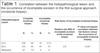
According to the departmental records review, 947 patients underwent their first resection of skin cancer at the surgical center. In this universe, eight hundred and eighty (93.3%) had complete excision (CE), and sixty-two (6.6%) had IE. Of those who had CE, five hundred and sixty-five (65%) had a BCC diagnosis, and two hundred and thirty (26%) had a diagnosis of SCC, the other sixty-seven (7.6%) had melanoma. Of the patients who had IE, forty-seven (75%) had BCC diagnosis while the other twelve (21.4%) had SCC, no case (0%) of melanoma had IE. The association between the exposure to SCC when compared to BCC leads to a relative risk of 2.8 and a p-value of 0.041, suggesting that it is a risk factor for the presence of surgical compromised margins (Table 1)
| Histopathological Lesion | Complete incisional biopsy | Incomplete incisional biopsy | Risk factor of incomplete incisional biopsy | |||
|---|---|---|---|---|---|---|
| N | % | N | % | Relative risk (RR) of incomplete excision at the incisional biopsy | P-value (chi-square correlation between risk factors versus the occurrence of incomplete incisional biopsy) | |
| SCC | 230 | 26 | 12 | 21.4 | 2.8 | 0.041 |
| BCC | 575 | 65 | 47 | 75 | ||
| Melanoma | 67 | 7.6 | 0 | 0 | ||
Second look (second surgery)
After the first surgery and based on the anatomopathological results, the team evaluated the cases with compromised margins, deciding each patient’s follow-up. Imaging examinations were necessary to stage the recurrent cases (6,6% IE) and decide the conduct.. We performed 28% of CT scans, 14% of nuclear magnetic resonances, and 28.5% of PET-Scans. From the staging, the need for the second surgical abortion was evaluated in 61.29% of the patients, 20.9% were under observation, 3.2% were absent from the service, 8% were direct to chemotherapy or radiotherapy, and 6.4 % rescheduled surgery.
The male prevalence was observed for IE. In a universe of 62 patients, 48 (77.4%) were men, and 14 (22.5%) were women. The mean age of the patients was 70.2 years in a range of 41 to 86 years old with a standard deviation of 12,9.
In a topographic evaluation of the lesions with an incomplete incisional biopsy, the face and neck areas were the most commonly affected (91,7%), precisely nose areas (23%) followed by ear areas (19,2%). The incidence of the tumors in occipital regions, frontal and neck were the same (11,5%). In the patients analyzed, 20,8% presented a single lesion, whereas 79,1% presented multiple lesions. Histologic subtypes were not identified in this study.
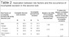
However, there is still a failure rate of 26,3% at the end of this second approach. Where 9.6% of the patients had metastasis, and 22.5% had to undergo lymph node dissection. (Table 2)
| Rick Factor of incomplete Excision (IE) at the Second Look | Complete Second look excision | Incomplete second look excision | Relative risk (RR) of incomplete excision at the second look | P-value (chi-square correlation between risk factors versus the occurrence of incomplete excision) | ||
|---|---|---|---|---|---|---|
| N | % | N | % | |||
| Male | 20 | 62.5 | 12 | 37.5 | 1.46 | 0.84 |
| Female | 4 | 66.6 | 2 | 33.3 | ||
| > 70 years | 24 | 60.0 | 16 | 40.0 | 0.75 | 0.49 |
| < 70 years | 8 | 50.0 | 8 | 50.00 | ||
| Size of tumor > 15mm | 6 | 50.0 | 4 | 50.0 | 1.8 | 0.07 |
| Size of tumor < 15mm | 8 | 28.5 | 20 | 71.4 | ||
| Safety margin < 15mm | 22 | 73.3 | 8 | 26.6 | 2.88 | 0.012 |
| Safety margim > 15 mm | 2 | 25.0 | 6 | 75.0 | ||
| Multiple lesions | 20 | 71.4 | 8 | 28.5 | 0.47 | 0.077 |
| Single lesion | 4 | 40.0 | 6 | 60.0 | ||
| IE - deep and peripheral | 4 | 66.6 | 8 | 33.3 | 4.44 | 0.013 |
| IE - deep | 6 | 60.0 | 4 | 40.0 | ||
| IE - peripheral | 14 | 87.5 | 2 | 12.5 | ||
| SCC | 8 | 80.0 | 2 | 20.0 | 3.80 | 0.001 |
| BCC | 22 | 78.5 | 6 | 21.4 | ||
| Perineural Invasion | 2 | 33.3 | 4 | 66.6 | 2.86 | 0.038 |
| Non invasive tumor | 6 | 23.0 | 20 | 76.9 | ||
DISCUSSION
Our case series was consistent with the current literature findings concerning cutaneous carcinomas, which will be described below. Data reveal that most incomplete excisions are in the head and neck12,13,14 and that the leading risk factor associated with skin cancer is chronic exposure to ultraviolet light. It is most often diagnosed in the body’s most exposed sites15,16 contributing to the association of direct exposure to ultraviolet light being a risk fator.17,18,19 We did not evaluate this criterion in this study. However, 91.5% of the lesions present and analyzed were from the face.
Tan et al., in 200720 have associated the higher rates of incomplete resection, the lesion’s invasion characteristic and an increased number of re-excisions. However, age, sex, tumor size, and surgery experience were not statistically significant risk factors. In our study using the chi-square test, the statistically significant risk factors for incomplete excision includes the diagnosis of SCC (p <0.01), perineural invasion (p = 0.038), safety surgical margin <15mm (p = 0.012) and characteristic of a deep and peripheral excision (p = 0.013). Other factors such as age (p = 0.49), tumor size> 15mm (p = 0.07) and sex (p = 0.84) were not found to be significant, in agreement with the literature findings.
CONCLUSION
The findings of this study are consistent with those in the literature. Locations of greater sun exposure are at all times increasing the incidence of the development of skin cancers. It is then necessary to improve our knowledge to make the procedure curable in a single approach. Knowing the risk factors for an ineffective approach, such as diagnosing SCCs, is possible to prepare for continued treatment. Statistically, significant risk factors were SCC diagnosis and previously incompletely excised lesions referred for re-excision.
The authors recommend more care with tumor markings, taking margins >15 mm, using deeper margins, and referring patients to more experienced centers. In addition to that, the significant number of patients with multiple lesions emphasizes the importance of periodic examination.
REFERENCES
1. Ribeiro LR, Marques EK. A importância da mutagênese ambiental na carcinogênese humana. In: Ribeiro LR, Salvadori DMF, Marques EK, orgs. Mutagênese ambiental. Canoas: ULBRA; 2003. p. 21-7.
2. Dazard JE, Piette J, Basset-Seguin N, Blanchard JM, Gandarillas A. Switch from p53 to MDM2 as differentiating human keratinocytes lose their proliferative potential and increase in cellular size. Oncogenese. 2000 Ago;19(33):3693-705.
3. Roewert-Huber J, Lange-Asschenfeldt B, Stockfleth E, Kerl H. Epidemiology and aetiology of basal cell carcinoma. Br J Dermatol. 2007 Dez;157(Supl 2):47-51.
4. Wong CSM, Strange RC, Lear JT. Basal cell carcinoma. BMJ. 2003 Out;327(7418):794-8.
5. Leiter U, Eigentler T, Garbe C. Epidemiology of skin cancer. Adv Exp Med Biol. 2014;810:120-40.
6. Na. Chronic stress and cancer risk. Cancer Biol Ther. 2005;4(1):6-12. DOI: https://doi.org/10.4161/cbt.4.1.1455
7. Brunssen A, Waldmann A, Eisemann N, Katalinic A. Impact of skin cancer screening and secondary prevention campaigns on skin cancer incidence and mortality: a systematic review. J Am Acad Dermatol. 2017 Jan;76(1):129-39.e10. DOI: https://doi.org/10.1016/j.jaad.2016.07.045
8. Culliford A, Hazen A. Dermatologia para cirurgiões plásticos. In: Thorne CH, Beasley RW, Aston SJ, Bartlett SP, Gurtner GC, Spear SL, eds. Grabb & Smith cirurgia plástica. 6a ed. Rio de Janeiro: Guanabara Koogan; 2009. p. 103-12.
9. Popim RC, Corrente JE, Marino JAG, Souza CA. Câncer de pele: uso de medidas preventivas e perfil demográfico de um grupo de risco na cidade de Botucatu. Ciênc Saúde Coletiva. 2008;13(4):1331-6.
10. Park SW, Heo EP, Choi JH, Cho HC, Kim SH, Xu L, et al. Reconstruction of defects after excision of facial skin cancer using a venous free flap. Ann Plast Surg. 2011 Dez;67(6):608-11.
11. Cautela JM, Mannocci A, Reggiani C, Persechino F, Ferrari F, Rossi E, et al. Identifying the factors that influence surgeon's compliance with excisional margins of non-melanoma skin cancer. PLoS One. 2018;13(9):e0204330. DOI: https://doi.org/10.1371/journal.pone.0204330
12. Ferrand PAS. Efectividad de um programa cognitivo social para prevenir el câncer de piel em mujeres adolescentes. Univ Psychol. 2006 Jun;5(3):585-97.
13. Tarallo M, Cigna E, Frati R, Delfino S, Innocenzi D, Fama U, et al. Metatypical basal cell carcinoma: a clinical review. J Exp Clin Cancer Res. 2008 Nov;27(1):65. DOI: https://doi.org/10.1186/1756-9966-27-65
14. Dallari S, Zaraca G, Giorgini S, Borgonzoni M. Close and positive margins in non-melanoma skin malignancies of the head and neck. What to do in patients over 75 years of age? A preliminary study. G Ital Dermatol Venereol. 2018 Jun;155(4):464-9. DOI: https://doi.org/10.23736/S0392-0488.18.05853-4
15. Silva SP, Dellon AL. Recurrence rate of positive margin basal cell carcinoma: results of a five-year prospective study. J Surg Oncol. 1985 Jan;28(1):72-4.
16. Howell JY, Ramsey ML. Cancer, squamous cell of the skin. Treasure Island, FL: StatPearls Publishing; 2020.
17. Nagore E, Grau C, Molinero J, Fortea JM. Positive margins in basal cell carcinoma: relationship to clinical features and recurrence risk. A retrospective study of 248 patients. J Eur Acad Dermatol Venereol. 2003 Mar;17(2):167-70.
18. Brantsch KD, Meisner C, Schönfisch B, Trilling B, Wehner-Caroli J, Röcken M, et al. Analysis of risk factors determining prognosis of cutaneous squamous-cell carcinoma: a prospective study. Lancet Oncol. 2008;9(8):713-20. DOI: https://doi.org/10.1016/S1470-2045(08)70178-5
19. Brandt MG, Moore CC. Nonmelanoma skin cancer. Facial Plast Surg Clin North Am. 2019 Fev;27(1):1-13. DOI: https://doi.org/10.1016/j.fsc.2018.08.001
20. Tan PY, Ek E, Su SY, Giorlando F, Dieu T. Incomplete excision of squamous cell carcinoma of the skin: a prospective observational study. Plast Reconstr Surg. 2007 Set;120(4):910-6. DOI: https://doi.org/10.1097/01.prs.0000277655.89728.9f
1. University Nove de Julho, UNINOVE, São Paulo,
SP, Brazil.
2. Royal Perth hospital, Plastic Surgery
Department, Perth, Austria.
JVPN Analysis and/or data interpretation, Conception and design study, Conceptualization, Data Curation, Final manuscript approval, Formal Analysis, Funding Acquisition, Investigation, Methodology, Project Administration, Realization of operations and/or trials, Resources, Supervision, Visualization, Writing - Original Draft Preparation, Writing - Review & Editing
GDM Final manuscript approval, Formal Analysis, Supervision, Visualization, Writing - Review &Editing
Corresponding author: João Vitor Pithon Napoli Rua Pamplona, 1119, Jardim Paulista, São Paulo, SP, Brazil. Zip Code: 01405-200 E-mail: joaovitorpithon@gmail.com
Article received: September 21, 2019.
Article accepted: January 10, 2021.
Conflicts of interest: none
 Read in Portuguese
 Read in English
 PDF PT
 Print
Print
 Send this article by email
 How to Cite
 Mendeley
 Pocket