


Case Report - Year 2021 - Volume 36 -
Reconstruction with a partial flap of the pectoralis major muscle, after complication of osteosynthesis of clavicle fracture
Reconstrução com retalho parcial de músculo peitoral maior, após complicação de osteossíntese de fratura de clavícula
FABIANE PINHEIRO1,* ; FABRICIO LUIS PINHEIRO1; DIEGO LOUREIRO DOS SANTOS1; CLAUDIO MESSIAS MORAES1; RICARDO PORTELLA PERRONE1; LEANDRO TUZUKI CAVALHEIRO1
ABSTRACT
Introduction: The pectoralis major is a muscle that covers the upper portion of the anterior chest wall and is the first option for reconstruction of the chest wall and aesthetic purposes.
Case Report: Male patient, 20 years old, presenting dehiscence of surgical wound, recurrent for three consecutive times, with exposure of the left clavicle osteosynthesis plate. Reconstruction was performed with the pectoralis major muscle to cover the plaque.
Conclusion: This flap showed to be an excellent option for covering synthetic material exposure after multiple dehiscences of surgical wounds. The reconstruction was effective, with no complications and a satisfactory aesthetic result.
Keywords: Myocutaneous flap; Pectoral muscles; Surgical flaps; Internal fracture fixation; Chest wall
RESUMO
Introdução: O peitoral maior é um músculo que recobre a porção superior da parede torácica anterior e é a primeira opção para reconstrução da parede torácica e fins estéticos.
Relato de Caso: Paciente masculino de 20 anos, apresentando deiscência de ferida operatória, recidivante por três vezes consecutivas, com exposição de placa de osteossíntese de clavícula esquerda. Realizado reconstrução com o músculo peitoral maior para cobertura de placa.
Conclusão: Este retalho mostrou excelente opção para cobertura de exposição de material de síntese após múltiplas deiscências de ferida operatória. A reconstrução foi efetiva, sem complicações e resultado estético satisfatório.
Palavras-chave: Retalho miocutâneo; Músculos peitorais; Retalhos cirúrgicos; Fixação interna de fraturas; Parede torácica.
INTRODUCTION
The pectoralis major is a muscle that covers the upper portion of the anterior chest wall and is the first option for reconstruction of the chest wall, especially for defects of the sternum and anterior chest. Based on the thoracoacromial blood supply, it can easily cover sternal and anterior chest wall defects as an island or advancement flap1.
The pectoralis major muscle flap has been widely used since the late 1970s when Ariyan described it in 19792.
In the literature, there are not many descriptions regarding this flap in reconstruction to cover synthesis material in the supraclavicular region.
OBJECTIVE
The work aims to report the partial applicability of the pectoralis major muscle to cover a clavicle osteosynthesis defect with synthesis material exposure.
CASE REPORT
Male patient, 20 years old, victim of a motorcycle fall, presented a fracture of the left clavicle, progressing with pain, limited movement, and deformity at the fracture site.
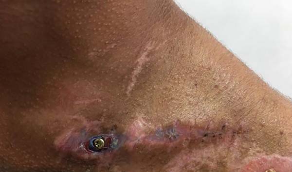
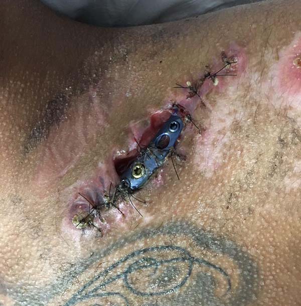
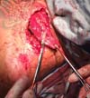
In the initial care, he underwent a surgical procedure with the orthopedics team, osteosynthesis of the left clavicle using a blocked plaque. After 14 days, he evolved with surgical wound dehiscence and exposure of synthetic material (Figure 1). Soft tissue re-approach and dehiscence closure were unsuccessful, with new material exposure five days after surgery (Figure 2).

In the preoperative evaluation and study, the defect was 6 cm wide and 4 cm deep, with exposure of synthesis material and absence of signs of infection. Reconstruction with muscle flap of the pectoralis was the best option for closure.
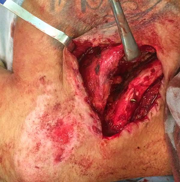
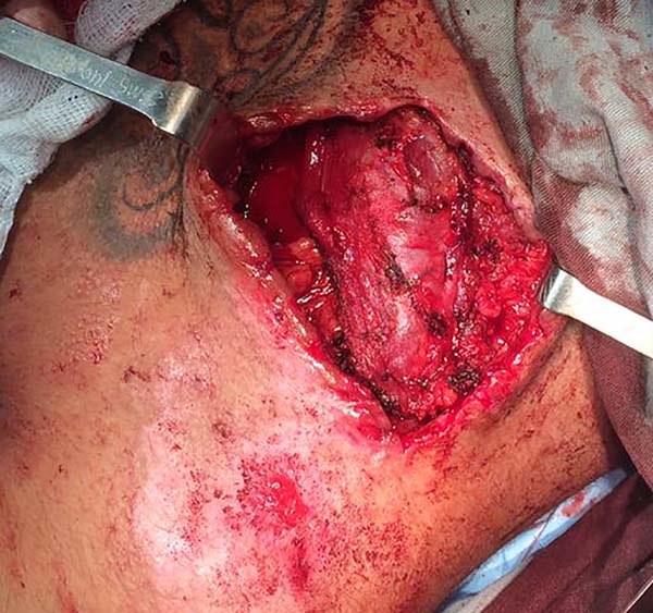
In the intraoperative period, the upper portion of the pectoralis major muscle was exposed through dissection (Figure 3). A skin flap is elevated inferiorly, exposing the pectoralis major at the fourth intercostal space level and laterally towards the border of the deltoid. The muscle fibers were divided longitudinally towards the muscle’s origin, and the muscle flap was dissected from the pectoralis minor muscle.

The medial dissection performed for the medial vascular perforators provided a divided pectoralis major flap, which can be easily rotated 45º to 60º degrees to fill the defect superiorly (Figure 4).

The flap was sutured superiorly to the platysma fascia and medially to the sternal fascia, maintaining good synthetic material coverage (Figure 5).

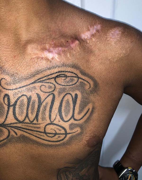
The patient evolved uneventfully in the postoperative period; the coverage was effective, without bruising, with a satisfactory aesthetic result. He is currently undergoing late postoperative outpatient follow-up, with preserved upper limb mobility (Figures 6 and 7).


DISCUSSION
The pectoralis major muscle flap is the most widely used pedicled muscle flap to cover defects in the sternal region, anterosuperior chest, intrathoracic, and neck regions3.
The standard flap development technique is the complete lateral mobilization of the pectoralis major muscle close to its insertion. This provides a large vascularized muscle mass adequate to fill a significant defect but results in the complete loss of the pectoral’s main function.
In October 1982, Nahai et al.4 described the technique that preserves the lateral third of the pectoralis major muscle with the dominant vascular pedicle and motor nerves, with the pectoralis’s muscle reconstruction major for wound closure in sternotomy with preservation of form and function.
It is known that there is no extreme morbidity due to loss of the pectoralis major muscle since the pectoralis major, serratus anterior, and latissimus dorsalis muscles can replace the function when working together-however, the loss of the pectoralis major muscle results in a significant decrease in humeral flexion strength.
According to the publication by Zehr et al., In 19995, in limited resections, reconstruction can be performed with a fraction of the pectoralis major muscle, with the technique being used in two patients, the first being after resection of a malignant sarcoma involving the head of the clavicle and, in the second case, resection was necessary for chronic osteomyelitis caused by Staphylococcus aureus in an intravenous drug user. No functional deficit was observed because the lower half of the pectoral muscle was preserved. The described technique preserves the function because half of the muscle remains intact, as we can see in our case report.
CONCLUSION
The pectoralis major muscle flap proved to be an excellent option for reconstructing a clavicle osteosynthesis defect with material exposure, maintaining the upper limb mobility preserved, thus obtaining a satisfactory result.
REFERENCES
1. Neligan PC. Cirurgia plástica: extremidade inferior, tronco e queimaduras. 3ª ed. Amsterdam: Elsevier; 2015. v. 4.
2. Ariyan S. The pectoralis major myocutaneous flap. A versatile flap for reconstruction in the head and neck. Plast Reconstr Surg. 1979 Jan;63(1):73- 81.
3. Clemens MW, Evans KK, Mardini S, Arnold PG. Introduction to chest wall reconstruction: anatomy and physiology of the chest and indications for chest wall reconstruction. Semin Plast Surg. 2011 Fev;25(1):5-15.
4. Nahai F, Morales Junior L, Bone DK, Bostwick J. Pectoralis major muscle turnover flaps for closure of the infected sternotomy wound with preservation of form and function. Plast Reconstr Surg. 1982 Out;70(4):471-4.
5. Zehr KJ, Reitmiller RF, Yang SC. Split pectoralis major muscle flap reconstruction after clavicular-manubrial resection. Ann Thorac Surg. 1999 Mai;67(5):1507-8.
1. Santa Casa da Misericórdia de Santos,
Plastic Surgery and Burns Service, Santos, SP, Brazil.
Institution: Santa Casa da Misericórdia de Santos, Plastic Surgery and Burns Service, Santos, SP, Brazil.
Corresponding author: Fabiane Pinheiro, Avenida Atlântica, 1144, Apto 2501, Centro, Balneário Camboriú, SC, Brazil. Zip Code: 88330-009. E-mail: dra.fabi.pinheiro@gmail.com
Article received: July 04, 2019.
Article accepted: January 10, 2021.
Conflicts of interest: none
 Read in Portuguese
 Read in English
 PDF PT
 Print
Print
 Send this article by email
 How to Cite
 Mendeley
 Pocket