


Original Article - Year 2021 - Volume 36 -
Evaluation of protein adsorption in rats submitted to nanotextured and polyurethane foam-coated silicone mini-implants
Avaliação da absorção de proteínas em ratas submetidas à colocação de mini-implantes de silicone nanotexturizados e revestidos pela espuma de poliuretano
Eduardo Nascimento Silva1,* ; Gisela Hobson Pontes3; Bárbara Zanon da Luz1; Adriana Yuriko Koga3; Lydia Masako Ferreira2; Leandro Cavalcante Lipinski3
ABSTRACT
Introduction: The control of protein absorption is necessary to define biomaterials' properties and their specific uses. Blood plasma contains several different proteins, including fibrinogen, which plays an important role in cell adhesion and biocompatibility results in implants. This study's objectives were to evaluate in the laboratory rats subjected to the placement of nano-textured silicone mini-implants and polyurethane foam-coated mini-implants based on the measurement of serum fibrinogen and plasma proteins.
Methods: Sixty albino rats were used, divided into two groups of 30 animals for each type of silicone mini-implant (nanotextured and polyurethane foam) and subdivided into three subgroups, according to the animals' euthanasia time (30, 60 and 90 days). The mini-implants were inserted in the animals' backs below the Panniculus carnosus. At the time of euthanasia, blood samples were obtained by cardiac puncture. The thermal precipitation technique was used to determine total and serum plasma proteins, and the difference between the latter two obtained the fibrinogen value.
Results: When the groups were compared, it was observed that the nanotextured group presented a higher amount of fibrinogen and plasma protein in the 90-day subgroup, with statistical significance (p=0.004). When comparing the subgroups among themselves, a significant difference was evidenced (p<0.001).
Conclusion: The nanotextured miniimplants showed a lower protein absorption concerning polyurethane foam-coated implants in the 90-day subgroup.
Keywords: Blood proteins; Experimental implants; Breast implants; Mammoplasty; Rats.
RESUMO
Introdução: O controle da absorção de proteínas é necessário para a definição das propriedades dos biomateriais e de seus usos específicos. O plasma sanguíneo contém diversas proteínas diferentes, dentre elas o fibrinogênio, que apresenta importante papel na adesão celular e nos resultados de biocompatibilidade em implantes. Os objetivos deste estudo foram avaliar laboratorialmente as ratas submetidas à colocação de mini-implantes de silicone nanotexturizados e revestidos por espuma de poliuretano a partir da aferição do fibrinogênio sérico e mensuração da proteína plasmática.
Métodos: Foram utilizadas 60 ratas albinas, divididas em dois grupos de 30 animais para cada tipo de mini-implante de silicone (nanotexturizado e espuma de poliuretano) e subdivididas em 3 subgrupos, conforme o tempo de eutanásia dos animais (30, 60 e 90 dias). Os mini-implantes foram inseridos no dorso dos animais abaixo do Panniculus carnosus. No momento das eutanásias, amostras de sangue foram obtidas por punção cardíaca. Utilizou-se a técnica de precipitação térmica para determinação das proteínas plasmáticas total e sérica, e o valor do fibrinogênio foi obtido mediante a diferença entre estas duas últimas.
Resultados: Quando comparados os grupos entre si, observou-se que o grupo nanotexturizado apresentou uma maior quantidade de fibrinogênio e da proteína plasmática no subgrupo de 90 dias, com significância estatística (p=0,004). Ao comparar os subgrupos entre si, em ambos os grupos, evidenciou-se uma diferença significativa (p<0,001).
Conclusão: Os mini-implantes nanotexturizados mostraram uma menor absorção de proteínas em relação aos implantes revestidos pela espuma de poliuretano, no subgrupo de 90 dias.
Palavras-chave: Proteínas sanguíneas; Implantes experimentais; Implantes de mama; Mamoplastia; Ratos
Introduction
The advancement of materials' functions and performance is the main objective of surface engineering, aiming to produce implants that trigger increasingly controlled biological responses1. When an alloplastic material is placed in contact with the living organism, the proteins present in the blood interact with the surface identified as foreign. Protein absorption in these biomaterials is one of the main points during the development of technologies in bio-devices2.
This is because, when a material is implanted, the absorption of proteins is one of the first steps in the biological process, as well as the coagulation cascade, which triggers immune and inflammatory reactions2,3. Therefore, the protein absorption phenomenon's control is necessary to define biomaterials' properties and their specific uses3. Blood plasma contains several different proteins, including fibrinogen, which is a signaling molecule with a wide spectrum of functions, which can lead from a balance between coagulation and protection against infections to processes of fibrosis and intense inflammation4. Thus, fibrinogen shows an important role in cell adhesion and, consequently, in the biocompatibility results of implants3.
During the evolution of breast implants, several coating surfaces were proposed to minimize tissue reactions5, of which nanotextured implants, through nanotechnology, proved to be safer concerning anaplastic large cell lymphoma6 and polyurethane foam if proved effective in reducing the rate of capsular contracture7.
Segmented polyurethane is a thermoplastic elastomer that has been used extensively in surgical procedures due to its excellent physical and mechanical properties, thermoplasticity and biocompatibility8-10.
Currently, with the advancement of nanotechnology, materials with surfaces of smaller and smaller sizes and more similar to biological structures have been proposed, thus allowing increasingly natural tissue reactions between the implant and adjacent tissues, which should decrease the intensity of the inflammatory response and influence the absorption of proteins 1,11.
OBJECTIVES
The present study aims to evaluate in the laboratory the rats submitted to the placement of nanotextured silicone implants and coated with polyurethane foam, with the following parameters:
METHODS
The research was carried out in the Universidade Estadual de Ponta Grossa (UEPG) experimental surgery vivarium after being approved by the Ethics Committee on the Use of Animals (CEUA) UEPG. CEUA process - 041/2018. UEPG Protocol: 16450/2018. All procedures strictly followed the existing regulations for animal research.
The study design was a primary study (randomized clinical trial), interventional, experimental in animals (rats), prospective, analytical, controlled, randomized, double-blind and single-centered.
A total of 60 albino rats (Rattus norvegicus albinus, Rodentia mammalia) weighing between 190 to 250 grams and 30 to 60 days old had free access to water and a species-specific diet, with room temperature and 12-hour circadian cycles.
They were randomly divided into two groups of 30 animals for each type of silicone mini-implant (nanotextured and polyurethane foam), and subdivided into 3 subgroups, according to the animals' euthanasia time (30, 60 and 90 days).
In the nanotextured group, n = 30, mini-implants with nanotextured surfaces (Silimed®), Rio de Janeiro, Brazil) were placed. In the polyurethane group, n = 30, mini-implants with polyurethane foam coating (Silimed®) were placed.
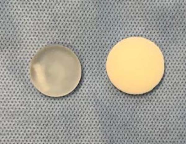
The implanted materials had the same layers as a human breast implant, discoid in shape, with 22 +/- 1 mm (mm) in diameter and 9 +/- 1 mm in height in mini-implants with a nanotextured surface, and with 24 + / - 1 mm in diameter and 11 +/- 1 mm high in mini-implants coated with polyurethane foam. The height was defined as the point of greatest implant projection on the vertical axis (Figure 1). Concerning the pores on the surface of the mini-implants, those with a nanotextured surface had the following dimensions: diameter 0.3 to 8.7 micrometers (300 to 8700 nanometers); average roughness (Ar) 4.12 micrometers (4120 nanometers); and depth 3.08 to 10.74 micrometers. The mini-implants coated with polyurethane foam had the following dimensions: diameter 120 to 320 micrometers; average roughness (Ar) 1500 micrometers; and pore depth 480 to 1200 micrometers. After distribution to groups, the rats were randomly removed from the cages and anesthetized by intraperitoneal injection, composed of an association of ketamine hydrochloride 1% (Dopalen®, Hertape, Belo Horizonte, Brazil) at a dose of 40mg/kg and hydrochloride of xylazine 2% (Dopasen®, Hertape) at a dose of 8mg/kg according to the guide for anesthesia and analgesia of laboratory animals - UNIFESP/CEUA (2017) 12. The effectiveness of anesthesia was assessed by the absence of movement, corneal-eyelid reflex, and motor reaction after grasping the fat pad of one of the hind legs, in addition to a good ventilatory pattern. With the rats positioned in the prone position, trichotomy was performed on the dorsal region, with subsequent antisepsis and sterile surgical field placement.
The incision's delimitation was performed regarding a subcostal horizontal line, following the posteroinferior costal edge, which was met with the middle sagittal line. With a scalpel cable no. 3, coupled with a blade no. 15, a horizontal incision was made, with an extension of 20 mm at the intersection of these reference lines.
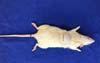
The pocket was made for the mini-implants in a retromuscular plane (below the Panniculus carnosus), and, later, the mini-implant was introduced vertically, being positioned horizontally according to the group (nanotextured or polyurethane). The skin's suture was intradermal with mononylon 5-0(Ethicon ®) with buried knots. There was no removal of the stitches in the postoperative period, and the surgical wound was kept exposed (Figure 2).

Postoperative analgesia was with a single intramuscular application of sodium dipyrone (20mg/kg) in the posterior limb's lateral region. No postoperative dressings or stitches were performed.
Euthanasia occurred according to subgroups of 30, 60 and 90 days by applying four times the therapeutic dose of Dopalen® and Dopasen® and subsequent cervical dislocation. There was no death, infection of the surgical site or extrusion of the implants, so no rats were excluded.
Evaluation methodology
Blood samples were obtained on the day of euthanasia of the animals, according to each subgroup, by intracardiac puncture performed by the veterinarian (Video 1), and were placed in tubes without and with anticoagulant ethylenediaminetetraacetic acid (EDTA) 13. The triplicate thermal precipitation technique was used for each animal, consisting of filling six capillary tubes with blood up to 3/4 of the capacity, properly closed at one end. After that, they were centrifuged at 8.0rpm in a microhematocrit centrifuge to separate the plasma for 5 minutes. After being centrifuged, three of the capillaries were randomly chosen and broken to obtain a drop, which was later placed on the Goldberg refractometer to measure total plasma protein (TPP) 13,14. The remaining three capillary tubes were taken to the water bath (temperature of 56-58 ° C, for three minutes) and afterward, again centrifuged, as previously described, obtaining this time the serum, which was measured in the refractometer, resulting in serum protein (SP) 13.
The fibrinogen value was obtained by the difference between the total plasma protein and the serum protein. The result was multiplied by 1,000, as fibrinogen is evaluated in mg.dL-¹13.
Statistical evaluation
The results were described by median, minimum and maximum values. For the comparison of groups (nanotextured and polyurethane), in each subgroup (30, 60 and 90 days), the Mann-Whitney non-parametric test was used. The comparisons between the subgroups for each group were made using the Kruskal-Wallis non-parametric test. Values of p <0.05 indicated statistical significance. The data were analyzed with the computer program Stata/SE v.14.1. StataCorpLP, USA.
RESULTS
The groups (nanotextured and polyurethane) were compared for the variables fibrinogen and plasma protein in the subgroups of 30, 60 and 90 days.
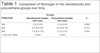
When the groups were compared to each other, it was observed that the nanotextured group had a greater amount of fibrinogen and plasma protein in the 90-day subgroup, with statistical significance (p = 0.004) (Tables 1 and 2 and Figures 3 and 4).
| Subgroups | Groups | p* | |
|---|---|---|---|
| Nanotextured median (min-max) | Polyurethane median (min-max) | ||
| 30d | 5.6 (5-5.6) | 5.5 (4.8-6) | 0.962 |
| 60d | 9.2 (8.8-9.8) | 9.2 (8.8-10) | 0.673 |
| 90d | 7.2 (6.4-7.8) | 6.3 (5-7.4) | 0.004 |
| p** (30 x 60 x 90d) | <0.001*** | <0.001*** | |
| Subgroups | Groups | p* | |
|---|---|---|---|
| Nanotextured median (min-max) | Polyurethane median (min-max) | ||
| 30d | 5.6 (5 - 6) | 5.6 (4.8 - 6) | 0.813 |
| 60d | 9.4 (9 - 10) | 9.4 (8.2 - 10) | 0.393 |
| 90d | 7.9 (6.4 - 8) | 7 (5.2 - 7.4) | 0.002 |
| p** (30 x 60 x 90d) | <0.001*** | <0.001*** | |
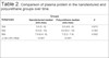

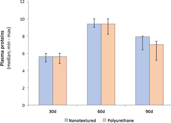
When comparing the subgroups, a significant difference was observed (p <0.001) (Tables 1 and 2 and Figures 3 and 4).
DISCUSSION
The foreign body reaction is the inflammatory sequence triggered by the implantation of biomaterials15, corresponding to the absorption of proteins on the implant surface, infiltration of inflammatory cells, fusion of macrophages and giant cells, activation of fibroblasts and, finally, formation of a fibrous capsule16.
The acute phase of healing is closely related to the activation of macrophages, which produce a variety of growth factors (IGF-1, VEGF-α, TGF-b and Wnt) that are proteins that regulate the proliferation of endothelial and epithelial cells, activate myofibroblasts, and can differentiate into progenitor cells and neovessel formation. Macrophages, therefore, recover tissue homeostasis by activating anti-inflammatory cells and regulating the deposition of collagen and fibrin17.
When there is inflammation, the presence of fibrinogen and fibrin is frequent. Like the macrophage, fibrinogen exerts its effects depending on the context in which it is, both in inflammation of the tissue and in its repair, in wound healing or in the development of fibrosis18.
Thus, the mechanisms that regulate these different activations of macrophages and fibrinogen have become active areas of research18. It is known today that these responses vary according to the characteristics of the implanted material, such as its size, biological behavior, pore size, surface topography and sterilization techniques17.
Biomaterial's engineering has focused on creating implants that more accurately simulate human tissues, both physically and chemically15. With the annual growth in the number of mammoplasty surgeries, the ideal implant's choice remains a challenge, aiming for natural results and safety procedures19.
Currently, breast implants can be classified according to their filling (silicone or saline), shape (round or anatomical) and surface texture (smooth, micro and macrotextured) 20. The surface texture is determined from the material's roughness: smooth (less than 10µm), microtextured (10-50µm) and macrotextured (greater than 50µm)5.
The silicone implant coated with polyurethane foam is considered a macrotextured implant. Its use began in 1970, motivated by a supposed reduction in capsular contracture21. The polyurethane foam forms a foamy mass in situ containing pores; this porosity allows for cell growth inside, leading to the incorporation of the implant lining into the adjacent tissue. After forming the capsule around the implant, the polyurethane coating degrades and merges with the capsule22.
More recently, with the advent of nanotechnology, greater tissue mimicry has become reality23, making it possible to build surfaces with specific nano protrusions, depending on the need, both for promotion and to prevent the absorption of proteins2.
Nanotextured surfaces demonstrated the ability to more efficiently control the interactions between the recipient tissue and the implant surface, decreasing the foreign body reaction, inflammation and scar tissue formation, in addition to greater control of colonization by pathogens24.
CONCLUSION
The nanotextured implants showed a lower protein absorption in relation to polyurethane foam-coated implants in the 90-day subgroup.
REFERENCES
1. Psarra E, Konig U, Ueda Y, Bellmann C, Janke A, Bittrich E, et al. Nanostructured biointerfaces: nanoarchitectonics of thermoresponsive polymer brushes impact protein adsorption and cell adhesion. ACS Appl Mater Interfaces. 2015 Jun;7(23):12516-29.
2. Garcia LEG, MacGregor-Ramiasa M, Visalakshan RM, Vasilev K. Protein interactions with nanoengineered polyoxazoline surfaces generated via plasma deposition. Langmuir. 2017 Jun;33(29):7322-31.
3. Kopf BS, Ruch S, Berner S, Spencer ND, Maniura-Weber K. The role of nanostructures and hydrophilicity in osseointegration: invitro proteinadsorption and bloodinteraction studies. J Biomed Mater Res A. 2015 Ago;103(8):2661-72.
4. Davalos D, Akassoglou K. Fibrinogen as a key regulator of inflammation in disease. Sem Immunopathol. 2012 Jan;34(1):43-62.
5. International Organization for Standardization (ISO). ISO 14607:2018: non-active surgical implants: mammary implants - particular requirements. Geneva: ISO; 2018. 48 p.
6. Collett DJ, Rakhorst H, Lennox P, Magnusson M, Cooter R, Deva AK. Current risk estimate of breast implant-associated anaplastic large cell lymphoma in textured breast implants. Plast Reconstr Surg. 2019 Mar;143(3S):30S-40S.
7. Barnsley GP, Sigurdson LJ, Barnsley SE. Textured surface breast implants in the prevention of capsular contracture among breast augmentation patients: a meta-analysis of randomized controlled trials. Plast Reconstr Surg. 2006 Jun;117(7):2182-90.
8. Stefanovic I, Djonlagic J, Tovilovic G, Nestrov J, Antic VV, Ostojic S, et al. Poly(urethanedimethylsiloxane) copolymers displaying a range of soft segment contents, noncytotoxic chemistry, and nonadherent properties toward endothelial cells. J Biomed Mater Res A. 2015 Abr;103(4):1459-75.
9. Silva EN, Ribas-Filho JM, Czeczko NG, Pachnicki JPA, Montemor Netto MR, Lipinski LC, et al. Histological evaluation of capsules formed by silicone implants coated with polyurethane foam and with a textured surface in rats. Acta Cir Bras. 2016 Dez;31(12):774-82.
10. Silva EN, Ribas-Filho JM, Tabushi FI, Silva MAP, Siqueira EBD, Noronha L, et al. Smooth muscle alpha actin immunoexpression (alfa-Sma) and CD-117 antibody (C-Kit) in capsules formed by polyurethane foam-coated silicone implants and with textured surface: a study on rats. Aesthetic Plast Surg. 2019 Fev;43(1):233-42.
11. Kang SH, Sutthiwanjampa C, Heo CY, Kim WS, Lee SH, Park H. Current approaches including novel nano/microtechniques to reduce silicone implant-induced contracture with adverse immune responses. Int J Mol Sci. 2018 Abr;19(4):1171.
12. Universidade Federal de São Paulo (UNIFESP). Comissão de Ética no Uso de Animais (CEUA). Guia de anestesia e analgesia de animais de laboratório. São Paulo (SP): UNIFESP/CEUA; 2017.
13. Tomaszewska E, Dobrowolski P, Kwiecien M. Intestinal alterations, basal hematology, and biochemical parameters in adolescent rats fed different sources of dietary copper. Biol Trace Elem Res. 2016 Mai;171(1):185-91.
14. Souza MV, Souza PC, Rodrigues BL, Júnior JIR, Cordeiro RR. Concentração do fibrinogênio no plasma sanguíneo de equinos da raça mangalarga marchador por diferentes métodos. Ceres. 2006;53(307):382-6.
15. Major MR, Wong VW, Nelson ER, Longaker MT, Gutner GC. The foreign body response: at the interface of surgery and bioengineering. Plast Reconstr Surg. 2015 Mai;135(5):1489-98.
16. Kastellorizios M, Tipnis N, Burgess DJ. Foreign body reaction to subcutaneous implants. Adv Exp Med Biol. 2005;865:93-108.
17. Boersema GSA, Grotenhuis G, Bayon Y, Lange JF, Bastiaansen-Jenniskens YM. The effect of biomaterials used for tissue regeneration purposes on polarization of macrophages. Biores Open Access. 2016;5(1):6-14.
18. Vanella KM, Wynn TA. Mechanisms of organ injury and repair by macrophages. Ann Rev Physiol. 2017 Fev;79:593-617.
19. Kaoutzanis C, Winocour J, Unger J, Gabriel A, Maxwell GP. The evolution of breast implants. Semin Plast Surg. 2014 Nov;134(1S):217-23.
20. Headon H, Kasem A, Mokbel K. Capsular contracture after breast augmentation: an update for clinical practice. Arch Plast Surg. 2015 Set;42(5):532-43.
21. Duxbury PJ, Harvey JR. Systematic review of the effectiveness of polyurethane-coated compared with textured silicone implants in breast surgery. J Plast Reconstr Aesthet Surg. 2016 Abr;69(4):452-60.
22. Laube T, Weisser J, Berger S, Borner S, Bischoff S, Schubert H, et al. In situ foamable, degradable polyurethane as biomaterial for soft tissue repair. Mater Sci Eng C. 2017 Set;78:163-74.
23. Barr S, Hill EW, Bayat A. Development, fabrication and evaluation of a novel biomimetic human breast tissue derived breast implant surface. Acta Biomater. 2016 Dez;49:260-71.
24. Prasad K, Zhou R, Zhou R, Schuessler D, Ostrikov KK, Bazaka K. Cosmetic reconstruction in breast cancer patients: opportunities for nanocomposite materials. Acta Biomater. 2019;86:41-65.
1. State University of Ponta Grossa, Ponta Grossa, PR, Brazil.
2. Federal University of São Paulo, Graduate Program in Translational Surgery, São
Paulo, SP, Brazil.
3. State University of Rio de Janeiro, Graduate Program in Pathophysiology and Surgical
Sciences, Rio de Janeiro, RJ, Brazil.
ENS Analysis and/or data interpretation, Conception and design study, Conceptualization, Data Curation, Final manuscript approval, Formal Analysis, Investigation, Methodology, Project Administration, Resources, Software, Validation, Visualization, Writing - Original Draft Preparation, Writing - Review & Editing
GHP Funding Acquisition, Realization of operations and/or trials, Resources
BZL Realization of operations and/or trials, Writing - Original Draft Preparation
Corresponding author: Eduardo Nascimento Silva, Avenida Doutor Francisco Burzio, 991, Centro, Ponta Grossa, PR, Brazil. Zip Code: 84010-200. E-mail: dr_eduardosilva@yahoo.com.br
Article received: December 01, 2020.
Article accepted: January 10, 2021.
Conflicts of interest: none
 Read in Portuguese
 Read in English
 PDF PT
 Print
Print
 Send this article by email
 How to Cite
 Mendeley
 Pocket