


Review Article - Year 2022 - Volume 37 -
Treatment of periorbital syringomas: review of scientific literature in the last 5 years
Tratamento de siringomas periorbitários: revisão da literatura científica nos últimos 5 anos
Karols Tatiana Vila Claro1,* ; Jorge Luis Hoyos Ramirez1; Alessandra Haddad1; Marisa Gonzaga da Cunha1; Miguel Francischelli1
ABSTRACT
Introduction: Syringomas are benign adnexal tumors with histopathological characteristics arising from the eccrine ducts, in yellowish or skin-colored papules, 1-3 mm, commonly in the lower periorbital region, which can cause important cosmetic problems. The goal of treatment is to improve appearance by destroying the tumor using minimally invasive methods and including surgery. There are multiple treatment options in the literature with varying degrees of success, but little is known about their effectiveness. Complete removal is unsuccessful, and side effects have been described, recurrence being the most frequent.
Methods: This is a narrative review of the literature of scientific publications in the period 2014-2019.
Results: After reviewing 45 articles and identifying those published in the last five years that had a record of the number of patients, treatment description, scales of evaluation of results and follow-up, six articles were selected. Of the total number of six articles, we found: a systematic review and five retrospective studies, one being a comparative one. A number was assigned to each article analyzed, and the number of patients included, treatment performed, assessment scales and results, complications and conclusions were collected.
Conclusions: Periorbital syringomas are still a therapeutic challenge, and so far, no treatment is consistently effective. The CO2 laser remains the first choice of treatment when used fractionally, and intralesional electrocoagulation represents a second alternative with moderate results and a lower risk of complications. New treatments such as Laser Erbium Yttrium Aluminum Garnet, Neodymium-Doped Yttrium Aluminum Garnet and botulinum toxin A monotherapy could be good alternatives. Comparative prospective studies are needed.
Keywords: Syringoma; Eyelids; Eccrine glands; Skin neoplasms; aesthetics.
RESUMO
Introdução: O siringomas são tumores anexais benignos com caraterísticas histopatológicas decorrentes dos ductos écrinos, em forma de pápulas amareladas ou cor da pele, de 1-3 mm, comumente na região periorbitária inferior, podendo causar problemas cosméticos importantes. O objetivo do tratamento é melhorar a aparência, através da destruição completa do tumor usando métodos minimamente invasivos e inclusa cirurgia. Existem na literatura múltiplas opções de tratamento com vários graus de sucesso, porém pouco se conhece sobre a eficácia. Em geral, a remoção completa não é bem-sucedida, e têm sido descritos efeitos colaterais, sendo a recorrência o mais frequente.
Métodos: Trata-se de uma revisão narrativa de literatura, de publicações científicas no período de 2014-2019.
Resultados: Após revisar 45 artigos, e identificar os publicados nos últimos cinco anos que tiveram registro de número de pacientes, descrição de tratamento, escalas de avaliação dos resultados e acompanhamento, foram selecionados seis artigos. Do número total de seis artigos, foram encontrados: uma revisão sistemática, e cinco estudos retrospectivos, sendo um comparativo. Foi designado um número para cada artigo analisado, e coletados o número de pacientes incluídos, tratamento realizado, escalas de avaliação e resultados, complicações e conclusões.
Conclusões: Os siringomas periorbitários ainda são um desafio terapêutico, e até agora nenhum tratamento demostrou ser consistentemente eficaz. O laser CO2 continua sendo a primeira escolha de tratamento quando usado fracionado, e a eletrocoagulação intralesional representa uma segunda alternativa com resultados moderados e menor risco de complicações. Novos tratamentos como Laser Erbium Laser Erbium Yttrium Aluminum Garnet, Neodymium-Doped Yttrium Aluminum Garnet e monoterapia com toxina botulínica A poderiam ser boas alternativas. Estudos prospetivos comparativos são necessários.
Palavras-chave: Siringoma; Pálpebras; Glândulas écrinas; Neoplasias cutâneas; Estética
INTRODUCTION
The term “syringoma” comes from the Greek word syrinx, which means tube. According to their histopathological characteristics, they are benign adnexal tumors arising from the eccrine ducts. The proliferation of cells in the lumen of the duct results in the development of spiral structures in which sweat can no longer move freely or come out to the surface of the skin1. Clinically, they comprise small, firm, yellowish or flesh-colored papules, 1 to 3 mm, commonly found in the lower periorbital region, causing cosmetic problems.
Although the variety on the eyelids in middle-aged women is the most frequent, many other clinical variants that differ in age at onset, location, and clinical appearance have been reported in the literature2,3. Friedman & Butler4 proposed a classification for syringomas with four variants: localized, familial, generalized, and associated with trisomy 21. In 2013, Lau & Haber5 published a new classification proposal based on hereditary patterns and clinical presentation.
Cho et al.6 and Kang et al.7 reported that the depth of periorbital syringomas varies, on average, from 1.06±0.34 mm and 0.70±0.20 mm, respectively, confirming that they are located in the dermis, but quite deep, and therefore are a challenge that involves the skin.
Treatment should be selective and should avoid injury to adjacent tissues. The goal is to improve the cosmetic appearance through complete tumor destruction using methods that may include topical tretinoin and atropine, chemical peels, surgical excision, electrodissection, electrosurgery, cryosurgery, laser therapy, use of botulinum toxin and combined therapies.
AtIn the literature, there are multiple treatment options with varying degrees of success, but little is known about the effectiveness of the different proposals. In 2016, Williams & Shinkail2 carried out a systematic review of the literature in which they described the clinical features, systemic associations and effective treatment strategies for syringomas. Of 826 cases, 215 were found that reported treatment, including conventional destructive methods, destructive methods such as lasers, peels, electrodissection, and surgical methods. A series of 18 cases were treated with low voltage electrocoagulation8. Intralesional electrodissection9 was described in 12 cases, with complete resolution and no recurrence report.
Seo et al.10 reported a retrospective series of 92 cases of patients with periorbital syringomas, in which they treated one group with CO2 laser and the other with botulinum toxin injection. Barzegar et al.11 published a case treated with electrodissection. Other treatments with Laser Erbium Yttrium Aluminum Garnet (YAG)12 and Neodymium-Doped Yttrium Aluminum Garnet (ND-YAG)13 have been described.
In 2019, two studies were published based on the treatment of syringomas: Ahn et al.14 reported a retrospective series of patients treated with intralesional electrocoagulation with microneedle alone; and Bae et al.15 reported a case of syringomas in the neck, treating one side with CO2 laser and the other with radiofrequency with a microisolated needle.
As mentioned above, periorbital syringomas remain a therapeutic challenge because, although they are benign tumors that cause only cosmetic complaints, we face a clinical scenario in which complete removal is usually not successful. Side effects have been described, such as prolonged erythema, scarring and changes in the treated skin’s pigment, and frequent recurrence.
As specialists, our responsibility is to know and better understand the various therapeutic options, considering the results and complications reported in the literature to avoid the greatest number of sequelae.
OBJECTIVE
The objective of this work was to review the national and international scientific production that addresses the treatment of periorbital syringomas, and secondarily to compare the various treatment techniques proposed during the last five years.
METHODS
This is a narrative review of the literature on journal publications. A bibliographic search was carried out through the search sources constituted by electronic resources in the following databases: Latin American and Caribbean Literature in Health Sciences (LILACS), Health Information from the National Library of Medicine (Medline), and in the Scientific Electronic Library Online (SciELO), published from 2014 to 2019.
The descriptors used were: Siringoma/Siringoma/Syringoma, Pálpebras/párpados/eyelids, Glândulas ecrinas/Glándulas ecrinas/Eccrine glands and Neoplasias cutâneas/Neoplasias cutáneas/Skin neoplasias. It should be noted that the descriptors mentioned above are found in the Descriptors in Health Sciences (DeCS).
Data collection was performed by the author, identifying articles in Portuguese, English, and Spanish that were published in the last five years and additional articles identified during the manual review of the references that had initially been captured in the primary review. The articles were analyzed and included in the present study.
RESULTS
The primary literature search yielded a total return of 45 articles, of which the abstract review excluded 32 articles. The references of the 13 articles identified in the primary search were reviewed, totaling 33 articles that included multiple treatments for periorbital syringomas published in the literature over time.
After a complete review of these 33 articles, those published in the last five years were selected, which had a record of the number of patients, description of the treatment performed, scales of evaluation of results and follow-up, totaling six articles.
A total number of six articles were found: a systematic review, and five retrospective studies, one being a comparative one. A number was assigned to each article analyzed, and the number of patients included in the study, treatment performed, assessment scales and results, complications and conclusions were collected.
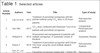
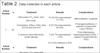
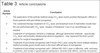
The results are described in Tables 1, 2 and 3.
| Article Number | Authors | Year | Title | Type of study |
|---|---|---|---|---|
| 1 | Lee SJ et al | 2015 | Treatment of periorbital syringomas with the pinhole method using CO2 laser in 29 Asian patients15 | Retrospective analysis |
| 2 | Seo HM, | 2015 | CO2 laser combined with Botulinum Toxin A for patients with periorbital syringomas9 | Comparative Restrospective analysis |
| 3 | Williams K, | 2016 | Assessment and management of patients with multiple syringomas: A systematic review of literature2 | Systematic Review |
| 4 | Kitano Y | 2016 | Treatment of periorbital syringomas with Erbium YAG laser using the ovoid multiple ablation method11 | Retrospective Analysis |
| 5 | Kim JY, Lee JW, Chung KY | 2017 | Periorbital syringomas treated with 1,444nm ND - YAG laser12 | Retrospective Analysis |
| 6 | Ahn GR et al | 2019 | Intralesional electrocoagulation with microneedle alone for the treatment of periorbital syringomas: A retrospective analysis13 | Retrospective Analysis |
YAG: Erbium Yttrium Aluminum Garnet; ND-YAG: Neodymium Doped - Yttrium Aluminum Garnet
| Article Number | n | Treatment | Results | Complications |
|---|---|---|---|---|
| 1 | 29 | Ultra pulse CO2 laser, char-free mode | 10 (34.5%) improvement 51-75% | Mild erythema: 5 patients |
| Parameters: 200µs, 50Hz, time of 0.04 and rest 0.01 | 8 (27.6%) improvement 26-50% | Prolonged erythema: 2 patients (Treated with Laser 595nm) | ||
| 2 sessions were held at two-month intervals | 7 (24.1%) improvement >75% | Post-inflammatory hyperpigmentation (PIH): 1 patient (Treated with ND-YAG) | ||
| 4 (13.8%) melhora 0-25% | ||||
| Article Number | n | Treatment | Results | Complications |
| 2 | 92 | 44 cases: CO2 laser with multiple holes (4-5w, 80mm) | Clinical improvement | PIH: 5 patients (11.4%) |
| Average treatments 3.89 | 12 (27.3%) Excellent | Recurrence: 50% | ||
| 19 (43.2%) Good | (2 years) | |||
| 12 (27.3%) Moderate | ||||
| 1 (2.3%) Poor | ||||
| Reduction of injuries | ||||
| 10 (22.7%) Excellent | ||||
| 16 (36.4%) Good | ||||
| 17 (38.6%) Moderate | ||||
| 1 (2.3%) Poor | ||||
| 48 cases: CO2 laser with multiple holes(4-5w, 80mm) and Botulinum Toxin A (100 IU diluted in 2.5ml and used from 5 to 10 iu PER SIDE) | Clinical improvement | PIH: 3 patients (6.3%) | ||
| Média de tratamentos 3.4 | 15 (31.3%) Excellent | Recurrence: 59% | ||
| 27 (56.3%) Good | (2 years) | |||
| 6 (12.5%) Moderate | ||||
| Reduction of injuries | ||||
| 14 (29.2%) Excellent | ||||
| 23 (47.9%) Good | ||||
| 11(22.9%) | ||||
| 3 | 215 | 169 cases (78.6%): CO2 laser alone or in combination | 71 (33%) Moderate resolution | Erythema resolved within 3 to 6 months |
| 51 (23.7%) Total resolution | Hypo- or Hyperpigmentation resolved between 3 and 9 months | |||
| 31 (14.4%) Close to full resolution | ||||
| 16 (7.4%) Poor response | ||||
| 18 cases (8.4%): Low voltage electrocoagulation | 11 (5.1%) Marked improvement | Redness, pain, swelling and Post-inflammatory hyperpigmentation resolved within 2 weeks | ||
| 7 (3.3%) Moderate improvement | ||||
| 12 cases (5.6%): Intralesional electrodissection | 12 (5.6%) Complete resolution | PIH resolved within 3 months | ||
| 02 cases (1%): Surgical excision | 02 (1%) Cosmetically acceptable results | Not Reported (NR) | ||
| 02 cases (1%): Intralesional needle | 02 (1%) High degree of satisfaction | NR | ||
| 02 cases (1%): Oral isotretinoin | 01 (0.5%) Discrete reduction of lesions | NR | ||
| 01 (0.5%) No response | ||||
| 02 cases: Fractional Photodermolysis | 02 (1%) Positive clinical results | NR | ||
| 01 case: Dermabrasion | 01 (0.5%) Moderate reduction in lesion size | NR | ||
| 01 case: Argon Laser | 01 (0.5%) Discreet reduction of injuries | Blisters heal within 1 week | ||
| 01 case: Topical atropine | 01 (0.5%) Discreet reduction of injuries | NR | ||
| 01 case (0.5%): Topical tretinoin | 01 (0.5%) Reduction in the size of lesions | NR | ||
| 03 case (1.5%): Tranilast | 01 (0.5%) Discreet reduction of injuries | Irritated bladder | ||
| 02 (1%) Good improvement | ||||
| 4 | 49 | Parameters: Irradiation diameter 1mm, 9J/cm2 and 250sec | Mean of 3.77 treatments in disseminated syringomas and 4.23 in clusters | Mild erythema: 1 patient |
| The tissue removed in ovoid shape (2-4mm) | 43 (87.7%) improvement >75% | PIH: 1 patient | ||
| Treatment interval: 2 months | 1 (2%) Improvement between 0-24% | Depression in the area: 1 patient | ||
| Maximum treatment: 10 injuries | 5 (10.2%) No recurrence | Improvement at 7 months | ||
| Closed hydrocolloid dressing for 2 weeks | ||||
| 5 | 19 | Parameters: Distance from lesion of 0,5cm, 160mJ, 1,6w, 10Hz. | 13 (68.4%) improvement in the first session | Ertyhema: 12 patients after 1st session |
| Fluency per point 1J, and 4 to 6 shots per point | 19 (100%) improvement > 50% in the second session | PIH: 1 patient | ||
| 2 sessions every 2 months | Patient satisfaction scale | Recurrence: 1 patient | ||
| 12 (63.2%) very satisfied after the first session | ||||
| 17 (89%) very satisfied after the second session | ||||
| 6 | 55 | Using a 1MHz monopolar radiofrequency device, the microneedle was inserted and the current was passed 2 to 3 times. | 2 (3.6%) Disappearance >75% | Edema,erythema and appearance of temporary crusts |
| Parameters: 4w and 100mseg. | 26 (47.3%) Improvement 55-75% | PIH: 1 patient spontaneously improved | ||
| Minimum of 3 treatments | 15 (27.3%) Improvement 25-55% | |||
| 7 (12.7%) Improvement 0-25% | ||||
| 5 (9.1%) Get worse | ||||
| The severity index of periorbital syringomas decreased after 1 treatment (mean 3.2) and 2 treatment (mean 1.7) |
PIH: Post Inflammatory Hyperpigmentation; NR: Not reported.
| Article Number | Conclusion |
|---|---|
| 1 | The application of the pinhole method using CO2 laser exerts positive therapeutic effects in Asian patients with periorbital syringomas. |
| 2 | The combined therapy treatment of CO2 laser and botulinum toxin A had better results than the one performed with CO2 laser. Studies of Botulinum Toxin A as monotherapy are needed. |
| 3 | Treatment of syringomas remains a challenge, although destructive methods - specifically CO2 laser and possibly intralesional electrocoagulation - may represent the best current options for surgical management. |
| 4 | Treatment of periorbital syringomas with Erbium YAG Laser using the ovoid multiple ablation method provides good results even in the most difficult types of syringomas. |
| 5 | Treatment of periorbital syringomas with 1,444nm ND-YAG Laser is effective, safe, and easy to perform, and results in good patient satisfaction, minimal side effects, and low recurrence rates. |
| 6 | Intralesional electrocoagulation with microneedle alone is an effective and safe treatment for periorbital syringomas |
YAG: Erbium Yttrium Aluminum Garnet; ND-YAG: Neodymium Doped - Yttrium Aluminum Garnet
DISCUSSION
The syringomas are benign adnexal tumors with histopathological features that demonstrate that they arise from the intradermal portion of the eccrine ducts1 and are often located in the orbital region but can be found on the vulva, penis, scalp and axillae2,3 in the form of small yellowish papules or skin color, with a size between 1 and 3 mm, which cause cosmetic problems and, in some cases, itching. The classification most used to date is the one proposed by Friedman & Butler4, with four variants: localized, familial, generalized, and associated with trisomy 21. There is a predominance in females, and the lesions begin to appear in adolescence or early adult age.
The objective of the treatment of periorbital syringomas is to improve the cosmetic appearance since these lesions are considered benign, non-progressive and typically asymptomatic.
Wheeland et al.16 described CO2 laser vaporization in a patient with no evidence of scarring or recurrence for 2 years. Castro et al.17 described the use of low-power super-pulsed CO2 laser, highlighting the reduction of thermal energy deposition with this modality, and had good results.
Gomez et al.18 performed the treatment with topical tretinoin in a case of eruptive syringomas, and the patient had moderate improvement due to the flattening of the lesions but had a recurrence in untreated areas. The use of topical atropine19 showed moderate efficacy, but there are few reported cases.
Karam & Benedetto20 presented a case report using intralesional electrodissection, with the total disappearance of syringomas and hyperpigmentation that resolved at 3 months post-treatment, and in 1997 they reported moderate efficacy in another series of 20 cases treated with the same technique and had two cases of post-inflammatory hyperpigmentation in Fitzpatrick IV phototype patients, which improved at 3 months9. In both publications, no recurrence was reported.
Kang et al.7 evaluated the histopathology and efficacy of placing 50% trichloroacetic acid (ATA) in the lesions after making three to four holes with a CO2 laser with low parameters in 20 patients and demonstrated the removal of deep syringomas, reducing the adverse effects. The presence of necrotic dermal collagen could explain the results for tissue regeneration and the stimulation of ATA to produce type I collagen. Intralesional ATA caused extra necrosis in the lesions, but a fixed CO2 laser protocol was used that did not project the size difference, and this could lead to an inconsistent result when applied to more patients. The benefits of the laser would be precisely to be able to adapt the controlled ablation of tissues of different depths. From our point of view, the fixed protocol ends up compromising the result.
The CO2 laser has been used in different modalities: Wang &Roenigk21reported the use of high-energy CO2 laser in 10 patients with Fitzpatrick phototype III and IV without sequelae; Sajben & Ross22 showed an efficiency of high energy pulsed CO2 laser using a 1 mm handpiece in the treatment of 4 patients; Frazier et al.23 combined ATA 35% (medium peel) and low-energy CO2 laser in an African-American patient, and achieved cosmetic improvement, even though the syringomas were not completely removed.
Park et al.24, after vaporizing periorbital syringomas with CO2 laser in 6 patients, introduced black ink into the dermis and later used Q-Switched Laser to remove the lesions, with no evidence of recurrence at 8 weeks of follow-up. Park et al.25 treated 11 patients using CO2 laser with multiple perforation method and observed good results without complications when the treatment was performed repeatedly. The fractional mode could be an option considered.
Bagatin et al.26 reported the experience in the surgical treatment of 38 patients using excision with Castroviejo scissors and healing by second intention. Thirty-six cases were described with excellent and good results, but 12 cases had hypochromia, 1 had a depressed scar, and 1 had a hypertrophic scar. Even if this method has shown good results, it would be important to evaluate the Fitzpatrick skin phototype due to the risk of hyperpigmentation in the healing process by the second intention, which can appear in larger phototypes. Some preventive dermatological protocols could be associated to avoid these complications.
Al Aradi8 carried out a pilot study of the efficacy of low-voltage electrocoagulation in 20 patients with phototype IV and V, placing the electrode superficially in each syringoma, with moderate clinical improvement after the third session. However, 40% of cases developed post-inflammatory hyperpigmentation and 10% hypopigmentation.
Akita et al.27 performed a fractional laser protocol using a 1550 nm Erbium laser in two Japanese patients. Cho et al.6 treated 35 patients, and Meesters et al.28 treated one patient with a 10,600 nm CO2 laser, with good cosmetic results, which would be a less invasive method and with a good neocollagenesis response; however, several treatments would be necessary to obtain full or near full resolution.
Hong et al.29 presented 2 cases treated with fine needle intralesional electrocoagulation, with excellent results and without complications or recurrence, due to the fact that introducing the needle into the lesion allows selectivity in the destruction of dermal lesions without damaging the epidermis. It is necessary to carry out this treatment with more patients and follow-up.
Lee et al.30 reported 29 cases treated with CO2 laser in the pinhole method. After the second session, they observed almost complete clinical improvement in 58.6% of patients, prolonged erythema in 5 patients and one case of post-inflammatory hyperpigmentation. Through this method, the CO2 laser can be directed on the lesion and provide high energy for its destruction, concomitantly stimulating neocollagenesis and dermal matrix remodeling, resulting in clinical improvement of the skin texture. This method proved to be effective, but it is operator-dependent, requires a longer procedure time depending on the number of lesions, and the treatment of invisible lesions cannot be performed. More prospective studies are needed.
Seo et al.10 performed a retrospective analysis of 92 cases comparing CO2 laser therapy with multiple perforations combined with botulinum toxin A. In a previous study carried out by the authors, they described the effectiveness of deep tumor removal using CO2 laser with multiple perforations, and in this new proposal, the authors decided to combine botulinum toxin A. The rate of improvement was significantly higher in the combination therapy, but the recurrence was similar in both groups. Post-inflammatory hyperpigmentation was described in 3 cases of combination therapy and 5 cases of CO2 laser therapy; other adverse effects were not reported.
The eccrine hidrocystomas have been treated with botulinum toxin A31-33, with good results. The results of this study could be explained by the similar origin of syringomas and the action of botulinum toxin, which blocks the cholinergic terminals of the autonomic nerve that regulate the secretion of eccrine sweat glands34; however, studies with transdermal or intralesional monotherapy are necessary.
A systematic review of the literature performed by Williams & Shinkail2 analyzed 215 cases treated in different medical and surgical modalities, such as treatment with topical isotretinoin and atropine, peels, dermabrasion, cryotherapy, electrodissection, intralesional electrocoagulation, laser therapy and laser treatment combinations. The authors noted that CO2 laser therapies and intralesional electrocoagulation might present the best treatment options.
CO2 laser has shown moderate efficacy and cosmetic improvement in most reported cases and, even though it does not offer complete resolution of the lesions, most adverse effects improve with time and are well tolerated by patients. Intralesional electrocoagulation decreased the size and number of lesions while sparing epidermal damage such as scarring and hyperpigmentation, and cases with post-inflammatory hyperpigmentation showed spontaneous improvement within 2 to 3 months.
Kitano12 described the treatment of periorbital syringomas with Erbium YAG laser using the multiple ablation method with an ovoid shape of 2 to 4 mm in 49 patients with disseminated and/or accumulated periorbital syringomas, with a mean of 3.77 and 4.23 sessions, respectively. Sessions were performed every 2 months, and lesions disappeared by more than 75% in 43 patients. A hydrocolloid dressing was used for 2 weeks.
The Erbium YAG, 2490 nm laser, has an absorption coefficient for water approximately 13 times higher than the CO2 laser, causing less thermal damage to underlying tissues and a very thin coagulation layer. It has been used for removing lesions, nevi, xanthomas and tattoos. In the case of syringomas, they do not infiltrate adjacent tissues and could be removed precisely, preserving deep tissue, as the thermal damage is confined to an extremely thin layer. The results were good; however, it was a retrospective study with variable goals according to the type of lesions of each patient, which limits the evaluation of their effectiveness.
Kim et al.13 treated 19 patients with Fitzpatrick IV skin phototypes with a 1,444 nm fiber optic ND-YAG laser with low parameters. All patients showed an improvement of more than 50% of satisfaction in the first session and 89.5% after the second session. Erythema was observed in 63.2% of patients, and post-inflammatory hyperpigmentation in 1 case. They reported worsening in 2 patients after the first session and 1 after the second.
The ND YAG wavelength 1,444 nm is well absorbed in water, making it a therapeutic option for treating cystic lesions such as eccrine hidrocystomas when fiber optic is used subdermally through a cannula 11 or 12. Due to its effectiveness for lipolysis and the low diffusion of heat from adjacent tissues, it is used to treat skin diseases related to adipose tissue. In the case of syringomas, it is believed that the absorption peaks for water and lipids target the lumen and the lipid membrane, which surround the syringomas, and in this way can be effective without causing damage to adjacent tissues. The 1,444 nm ND YAG laser is considered a good treatment option when used with low parameters. However, it is necessary to carry out prospective studies with a larger number of patients and in other skin phototypes.
Ahn et al.14 performed a retrospective analysis of a case series with 55 patients who underwent 3 sessions of intralesional electrocoagulation, using monopolar radiofrequency with an isolated needle. The evaluators, dermatologists, did not know the number of treatments performed. A scale created by the authors, called Periorbital Syringoma Severity Index, was used, which evaluated number, size, involvement and density. Half of the patients had a marked improvement greater than 50% after the first session, and the severity of the injury decreased with the remaining sessions. The erythema improved until the fourth day, and there was one case of post-inflammatory hyperpigmentation improved with time.
With this method, a thermal coagulative zone is induced within the ductal structure through the microneedle, and the difference in electrical impedance between the epidermis (high) and dermis (low) allows radiofrequency energy to flow into the target dermal tissue. The treatment described is a safe and effective treatment for periorbital syringomas, and the evaluation scale represents an objective way of describing the involvement. However, it is necessary to carry out prospective, comparative studies with different skin phototypes.
Bae et al.15 reported a case of syringomas in the neck, treating one side with a CO2 laser and the other with radiofrequency with a microisolated needle, finding better results on the radiofrequency side. The lesions disappeared after 3 sessions, and there was no record of recurrence on this side. It was necessary to use corticosteroids on the side of the CO2 laser due to the presence of hypertrophic scars. This study is a case report from another location, but it is worth naming as it is the only one that compares these forms of treatment in the same patient.
CONCLUSIONS
Periorbital syringomas remain a therapeutic challenge, and so far, no treatment has been shown to be consistently effective. The CO2 laser is considered the first choice of treatment for syringomas due to its penetration power and the demonstrated success: when performing a deep ablation, it can completely remove lesions and those that are hidden, but post-procedure care must be taken into account, mainly in phototypes IV, V and VI, due to the high risk of side effects such as post-inflammatory hyperpigmentation and inadequate healing; when it is performed in the fractionated modality or the multiple-hole technique and/or pinhole method, with low parameters, the pulse energy is delivered more safely; however, the resolution is partial, and several treatments are necessary.
Intralesional electrocoagulation represents a second alternative with moderate results and lower risk of complications; however, it has a small number of cases reported in the literature, and it is necessary to carry out studies with a greater number of cases, establish treatment parameters and that they are performed in different skin phototypes.
New treatments such as Erbium YAG and ND YAG laser and monotherapy with botulinum toxin A could be good treatment alternatives with a lower risk of sequelae. However, further studies are needed to demonstrate their effectiveness.
Prospective, comparative studies with various skin phototypes are needed to assess the effectiveness and determine the best option.
REFERENCES
1. Langbein L, Cribier B, Schirmacher P, Praetzel-Wunder S, Peltre B, Schweizer J. New concepts on the histogenesis of eccrine neoplasia from keratin expression in the normal eccrine gland, syringoma and poroma. Br J Dermatol. 2008;159(3):633-45.
2. Williams K, Shinkai K. Evaluation and management of the patient with multiple syringomas: A systematic review of the literature. J Am Acad Dermatol. 2016;74(6):1234-40.e39.
3. Ciarloni L, Frouin E, Bodin F, Cribier B. Syringoma: A clinicopathological study of 244 cases. Ann Dermatol Venereol. 2016;143(8-9):521-8.
4. Friedman SJ, Butler DF. Syringoma presenting as milia. J Am Acad Dermatol. 1987;16(2 Pt 1):310-4.
5. Lau J, Haber RM. Familial eruptive syringomas: case report and review of the literature. J Cutan Med Surg. 2013;17(2):84-8.
6. Cho SB, Kim HJ, Noh S, Lee SJ, Kim YK, Lee JH. Treatment of syringoma using an ablative 10,600-nm carbon dioxide fractional laser: a prospective analysis of 35 patients. Dermatol Surg. 2011;37(4):433-8.
7. Kang WH, Kim NS, Kim YB, Shim WC. A new treatment for syringoma. Combination of carbon dioxide laser and trichloroacetic acid. Dermatol Surg.1998;24(12): 1370-4.
8. Al Aradi IK. Periorbital syringoma: a pilot study of the efficacy of low- voltage electrocoagulation. Dermatol Surg. 2006;32(10):1244-50.
9. Karam P, Benedetto AV. Intralesional electrodesiccation of syringomas. Dermatol Surg. 1997;23(10):921-4.
10. Seo HM, Choi JY, Min J, Kim WS. Carbon dioxide laser combined with botulinum toxin A for patients with periorbital syringomas. J Cosmet Laser Ther. 2016;18(3):149-53.
11. Barzegar M, Abdollahimajd F, Golfeshan A, Nikvar M. Syringomas Accentuated on the Upper Lip. Pediatr Dermatol. 2016;33(2): e172-3.
12. Kitano Y. Erbium YAG Laser treatment of periorbital syringomas by using the multiple ovoid-shape ablation method. J Cosm Laser Ther. 2016;18(5):280-5.
13. Kim JK, Lee JW, Chung KY. Periorbital Syringomas Treated With an Externally Used 1,444 nm Neodymium-Doped Yttrium Aluminum Garnet Laser. Dermatol Surg. 2017;43(3):381-8.
14. Ahn GR, Jeong GJ, Kim JM, Hong JY, Li K, Lee CK, et al. Intralesional Electrocoagulation With Insulated Microneedle for the Treatment of Periorbital Syringomas: A Retrospective Analysis. Aesthet Surg J. 2021;41(4):490-8.
15. Bae JY, Jang DH, Lee JI, Jung HJ, Ahn JY, Park MY. Comparison of microinsulated needle radiofrequency and carbon dioxide laser ablation for the treatment of syringoma. Dermatol Ther. 2019;32(3):e12912.
16. Wheeland RG, Bailin PL, Reynolds OD, Ratz JL. Carbon dioxide (CO2) laser vaporization of multiple facial syringomas. J Dermatol Surg Oncol. 1986;12(3):225-8.
17. Castro DJ, Tartell PB, Soudant J, Saxton RE. The surgical management of facial syringomas using the superpulsed CO2 laser. J Clin Laser Med Surg. 1993;11(1):33-7.
18. Gómez MI, Pérez B, Azaña JM, Núñez M, Ledo A. Eruptive syringoma: treatment with topical tretinoin. Dermatology. 1994;189(1):105-6.
19. Sánchez TS, Daudén E, Casas AP, García-Díez A. Eruptive pruritic syringomas: treatment with topical atropine. J Am Acad Dermatol. 2001;44(1):148-9.
20. Karam P, Benedetto AV. Syringomas: new approach to an old technique. Int J Dermatol.1996;35(3):219-20.
21. Wang JI, Roenigk HH Jr. Treatment of multiple facial siringomas with the carbon dioxide (CO2) laser. Dermatol Surg.1999;25(2):136-9.
22. Sajben FP, Ross EV. The use of the 1.0 mm handpiece in high energy, pulsed CO2 laser destruction of facial adnexal tumors. Dermatol Surg.1999;25(1):41-4.
23. Frazier CC, Camacho AP, Cockerell CJ. The treatment of eruptive syringomas in an African American patient with a combination of trichloroacetic acid and CO2 laser destruction. Dermatol Surg. 2001;27(5):489-92.
24. Park HJ, Lim SH, Kang HA, Byun DG, Houh D. Temporary tattooing followed by Q- switched alexdrite laser for treatment of syringomas. Dermatol Surg. 2001;27(1):28-30.
25. Park HJ, Lee DY, Lee JH, Yang JM, Lee ES, Kim WS. The treatment of syringomas by CO(2) laser using a multiple-drilling method. Dermatol Surg. 2007;33(3):310-3.
26. Bagatin E, Enokiahara MY, Souza PK. Periorbital syringomas - Excision with Castroviejo scissors. Experience in 38 patients and literature review. An Bras Dermatol. 2006;81(4):341-6.
27. Akita H, Takasu E, Washimi Y, Sugaya N, Nakazawa Y, Matsunaga K. Syringoma of the face treated with fractional photothermolysis. J Cosmet Laser Ther. 2009;11(4):216-9.
28. Meesters A, De Rie M, Wolkerstorfer A. Timed exposure 10,600nm CO2 Laser drilling in various benign dermal tumours. Eur J Dermatol. 2015;25(4):358-9.
29. Hong SK, Lee HJ, Cho SH, Seo JK, Lee D, Sung HS. Syringomas treated by intralesional insulated needles without epidermal damage. Ann Dermatol. 2010;22(3):367-9.
30. Lee SJ, Goo B, Choi MJ, Oh SH, Chung WS, Cho SB. Treatment of periorbital syringoma by the pinhole method using a carbon dioxide laser in 29 Asian patients. J Cosmetic Laser Ther. 2015;17(5):273-6.
31. Gandhi V, Naik G, Verma P. Eccrine hydrocistoma successfully treated with topical synthetic botulinum peptide. J Cutan Aesthet Surg. 2011;4(2):154-5.
32. Blugerman G, Schavelzon D, D’Angelo S. Multiple Eccrine hidrocistomas: a new therapeutic option with botulinum toxin. Dermatol Surg. 2003;29(5):557-9.
33. Correia O, Duarte AF, Barros AM, Rocha N. Multiple eccrine hidrocystomas--from diagnosis to treatment: the role of dermatoscopy and botulinum toxin. Dermatol. 2009;219(1):77-9.
34. Guida S, Farnetani F, Nisticò SP, Mariarosaria CG, Babino G, Pellacani G, et al. New trends in botulinum toxin use in dermatology. Dermatol Pract Concept. 2018;8(4):277-82.
1. Hospital Albert Einstein Israelita, Institute of Education and Research, São Paulo,
SP, Brazil
| COLLABORATIONS | |
|---|---|
| KTVC | Analysis and/or interpretation of data, Data Collection, Conception and design of the study, Project Management, Methodology, Writing - Preparation of the original |
| JLHR | Methodology, Writing - Preparation of the original |
| AH | Final approval of the manuscript, Writing - Review and Editing, Supervision |
| MGC | Final approval of the manuscript, Writing - Review and Editing, Supervision |
| MFN | Final approval of the manuscript, Writing - Review and Editing, Supervision |
Corresponding author: Karols Tatiana, Vila-Claro Irmão Norberto Francisco Rauch 755 1418B - Jardim Carvalho, Porto Alegre, RS, Brazil, Zip Code 91450-147, E-mail: dratatianavila@gmail.com
Article received: January 04, 2021.
Article accepted: October 15, 2021.
Conflicts of interest: none.
 Read in Portuguese
 Read in English
 PDF PT
 Print
Print
 Send this article by email
 How to Cite
 Mendeley
 Pocket