


Case Report - Year 2022 - Volume 37 -
Use of a flap based on the superior lateral genicular artery to cover lower limb injuries: case reports
Utilização de retalho baseado na artéria genicular lateral superior na cobertura de lesões de membros inferiores: relato de casos
Pedro Henrique Silva Benevides1* ; Renan Correia Arcanjo1; Lauro César Brito Rezende1; Anderson Cleyton Moreira Oliveira1; Gustavo Teixeira Leão1; Sérgio José de Lima1; João Emílio Hanum Paes1; Daniel Ribeiro de Resende1
ABSTRACT
The knee is one of the main joints in the human body, and due to its conformation and functionality, it is easily susceptible to injuries. Surgical reconstruction of injuries around the knee and proximal region of the leg represents a major challenge due to the exposure of osteoarticular, tendinomuscular and neurovascular structures and has been performed using various muscle and musculocutaneous flaps. Other flaps are also described for treating these lesions, although less frequently in the literature, such as the cutaneous flap based on the pedicle of the superior lateral genicular artery. Next, It presents a report of five cases of patients who were victims of motorcycle accidents in which the lesion was covered around the knee and proximal region of the leg using a skin flap based on the pedicle of the superior lateral genicular artery, in which good coverage of lesions, high flap survival rate, good clinical and aesthetic results. The use of the superior lateral genicular artery presents aesthetic advantages since the color and texture of the flap are similar to those of the knee region and provide a better quality appearance, in addition to not causing joint mobility problems.
Keywords: Knee; Thigh; Surgical Flaps; Lower extremity; Reconstructive surgical procedures.
RESUMO
O joelho é uma das principais articulações do corpo humano e, em função de sua conformação e funcionalidade, é facilmente suscetível a lesões. A reconstrução cirúrgica de lesões ao redor do joelho e região proximal da perna representa um grande desafio devido à exposição de estruturas osteoarticulares, tendinomusculares e neurovasculares e tem sido realizada com uso de diversos retalhos musculares e musculocutâneos. Outros retalhos também são descritos para tratamento dessas lesões, embora em menor frequência na literatura, como o retalho cutâneo baseado no pedículo da artéria genicular lateral superior. A seguir, é apresentado o relato de cinco casos de pacientes vítimas de acidentes motociclísticos nos quais realizou-se a cobertura da lesão ao redor do joelho e região proximal da perna com o uso do retalho cutâneo baseado no pedículo da artéria genicular lateral superior, nos quais foram obtidos uma boa cobertura das lesões, alta taxa de sobrevida do retalho, bons resultados clínicos e estéticos. O uso do artéria genicular lateral superior apresenta vantagens estéticas, visto que a cor e a textura do retalho são semelhantes às da região do joelho e conferem uma aparência de melhor qualidade, além de não causarem problemas de mobilidade articular.
Palavras-chave: Joelho; Coxa da perna; Retalhos cirúrgicos; Extremidade inferior; Procedimentos cirúrgicos reconstrutivos.
INTRODUCTION
The main causes of tissue injuries around the knee include traumatic wounds, oncological surgeries, invasive infections or burns1,2. Surgical reconstruction of these injuries represents a major challenge due to the exposure of osteoarticular, tendinomuscular and neurovascular structures and has been performed using different muscular and musculocutaneous flaps3,4. The use of a skin flap based on the pedicle of the superior lateral genicular artery (SLGA) is less frequently reported in the scientific literature5.
OBJECTIVE
This article aimed to report the feasibility of using the SLGA-based flap to cover injuries around the knee and proximal region of the leg in patients who were victims of motorcycle accidents.
CASE REPORTS
The Ethics Committee approved this study under CAAE number: 52573721.3.0000.0033. Five patients, four males (80%) and one female (20%), who were victims of motorcycle accidents, were treated for injuries around the knee. Patient 1, male, 22 years old, 15 cm soft tissue lesion in the lateral region of the right knee. Patient 2, male, 19 years old, 16 cm soft tissue lesion in the lateral region of the right knee. Patient 3, male, 35 years old, 13 cm soft tissue lesion in the lateral region of the right knee. Patient 4, male, 30 years old, 14 cm soft tissue lesion in the lateral region of the left knee. Patient 5, female, 27 years old, 12 cm soft tissue lesion in the lateral region of the right knee.
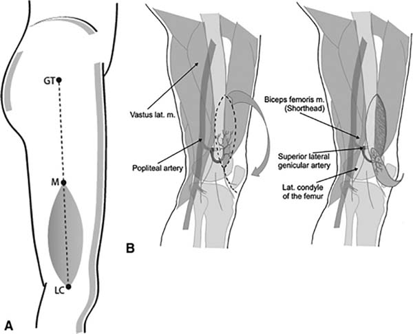
The flap designed to adjust to the defect resulting from the injury was based on the SLGA vascular pedicle in all cases, and the donor areas were closed in the same surgical procedure or supplemented with a skin graft. Figure 1 shows the schematic planning of the flap based on the SLGA.

Surgical technique
The patient was under spinal anesthesia and placed in lateral decubitus. After demarcation of the anatomical parameters, which have as reference the projection of the greater trochanter of the femur, the lateral condyle of the femur, the posterior border of the vastus lateralis muscle and the anterior border of the biceps femoris. In this previously designed area is found the SLGA. Next, exsanguination of the limb was performed by gravity, followed by a tourniquet with a smarch band at the proximal level of the thigh.
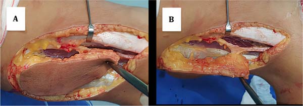
After making an incision on the skin and subcutaneous tissue following the line of the lateral edge of the patella, the muscle fascia was incised after identifying the edge of the vastus lateralis muscle, extending from the lateral projection of the gluteal fold to the lateral topography of the knee. Careful dissection was then performed in the subfascial region, delimited by the vastus lateralis muscle and posteriorly by the biceps femoris muscle, continuing until it met the perforating branch of the SLGA, increasing the dissection to deeper planes.
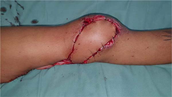
Subsequently, the necessary extension of the flap was defined, completing its dissection towards the posterior aspect of the femur up to its origin in the popliteal artery. The tourniquet was released to assess perfusion in the flap. After being perfused, the flap was rotated towards the receiving area and fixed using simple stitches. Then, in the donor area, the three tissues were brought together with a simple suture and, when necessary, supplemented with a second skin graft.
RESULTS
Patients were discharged after five days of hospitalization with a prescription of rivaroxaban 10mg, once a day, for 15 days and returned for dressing change twice a week in the first two weeks and then twice every 15 days. In the periods of 5 and 14 days after hospital discharge, all patients showed good healing and good knee range of motion. After the 15th postoperative day, the patients started physiotherapy to recover joint range of motion, which occurred after 20 physiotherapy sessions.
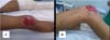
The age group ranged from 19 to 35 years old, with an average of 26 years old. The mean dimensions of the patients’ flaps were 13×7cm. There was complete flap survival in all patients. Only patient 1 had necrosis of the skin graft three weeks after the operation, which was resolved later with a new graft. The donor areas of all patients healed well with no restriction in knee joint mobility. Hospitalization time was 14 days, and follow-up time was 12 months. Figures 2 to 5 show the photographic records of one of the cases (patient 5).


DISCUSSION
Most cases of lower limb soft tissue injuries occur in males young adults and result from motorcycle accidents. Soft tissue defects around the knee are reconstructed with pedicled perforator flaps, with the SLGA being a reliable flap option6.
Moktader et al.7 evaluated the reliability of the SLGA flap in 15 patients with injuries around the knee, succeeding in 14 cases, and only one patient had necrosis at the distal margin of the flap.
Wiedner et al.8 analyzed six cases of using the SLGA flap for soft tissue reconstruction around the knee and observed flap survival in all patients, and only one patient had partial necrosis at the distal tip of the flap without late complications.
Mahipathy et al.9 evaluated five patients with a mean age of 42 years, in whom the SLGA-based flap was used for reconstruction of defects around the knee, and observed complete survival of the flap in all patients, with distal necrosis in one patient who was treated conservatively.
The use of AGLS has aesthetic advantages, as the color and texture of the flap are similar to those of the knee region and provide a better quality appearance compared to muscle or musculocutaneous flaps, in addition to not causing problems in knee joint mobility9.
CONCLUSION
The use of the SLGA-based flap is a viable technique and provides good coverage of lesions around the knee and proximal region of the leg, with a high flap survival rate and good clinical results consistent with those reported in the literature.
REFERENCES
1. Valente AS, Borba DF, Resende DR, Resende MR, Goulart RG, Lima SJ. Utilização de retalho em hélice para cobertura de lesões de partes moles em membro inferior. Rev Bras Ortop. 2021;56(2):192-7.
2. Warner SJ, Garner MR, Schottel PC, Fabricant PD, Thacher RR, Loftus ML, et al. The effect of soft tissue injuries on clinical outcomes after tibial plateau fracture fixation. J Orthop Trauma. 2018;32(3):141-7.
3. Macedo JLS, Rosa SC, Silva AA, Filho Neto AVR, Ruguê PHS, Scartazzini C. Versatilidade do uso do retalho do músculo gastrocnêmio medial na reconstrução de lesões de partes moles de membros inferiores. Rev Bras Cir Plást. 2016;31(4):527-33.
4. Vendramin FS, Santos FA, Fonseca ANN, Sá JP, Morikawa LS. Análise epidemiológico-evolutiva de pacientes submetidos a cirurgia plástica reparadora em um hospital de referência em trauma. Rev Bras Cir Plást. 2019;34(1):101-7.
5. Utiyama DMO, Santos HM, del Papa LGA, Silva NM, Sales VC, Ayres DVM, et al. Características do perfil de indivíduos amputados atendidos em um instituto de reabilitação. Acta Fisiatr. 2019;26(1):14-8.
6. Elsahar H, Sadek K, Reda WE. Reconstruction of acute traumatic defects around the knee; our experience with the lateral superior genicular flap. Kasr El Aini J Surg. 2017;18(3):1-8.
7. Moktader MA, Hassan M, Taman E, Taha A, Elaw S. Lateral superior genicular flap for reconstruction around the knee. J Plast Reconstr Surg. 2010;34(2):223-6.
8. Wiedner M, Koch H, Scharnagl E. The superior lateral genicular artery flap for soft-tissue reconstruction around the knee: clinical experience and review of the literature. Ann Plast Surg. 2011;66(4):388-92.
9. Mahipathy SRRV, Durairaj AR, Sundaramurthy N, Jayachandiran AP. Lateral genicular artery flap for reconstruction of defects around the knee: a series of 5 cases. Int Surg J. 2020;7(10):3411-3.
1. Hospital Estadual de Urgências de Goiás - Dr. Valdemiro Cruz, Goiânia, GO, Brazil
ACMO Data Curation, Final manuscript approval, Writing - Review & Editing.
DRR Final manuscript approval, Project Administration, Supervision.
GTL Analysis and/or data interpretation, Writing - Original Draft Preparation, Writing - Review & Editing.
JEHP Analysis and/or data interpretation, Writing - Original Draft Preparation.
LCBR Methodology, Writing - Review & Editing.
PHSB Data Curation, Final manuscript approval, Writing - Review & Editing.
RCA Final manuscript approval, Writing - Original Draft Preparation, Writing - Review & Editing.
SJL Project Administration, Supervision, Writing - Original Draft Preparation.
Corresponding author: Pedro Henrique Silva Benevides Avenida 31 de março esq. c/ 5ª Radial, Setor Pedro Ludovico, Goiânia, GO, Brazil. Zip Code: 74820-300, E-mail: phsilvabenevides1@gmail.com
Article received: March 11, 2022.
Article accepted: July 13, 2022.
Conflicts of interest: none.
 Read in Portuguese
 Read in English
 PDF PT
 Print
Print
 Send this article by email
 How to Cite
 Mendeley
 Pocket