


Original Article - Year 2023 - Volume 38 -
Treatment of complex wounds with PVC prosthesis and autologous partial skin graft: Accelerated and low-cost third-intention healing protocol
Tratamento de feridas complexas com prótese de PVC e enxerto parcial de pele autólogo: Protocolo acelerado e de baixo custo de cicatrização por terceira intenção
Diego Ariel-de-Lima1,2* ; Lana Lacerda de-Lima1,2; Renata Clazzer3; Kêilerte Renes Gurgel Paiva2; Antônio Vicente Dias de-Andrade2; Jorge Edson Pinheiro dos-Santos1; João Firmino da Silva2
ABSTRACT
Introduction: Complex wounds are serious tegumentary injuries that are difficult to resolve with conventional dressings. This study aimed to describe a third-intention wound healing technique, reproducible and low cost, applicable to complex wounds, using polyvinyl chloride (PVC) prosthesis temporarily placed in the injured area to promote the protection and stimulate its "granulation," followed by autologous partial-thickness skin grafting.
Method: Consecutively, 20 patients with complex wounds resulting from external causes were selected and divided into 2 groups: A - patients who underwent the coverage technique with PVC prosthesis, followed by grafting, and B - patients submitted to the care of the dressing team, with daily changes until wound granulation, standard in our institution. Patients were evaluated regarding length of stay; costs; local pain; complications; the time until medical discharge; and patient satisfaction.
Results: The length of hospital stay, its costs, and the time until medical discharge were shorter in group A (p<0.05). However, there was no statistically significant difference in local pain between techniques A and B.
Conclusion: The technique using PVC prosthesis and graft has good efficacy for treating complex wounds, being reproducible and inexpensive.
Keywords: Wounds and Injuries; Wound healing; Polyvinyl chloride; Skin transplantation; Therapeutics
RESUMO
Introdução: Feridas complexas são lesões tegumentares graves, de difícil resolução com
curativos convencionais. O objetivo deste estudo foi descrever uma técnica
de cicatrização de feridas por terceira intenção, reprodutível e de baixo
custo, aplicável a feridas complexas, utilizando uma prótese de policloreto
de vinila (PVC) colocada temporariamente na área da lesão para promover
proteção e estimular sua "granulação", seguida de enxerto autólogo de pele
de espessura parcial.
Método: De forma consecutiva, foram selecionados 20 pacientes com feridas complexas,
decorrentes de causas externas, divididos em 2 grupos: A - pacientes que
foram submetidos à técnica de cobertura com prótese de PVC, seguida de
enxerto; e B - pacientes submetidos aos cuidados da equipe de curativo, com
trocas diárias até granulação da ferida, padrão da nossa instituição. Os
pacientes foram avaliados quanto ao tempo de internação; custos; em relação
à dor local; à presença de complicações; ao tempo até a alta médica; e à
satisfação do paciente.
Resultados: O tempo de internamento e seus custos, assim como o tempo até a alta médica,
foram menores no grupo A (p<0,05). Todavia, não houve
diferença estatisticamente significativa em relação à dor local entre as
técnicas A e B.
Conclusão: A técnica utilizando prótese de PVC e enxerto possui boa eficácia para o
tratamento de feridas complexas, sendo reprodutível e de baixo custo.
Palavras-chave: Ferimentos e lesões; Cicatrização; Cloreto de polivinila; Transplante de pele; Terapêutica
INTRODUCTION
A wound is defined as the loss of skin coverage, representing a break in the continuity of normal tissue structures and functions, and may affect not only the skin but also the subcutaneous tissue, muscles, and bones.1 Many of these wounds represent a challenge for medical and nursing teams, being difficult to resolve using conventional treatments and simple dressings. These cases are classified as ”complex wounds” and must be treated in a specialized hospital center and by a multidisciplinary team.2 Complex wounds cause high morbidity and mortality and have been identified as a serious public health problem in many centers.3
There are numerous techniques described for the treatment of complex wounds. However, several of them are not fully reproducible in many centers due to the complexity of their execution and/or cost.4
OBJECTIVE
This study aimed to describe a third intention wound healing technique, reproducible and inexpensive, applicable to complex wounds, using a polyvinyl chloride (PVC) prosthesis temporarily placed in the area of injury to promote the protection and stimulate its ”granulation,” followed by autologous partial-thickness skin grafting.
METHOD
After approval by the Research Ethics Committee (CEP) (CAAE 53971021.8.0000.5294), a prospective clinical study was carried out in the Trauma sector of a tertiary regional hospital (Hospital Tarcísio de Vasconcelos Maia - HRTVM -, Mossoró, RN, Brazil), between February and September 2022. Consecutively, 20 patients with complex wounds resulting from external causes, which mainly affect the skin/tegument, subcutaneous tissue, aponeurosis/fascia, and muscle, were selected. These patients were divided into 2 groups: A - patients who underwent the PVC prosthesis coverage technique, followed by grafting, and B - patients submitted to the care of the dressing team, with daily changes until wound granulation, standard in our institution.
The following patients were excluded: with mucosal involvement; lesion in the genital region; injury to the face and skull; injury with tendon exposure; injury with bone exposure; injury with exposure of the peritoneal and/or pelvic cavity; injury with exposure of the pleural and/or mediastinal cavity; presence of infections; previous surgeries in the injured region; systemic diseases that significantly compromise immunity, such as decompensated diabetes, acquired immunodeficiency syndrome, psoriasis, lupus erythematosus, rheumatoid arthritis, tumors, among others.
Patients were evaluated regarding length of stay; hospitalization costs; concerning local pain according to the visual analog scale for pain5, graduated from 0 to 10; the presence of complications; the time until medical discharge; and patient satisfaction (measured by a Likert-type scale6: In general, what is your level of satisfaction or dissatisfaction with the evolution of your injury? 5 - Very satisfied; 4 - More or less satisfied; 3 - Neither satisfied nor dissatisfied; 2 - More or less dissatisfied; and 1 - Very dissatisfied).
Description of technique A
First part: coverage with PVC prosthesis
After anesthesia, asepsis, and antisepsis, the wound is carefully debrided to leave it with a minimum of devitalized tissue and as clean as possible. The extent of the wound is determined immediately after proper debridement.
Then, the wound is covered with polyvinyl chloride (PVC) prosthesis obtained from a sterile closed-system urine collection bag (Figure 1A). This collection bag is made of flexible, double-sided PVC, with the front side usually transparent and the back white. This material is easily accessible in surgical centers (Figure 1B).
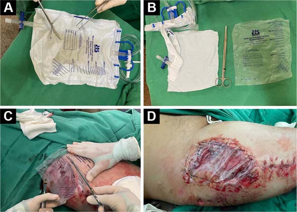
The bag (PVC prosthesis) is then cut in a similar shape and 0.5cm larger than the debrided area of the wound. Then, the prosthesis is sutured to the healthy edges of the lesion with simple stitches (Nylon 3-0 thread) to perfectly fit the prosthesis without exerting pressure on the wound, that is, functioning, more or less, as a semi-occlusive dressing.7.
The dressing comprises sterile gauze and a crepe bandage covering the prosthesis. Liquid exudate forms in this initial phase, slightly wetting the dressing. After the first week, the exudate decreases significantly, forming fibrin tissue, which will be gradually replaced by granulation tissue, thus filling the area lost in the original wound format (healing by second intention). Dressings are changed daily (local cleaning of adjacent skin and PVC prosthesis with chlorhexidine, later covered with sterile gauze and crepe dressing). The patient is discharged after three days, and the dressings are changed in outpatient consultations.
Second part: partial skin graft
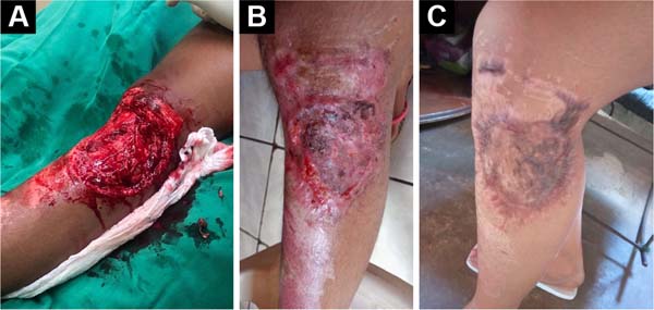
The prosthesis is removed after six to eight weeks, with granulation tissue filling the initial wound (Figure 2). The patient is admitted without the prosthesis, and the wound is covered with a partial-thickness skin graft.
After anesthesia, asepsis, and antisepsis, the granulation tissue of the wound is carefully debrided, leaving it at the same height as the adjacent skin.
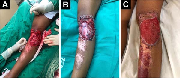
The autologous graft is removed from the anterolateral thigh donor area with a 32 cm Blair blade (Montserrat®), with a graft size sufficient to cover the wound, with a thickness of 0.3 to 0.4 mm (Figure 3A).
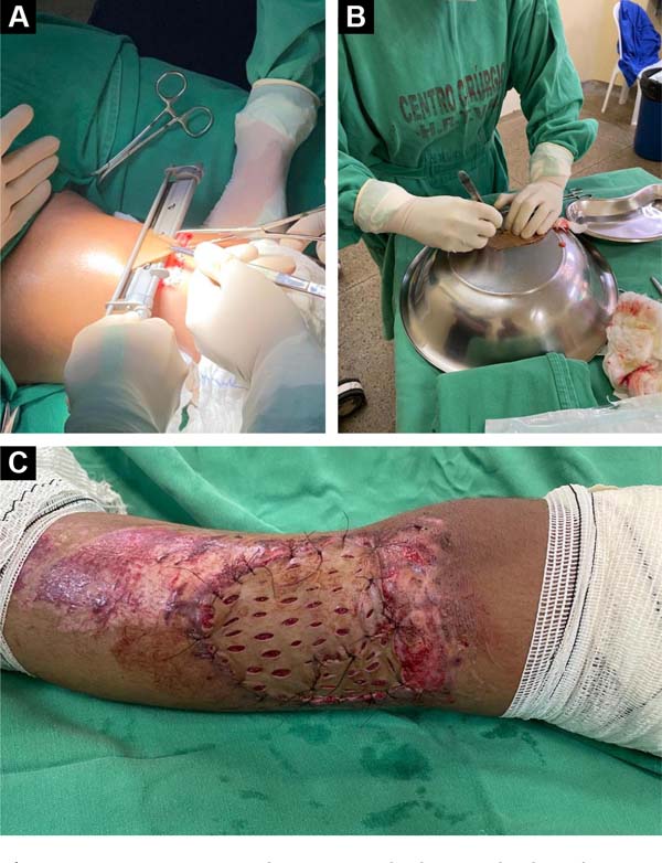

Immediately after removing the skin, the donor area is covered with rayon-type gauze soaked in an adrenaline solution at a concentration of 1:200,000 for 10 minutes for hemostasis, and then a dressing is made with rayon-type gauze maintained in occlusion by sterile cotton gauze and a bandage. The split-thickness skin graft is placed on a sterile metal surface and subjected to multiple parallel incisions of approximately 5-10mm. These incisions help increase the graft area and drain secretions, preventing secretions from forming below the graft and making it difficult to integrate with the grafted area (Figure 3B).
The graft is cut to the shape of the granulated area of the wound and sutured at its healthy edges. Simple sutures (Nylon 4-0 thread) are sufficient to fix the graft to the edge of the wound, providing a perfect fit (Figure 3C). The dressing is made of sterile gauze and crepe bandage, the first change only after five days, and daily in the donor area. The patient is discharged after the first dressing change in the grafted area (five days), with the remaining dressings performed daily on an outpatient basis. The stitches are removed after two weeks. Follow-ups occur at 15, 30, 45, 60, 90, and 180 days after split-thickness skin grafting (Figure 4).

Description of technique B
The patients selected for group B underwent standard care at our institution, performed by the dressing team, with daily changes until the wound granulated. The dressings were changed twice a day according to the following protocol:
- Gentle cleaning with heated 0.9% saline solution and cleaning solution with PHMB;
- Removal of devitalized tissue through mechanical debridement;
- Cleaning the skin in the area around the lesion (perilesional) with a PHMB cleaning solution;
- Cover with sterile gauze.
This procedure is performed until wound granulation, epithelialization, or the plastic surgery team indicates intervention.
Data analysis
Categorical and numerical variables were tabulated and analyzed using the R software for Mac OS X GUI 1.73 (7892 Catalina build), which provided central tendency, percentile values, and dispersion measures.
Data normality was verified using the Shapiro-Wilk test. The homogeneity of the variances of the groups was verified using the Levene test. To reject or reject a null hypothesis, the comparison of the means of the groups was performed using the t-test for independent samples. The presence of outliers was verified through the construction of boxplots. Homoscedasticity was tested by building a linear regression model between the variables.
Analyzes with a confidence interval of 95% and p less than 0.05 were considered statistically significant.
RESULTS
The mean age of patients was 38.6±15.12 years, 80% male and 20% female. The length of hospital stay, its costs, and the time until medical discharge were shorter in group A (P<0.05). Patients submitted to technique A had an average hospital stay of 9.9±0.7 days, while patients submitted to technique B had an average of 37.3±2.0 days. Regarding the length of stay, the t-test showed that there was a statistically significant difference between them (t = -40.596, df=18, p<0.0001), with an advantage for technique A (Table 1).
| Patients | Technique | VAS | Likert | Hospitalized days |
|---|---|---|---|---|
| 1 | A | 4 | 4 | 9 |
| 2 | A | 5 | 5 | 10 |
| 3 | A | 4 | 4 | 10 |
| 4 | A | 6 | 4 | 9 |
| 5 | A | 6 | 4 | 11 |
| 6 | A | 5 | 3 | 10 |
| 7 | A | 5 | 3 | 10 |
| 8 | A | 6 | 4 | 10 |
| 9 | A | 4 | 4 | 9 |
| 10 | A | 5 | 4 | 11 |
| Average | 5.0±0.8 | 3.9±0.5 | 9.9±0.7 | |
| 11 | B | 5 | 3 | 38 |
| 12 | B | 6 | 3 | 40 |
| 13 | B | 4 | two | 41 |
| 14 | B | 5 | two | 35 |
| 15 | B | 4 | two | 36 |
| 16 | B | 5 | 3 | 38 |
| 17 | B | 4 | two | 37 |
| 18 | B | 6 | 3 | 35 |
| 19 | B | 5 | 3 | 37 |
| 20 | B | 5 | 3 | 36 |
| Average | B | 4.9±0.7 | 2.6±0.5 | 37.3±2.0 |

Four weeks after admission, none of the patients who underwent technique B evolved with complete wound granulation or epithelialization. All those who underwent technique B were approached by the plastic surgery team, with subsequent use of flaps and/or grafts.
However, after 180 days, there was no difference in local pain between techniques A and B (VAS 5.0±0.8 for A and VAS 4.9±0.7 for B). Regarding the VAS, the t-test showed no statistically significant difference (t=0,28735, df=18, p=0.7771). In this evaluation, we had a p>0.05, confirming the null hypothesis (H0) of no difference between the two groups (Table 1).
Neither group had complications; however, the degree of satisfaction at the end of the follow-up (180 days) was higher in technique A, with a Likert of 3.9±0.5 for A and 2.6±0.5 for B. Regarding the Likert scale, the t-test showed that there was a statistically significant difference between them (t=5.3571, df =18, p<0.0001), with advantage for technique A (Table 1).
DISCUSSION
The tissue repair process of complex wounds is typically inadequate, preventing the integrity of the integument, often requiring specialized intervention.8
The treatment of skin wounds is dynamic, depends on the evolution of the tissue repair phases, and is initially clinical, using mainly dressings or coverings. Surgery is indicated when initial treatment is ineffective or prolonged9. The standard for reconstruction of the cutaneous tegument is the autogenous skin graft10.
In the present article, the semi-occlusive dressing with PVC provided a constantly humid environment through the accumulation of serous transudate, conducive to tissue granulation. There are precedents for the medical use of PVC as a dressing, having already been used temporarily in burns.11, as well as after rhytidectomy12.
Numerous devices/materials have been used in recent decades, creating an environment conducive to tissue granulation. Figueiredo et al.4 reported satisfactory results of the treatment of fingertip lesions, reproducible and low cost, which uses a polypropylene prosthesis that temporarily replaces the nail and is placed over the area of the lesion, promoting protection and stimulus for its healing by second intention. Zook13 described using a silicone blade; Dumontier et al.14 reported using a portion of the X-ray film or the suture envelope itself. These materials are easily available and adaptable, especially PVC.
Poonyakariyagorn et al.15 compared the use of a PVC dressing in partial skin graft donor areas, comparing this material with the ready-made Op-site dressing and sterile gauze. The authors found no difference between Op-site and PVC film regarding healing time and pain. Both were better than gauze. The results demonstrate the usefulness of PVC film as a dressing for the donor area, as it promises relatively fast healing, less pain, and is inexpensive.15.
Similarly, Meyer16 described a modality of dressing for donor areas of partial skin grafts using PVC plastic film. Good results have been reported without infection, and using this material is recommended, as it has several advantages: being easily found, easy to handle, having a very low cost, and, above all, allowing good results.16.
Many studies in the literature have shown the importance of wound coverage (occlusive dressings) in the initial repair to avoid pain, prevent fluid loss and protect against infection.17. Partial skin grafting is a reconstructive technique that has many benefits, including accelerating the healing of burns, trauma, ulcers, and other wounds, and reducing the occurrence of extensive scarring.18. In this context, there are well-established techniques for managing the skin graft site to ensure an adequate result and promote wound healing. However, the best results are obtained when there is already an area of granulation tissue as a bed for the graft. This is the great challenge for managing a complex wound: creating the granulation bed for the graft19.
As seen in the present study (patients submitted to technique B), the simple dressing is a time-consuming and expensive technique, especially due to the length of hospital stay. Creating a humid ”microclimate” with PVC helped speed up the process. The use of PVC in medical equipment has been contested in the literature, mainly in the manufacture of catheters and serum and blood bags.20. Phthalate esters, primarily diethylhexyl phthalate (DEHP), represent a class of chemicals used primarily as plasticizers for polyvinyl chloride in a wide range of domestic and industrial applications. These phthalate esters are low-toxicity environmental contaminants21. However, with the evolution of the chemical industry, alternatives such as Medical Grade PVC and DEHP-free PVC plasticizers are safer alternatives.22. Thus, although questionable, the use of collection bags with this type of PVC may prove to be a relatively safe alternative.
Thus, this manuscript demonstrates a reproducible, low-cost, and effective technique for treating complex wounds. We use a combination of inert PVC prosthesis (easily obtained from the sterile urine collection bag), followed by the gold standard (skin grafting), to accelerate and optimize the healing of complex wounds.
Limitations
Abiotic and biotic factors can degrade plastics. However, when degraded, particles of micro and nanoplastic dimensions can be absorbed, generating a series of factors hostile to the organism. This confirms that oxidative stress is one of the mechanisms of cytotoxicity at the cellular level of exposure to micro (nano) plastics. Furthermore, a study carried out by Revel et al.23 showed that in rats, when exposed to microplastics, it induces oxidative stress, alters energy and lipid metabolism, and has neurotoxic effects.
In addition, due to the lack of information on the toxicology of nanoplastics, their use is restricted to certain applications that are directly in contact with humans, such as inclusion in cosmetics, detergents, and foods, in order to prevent their potential toxicity and long-term side effects24.
Thus, the major flaw and limitation of the present study is the failure to measure the concentration of micro (nano) plastics in patients submitted to technique A with the use of PVC. Many countries do not have clear legislation on maximum tolerable, safe health values. Thus, in future studies, the dosage of such polymers is of great value, further validating, or not, the present technique.
CONCLUSION
The technique using PVC prosthesis and partial skin graft has good efficacy for treating complex wounds, being reproducible and inexpensive.
1. Universidade Federal Rural do Semi-Árido,
Departamento de Ciências da Saúde, Mossoró, RN, Brazil
2. Hospital Tarcísio de Vasconcelos Maia,
Departamento de Ortopedia e Traumatologia, Mossoró, RN, Brazil
3. Hospital Otávio de Freitas, Departamento de
Ortopedia e Traumatologia, Recife, PE, Brazil
Corresponding author: Diego Ariel de Lima Rua Francisco Mota, 572, Pres. Costa e Silva, Mossoró, RN, Brazil, Zip Code: 59625-900, E-mail: arieldelima.diego@gmail.com
 Read in Portuguese
 Read in English
 PDF PT
 Print
Print
 Send this article by email
 How to Cite
 Mendeley
 Pocket