


Ideas and Innovation - Year 2024 - Volume 39 -
Silicone explant: the importance of breast magnetic resonance imaging in preoperative planning
Explante de silicone: a importância da ressonância nuclear magnética das mamas no planejamento préoperatório
Patricia Jackeline Maciel1,* ; Luan Aguiar Ferretti1; Jessica Sierra Ferraz de Campos1; Barbara Valenca Pereira Conde1
ABSTRACT
Introduction: Recent concerns about the safety of silicone implants have led many women to seek the removal of their implants, even without apparent breast complications. On the other hand, many surgeons do not feel comfortable performing the explant for fear that the patient will not like the aesthetic result after surgery. Magnetic resonance imaging (MRI) is a valuable resource for diagnostic evaluation of the breast and can be used in explant planning. The objective is to demonstrate how the systematic analysis of breast MRI images can assist in planning silicone explantation.
Method: A detailed analysis of the axial and sagittal MRI sections was performed to assess the amount of tissue in each breast. These images were presented to patients during the preoperative consultation so that they could clearly understand how much the implant influences the size of their breasts. At the same time, post-operative photos of patients with similar characteristics were presented so that the patient could analyze, more objectively, whether or not she would be satisfied with the aesthetics of her breasts after explantation.
Results: The patients demonstrated a high degree of understanding of the images presented and were satisfied with this detailed analysis of expected results.
Conclusion: The comparison of breast MRI images and postoperative results images provides greater objectivity to the preoperative dialogue, favoring the understanding of the expected result and bringing greater clarity to the decision for explantation.
Keywords: Breast implants; Breast diseases; Preoperative care; Magnetic resonance imaging; Silicone elastomers
RESUMO
Introdução: A recente preocupação sobre a segurança dos implantes de silicone tem levado muitas mulheres a buscarem a retirada de seus implantes, mesmo sem aparente complicação nas mamas. Por outro lado, muitos cirurgiões não se sentem confortáveis em realizar o explante por receio de que a paciente não gostará do resultado estético após a cirurgia. A ressonância nuclear magnética (RNM) é um recurso valioso para avaliação diagnóstica das mamas e pode ser usada no planejamento do explante. O objetivo é demonstrar como a análise sistematizada das imagens da ressonância magnética das mamas pode auxiliar no planejamento do explante de silicone.
Método: Uma análise detalhada dos cortes axial e sagital da RNM foi feita para avaliar a quantidade de tecido em cada mama. Essas imagens foram apresentadas às pacientes durante a consulta pré-operatória para que elas pudessem perceber, com clareza, o quanto o implante influencia no tamanho de suas mamas. No mesmo momento, foram apresentadas fotos de pós-operatório de pacientes com características semelhantes para que a paciente pudesse analisar, de forma mais objetiva, se ficaria satisfeita ou não com a estética das mamas após o explante.
Resultados: As pacientes demonstraram alto grau de compreensão das imagens apresentadas e se mostraram satisfeitas com esta análise detalhada de expectativa de resultado.
Conclusão: A comparação das imagens da RNM das mamas e das imagens de resultados de pós-operatório confere maior objetividade ao diálogo pré-operatório, favorecendo a compreensão do resultado esperado e trazendo maior clareza à decisão pelo explante.
Palavras-chave: Implante mamário; Doenças mamárias; Cuidados pré-operatórios; Imageamento por ressonância magnética; Elastômeros de silicone
INTRODUCTION
The recent concern about the safety of silicone implants has led many women to seek the removal of their implants, even without rupture, contracture, or any other complication in the breasts1-3.
Since their creation, silicone implants have gone through several moments of discussion about their safety1,4,5. Currently, social networks have made it easier to share information among patients2,4.
When public figures remove their implants and share their anxieties on social media, these anxieties spread quickly and their followers begin to wonder whether their implants are also compromising their quality of life2,3.
Publications about anaplastic giant cell lymphoma (BIA-ALCL) and the growing belief in diseases that are difficult to diagnose, such as ASIA syndrome and silicone disease, have generated fear in patients with silicone implants3-5. Furthermore, the use of large-volume implants in recent years has caused early ptosis and aesthetic dissatisfaction even in relatively recent postoperative periods.
For all these reasons, many women have wanted to remove their implants, even if this means that their breasts may no longer please them aesthetically.
On the other hand, many surgeons do not feel comfortable performing explantation due to the lack of scientific evidence regarding the possible insecurities of implants associated with the fear that the patient will not like the aesthetic result after removing the implants3.
Magnetic resonance imaging (MRI) is a valuable resource for the diagnostic evaluation of the breasts. It has a high sensitivity for identifying oncological changes in breast tissue and is capable of showing changes in implants and the peri-implant capsule6,7.
To perform the exam, the patient is positioned face down and the images are captured in axial, sagittal, and coronal sections, preferably using contrast (to assess the health of the breast tissue)6.
Careful analysis of the axial and sagittal sections allows the surgeon to determine whether there are areas of thinning of the breast tissue (with a tendency to depression after removal of the implants), breast asymmetry, differences in the positioning of the implants, in addition to showing changes in the implant.
OBJECTIVE
This work aims to demonstrate how the systematic analysis of breast MRI images can help in planning silicone explants, facilitating communication between surgeon and patient, making the decision for explantation more conscious, and favoring the chance of post-operative satisfaction. operative.
METHOD
The author has been using this resource in the pre-operative planning of patients intending to undergo explantation since November 2020, in São Paulo, SP.
The patients showed no resistance to undergoing the MRI, even though it was an uncomfortable and expensive exam. An analysis of the axial and sagittal MRI sections was performed to assess the amount of tissue in each breast. These images were presented to patients during the preoperative consultation so that they could clearly understand how much the implant influences the size of their breasts.
At the same time, post-operative photos of patients with similar characteristics (size and implant placement plan, body type, weight, height, technique used in post-explant reconstruction) were presented so that the patient could analyze them more objectively, whether or not she would be satisfied with the aesthetics of her breasts after explantation.
RESULTS
The patients demonstrated a high degree of understanding of the images presented, both the MRI and the results photos of other patients, chosen based on similarity with the case under analysis.
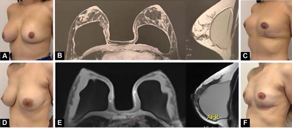
Regardless of whether or not they decided to proceed with the explant, all patients felt more confident with their choice after this individualized prediction of post-explant results. All those who chose to proceed with the explant considered the result consistent with the expectations set in the preoperative period (Figure 1A-F).
DISCUSSION
When the patient begins to consider permanently removing their implants, insecurity arises regarding the possible aesthetic appearance of the breasts after surgery. The fear of a bad result is fueled by doctors, friends, and family, but even so, many patients follow their desire to explant seeking the benefits of avoiding future surgeries and possible complications related to the presence of a foreign body in the body.
Often, the aesthetic result is unsatisfactory, as the patient idealized a breast similar to the one she had before the implant, but distortion of the breast tissue and distension of the skin are inevitable.
As in any plastic surgery, aligning the expectation of results is essential for patient satisfaction, and analyzing the breast MRI image together with postoperative photos of other patients makes this dialogue more intelligible.
It is important to make it clear that this is not a promise of results, but rather a tool to facilitate communication between surgeon and patient and to document the characteristics of the breast/implant relationship before explantation.
To avoid errors in interpreting the amount of tissue in each breast, it is important to compare MRI images respecting the implant placement plan, as the expansion of breast tissue is greater when the implant is retroglandular than when it is retropectoral.
It is worth noting that, to be coherent, comparisons must be made between similar cases. Therefore, it is necessary to organize an image bank with data on the implants (size, shape, placement plan) and patients (weight, height, technique used in post-explant reconstruction) to make analyses more reliable and reproducible.
CONCLUSION
Numerous factors can lead the patient to seek explantation, but, regardless of the motivation, we cannot help but worry about the patient’s emotional capacity to live well with her new self-image after the implants are removed.
The comparison of breast MRI images and postoperative results images brings valuable information to align the expectations of patients seeking to remove their implants, as it provides greater objectivity to the preoperative dialogue, favoring understanding of the expected result and bringing greater clarity to the decision for explantation.
REFERENCES
1. Calobrace MB. Elective Implant Removal and Replacement in Asymptomatic Aesthetic Patients with Textured Devices. Plast Reconstr Surg. 2021;147(5S):14S-23S.
2. Magnusson MR, Cooter RD, Rakhorst H, McGuire PA, Adams WP Jr, Deva AK. Breast Implant Illness: A Way Forward. Plast Reconstr Surg. 2019;143(3S):74S-81S.
3. Tanna N, Calobrace MB, Clemens MW, Hammond DC, Nahabedian MY, Rohrich RJ, et al. Not All Breast Explants Are Equal: Contemporary Strategies in Breast Explantation Surgery. Plast Reconstr Surg. 2021;147(4):808-18.
4. Rohrich RJ, Bellamy JL, Alleyne B. Assessing Long-Term Outcomes in Breast Implant Illness: The Missing Link? A Systematic Review. Plast Reconstr Surg. 2022;149(4):638e-45e.
5. Rohrich RJ, Kaplan J, Dayan E. Silicone Implant Illness: Science versus Myth? Plast Reconstr Surg. 2019;144(1):98-109.
6. Rossi AJRE, Kluthcovsky ACGC, Mansani FP. Comparison between magnetic resonance imaging and ultrasonography as the best examination to measure malignant breast tumors in surgical planning. Mastology. 2018;28(3):176-81.
7. Schmitt W, Coelho JM, Lopes J, Marques JC. O Papel da Radiologia na Monitorização das Complicações Relacionadas com as Próteses Mamárias. Acta Radiol Port. 2018;30(1):23-34.
1. Instituto de Cirurgia Plástica Santa Cruz, São
Paulo, SP, Brazil
Corresponding author: Patrícia Jackeline Maciel Sales Rua Borges Lagoa, 1070, cj 62, Vila Clementino, São Paulo, SP, Brazil, Zip Code: 04038-002, E-mail: patricia.cirurgia@gmail.com
 Read in Portuguese
 Read in English
 PDF PT
 Print
Print
 Send this article by email
 How to Cite
 Mendeley
 Pocket