


Case Report - Year 2024 - Volume 39 -
Association of pectoralis major myocutaneous flap and inverted T mammoplasty for reconstruction of extensive chest wall defect: case report
Associação do retalho miocutâneo do músculo peitoral maior e da mamaplastia em T invertido para reconstrução de extenso defeito da parede torácica: relato de caso

ABSTRACT
Introduction: In 1977, based on anatomical studies by McCraw et al., the pectoralis major muscle began to be used as an island myocutaneous flap. The present article describes a case of reconstruction of a defect in the anterior wall of the right hemithorax using the pectoralis major myocutaneous flap in an ipsilateral island.
Case Report: AES, 66 years old, male, underwent wide resection of a recurrent infiltrative basal cell carcinoma measuring 13.0 x 8.0 cm in the right parasternal region. The myocutaneous flap was transposed through subcutaneous tunneling and the scars were positioned in the shape of an inverted T mammoplasty.
Conclusion: This surgical tactic is easy to perform for surgeons accustomed to breast reconstruction, has a short surgical time, and has satisfactory aesthetic-functional results.
Keywords: Skin neoplasms; Myocutaneous flap; Mammaplasty; Pectoralis muscles; Carcinoma, basal cell
RESUMO
Introdução: Em 1977, a partir dos estudos anatômicos de McCraw et al., passou-se a utilizar o músculo peitoral maior como retalho miocutâneo em ilha. O presente artigo descreve um caso de reconstrução de um defeito da parede anterior do hemitórax direito através do retalho miocutâneo peitoral maior em ilha ipsilateral.
Relato do Caso: A.E.S., de 66 anos, sexo masculino foi submetido a ressecção ampla de um carcinoma basocelular infiltrativo recidivante de 13,0 x 8,0cm da região paraesternal direita. O retalho miocutâneo foi transposto através de tunelização subcutânea e as cicatrizes posicionadas em forma de mamaplastia em T invertido.
Conclusão: A presente tática cirúrgica é de fácil execução para cirurgiões habituados com reconstrução mamária, apresenta tempo cirúrgico curto e resultado estético-funcional satisfatório.
Palavras-chave: Neoplasias cutâneas; Retalho miocutâneo; Mamoplastia; Músculos peitorais; Carcinoma basocelular.
INTRODUCTION
In 1947, Pickrell et al. used the pectoralis major muscle to reconstruct a post-mastectomy chest wall defect1. However, it was only in 1977 that McCraw et al.2,3 carried out anatomical studies that enabled its use as a myocutaneous flap. The accumulated experience in using the flap and greater knowledge of its anatomy led to the transfer of increasingly larger skin segments4. The pedicle, which initially included the skin, now consists only of muscles and thoracoacromial vessels in the proximal part. This allowed the use of island flap5.
In 2019, Rauchenwald et al.6 published a retrospective study with 23 patients who underwent reconstruction with a pectoralis major island myocutaneous flap for fistula prophylaxis after rescue laryngectomy, demonstrating great evolution in the operative technique and its indications.
On the other hand, the idea of combining mammaplasty techniques to access the myocutaneous flap of the pectoralis major muscle to cover chest wall defects began to be described in 1996 by some authors such as de Fontaine et al.7 and Griffin8. In 2023, Boodhun & Zinn9 also used the myocutaneous flap of the pectoralis major muscle in association with reduction mammaplasty to cover a defect in the anterior cervical region.
In the present article, we describe the case of a male patient who underwent reconstruction of a large defect of the anterior chest wall using the islanded myocutaneous flap of the pectoralis major muscle in association with mammoplasty techniques.
CASE REPORT
AES, 66 years old, male, attended the Hospital Governador Celso Ramos, in Florianópolis, SC, and presented with a skin tumor that had evolved over approximately 10 years in the right parasternal region. During this period, he had already undergone several cryotherapy sessions and four resections of the same tumor in other Services. A biopsy of the lesion revealed the diagnosis of infiltrative basal cell carcinoma.
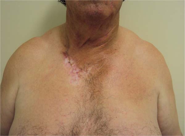
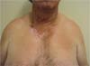
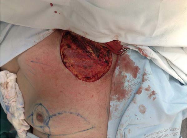
On physical examination, the skin tumor affected the region corresponding to the medial portion of the right clavicle, running inferiorly and parallel to the right of the sternum. The tumor measured 13.0cm on its longest vertical axis and 8.0cm on its longest horizontal axis. The patient also had skin retraction in the anterior region of the neck (Figure 1). However, there was no cervical lymph node enlargement.
Surgical technique
Scheduling tumor resection
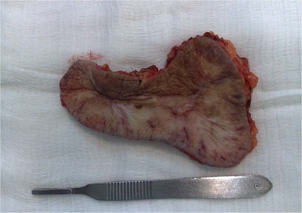
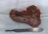
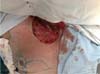
The marking of the tumor piece to be resected was carried out with lateral safety margins of 2.0cm (Figure 2). The resulting wound exposed the medial portion of the right clavicle and the ipsilateral pectoralis major muscle (Figure 3).
Marking the flap donor area
It was performed with the patient in an upright position. Point A of the breast was defined and from there, a periareolar semicircle was drawn, keeping the papilla in its center. The semicircle was completed at points defined as B and C, 1.0cm from the lower limit of the areola.
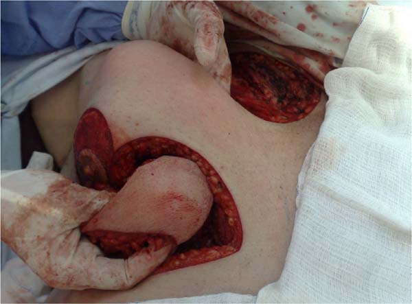
The skin island of the myocutaneous flap corresponded to the medial triangular area of the breast that would normally be resected and discarded in a reduction mammaplasty (Figure 4).

Surgical description
Wide resection of the tumor was performed with the patient in the supine position and under general anesthesia.
The skin of the right breast was infiltrated with a solution containing 200ml of 0.9% SF, 40ml of 2% xylocaine without vasoconstrictor, and 1ml of adrenaline.
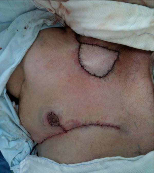
The Schwarzmann10 maneuver was performed and the medial triangle of skin and fat was sectioned. Subcutaneous detachment of the right pectoral region was performed, exposing the entire anterior surface of the pectoralis major muscle up to its origin. Afterwards, its insertions on the costal arches and sternum were sectioned and its posterior surface was also detached to its origin. In this way, the myocutaneous flap was completely released, allowing its subcutaneous rotation and coverage of the defect in the right parasternal region (Figures 4 and 5).
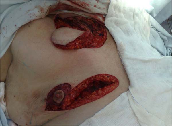

After the small rise of the nipple-areola complex (NAC), the male breast is sutured in layers. A lateral triangle of skin and fat was resected to adjust the inverted T mammaplasty scar11,12. In the detachment area, a 4.8 suction drain was left (Figure 6)13.

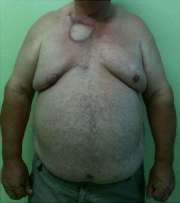
Postoperatively, the drain was removed after 7 days and there were no complications13. The pathological examination confirmed the diagnosis of infiltrative basal cell carcinoma and surgical margins free of neoplasia. The patient underwent outpatient follow-up and did not experience any further tumor recurrence (Figure 7).
DISCUSSION
Although skin grafting was a simpler treatment option, in this case, this technique had some disadvantages. The majority of the defect bed was made up of the muscle belly, which favored the integration of the skin graft, but there were other areas of exposed bone (clavicle) and subcutaneous cellular tissue. There would also be greater morbidity in the skin donor area. Furthermore, the thickness and texture of the graft skin would be different from that found in the pectoral region.
Some local skin flaps could be planned, but they would result in large visible scars14. In the case of other pedicled myocutaneous flaps, there would be the latissimus dorsi and rectus abdominis alternatives. However, they would result in more scars, longer surgical time, and greater postoperative morbidity.
In turn, free cutaneous flaps based on the deep or superficial epigastric artery would be good alternatives for covering the thoracic defect. However, the patient was obese and had a bulky and protruding abdominal apron. Therefore, dissection of the lower abdominal region would be difficult to perform and also pose a greater chance of local postoperative complications.
On the other hand, the simplicity and low morbidity of the myocutaneous pectoral island flap motivated its choice. Detachment of the pectoralis major muscle in its anterior and posterior planes was simple to perform. The pivot point of subcutaneous transposition of the skin island was the acromion and the thoracic defect was within its arc of rotation. To repair the flap donor area, mammaplasty techniques were used to avoid scars in unfavorable locations or causing any unacceptable asymmetry.
Although the inverted T scar could produce the stigma of female mammaplasty, in this case, this did not occur. As the patient had grade 4 gynecomastia and the mammoplasty was not intended to treat this condition, the final scars on the flap’s donor breast were not as prominent.
CONCLUSION
With the association of pectoral myocutaneous flap techniques and the closure of the flap donor area in the form of an inverted T mammaplasty, it was possible to obtain a satisfactory aesthetic and functional result, in a single surgical procedure and with low morbidity.
This surgical tactic is a simple alternative for the treatment of defects located in the arc of rotation of the pectoralis major flap when performed by surgeons accustomed to breast reconstruction.
| COLLABORATIONS | |
|---|---|
| FBW | Final manuscript approval, Realization of operations and/or trials, Writing - Original Draft Preparation. |
| VE | Final manuscript approval, Supervision. |

REFERENCES
1. Pickrell KL, Baker HM, Collins JP. Reconstructive surgery of the chest wall. Surg Gynecol Obstet. 1947;84(4):465-76.
2. McCraw JB, Dibbell DG, Carraway JH. Clinical definition of independent myocutaneous vascular territories. Plast Reconstr Surg. 1977;60(3):341-52.
3. McCraw JB, Dibbell DG. Experimental definition of independent myocutaneous vascular territories. Plast Reconstr Surg. 1977;60(2):212-20.
4. Freeman JL, Walker EP, Wilson JS, Shaw HJ. The vascular anatomy of the pectoralis major myocutaneous flap. Br J Plast Surg. 1981;34(1):3-10.
5. Wei WI, Lam KH, Wong J. The true pectoralis major myocutaneous island flap: an anatomical study. Br J Plast Surg. 1984;37(4):568-73.
6. Rauchenwald T, Dejaco D, Morandi EM, Djedovic G, Wolfram D, Riechelmann H, et al. The Pectoralis Major Island Flap: Short Scar Modified Muscle-Sparing Harvesting Technique Improves Aesthetic Outcome in Reconstructive Head and Neck Surgery. ORL J Otorhinolaryngol Relat Spec. 2019;81(5-6):327-37. DOI: 10.1159/000503008
7. de Fontaine S, Devos S, Goldschmidt D. Reduction mammaplasty combined with pectoralis major muscle flaps for median sternotomy wound closure. Brit J Plast Surg. 1996;49(4):220-2.
8. Griffin P. Reduction mammaplasty combined with pectoralis major muscle flaps for median sternotomy wound closure. Brit J Plast Surg. 1996;49(8):575-6.
9. Boodhun WS, Zinn R. Using a breast reduction to access a pedicled myocutaneous pectoralis major flap for anterior neck defect reconstruction: a case report. Australas J Plast Surg. 2023;6(1):71409. DOI: 10.34239/ajops.71409
10. Schwarzmann E. Die technik der mammaplastik. Chirurg. 1930;2:932-43.
11. Pitanguy I. Une nouvelle technique de plastie mammaire. Étude de 245 cas consécutifs et présentation d’une technique personnelle. Ann Chir Plast (Marseille). 1962;7(3):199-208.
12. Duan W, Cao C, Wu J, Cen Y, Xu X, Liu Y. Application of modified inferior pedicle technique with inverted T pattern for severe breast hypertrophy. Zhongguo Xiu Fu Chong Jian Wai Ke Za Zhi. 2019;33(3):341-4. DOI: 10.7507/1002-1892.201811076
13. Scomacao I, Cummins A, Roan E, Duraes EFR, Djohan R. The use of surgical site drains in breast reconstruction: A systematic review. J Plast Reconstr Aesthet Surg. 2020;73(4):651-62. DOI: 10.1016/j.bjps.2019.11.019
14. Jo GY, Yoon JM, Ki SH. Reconstruction of a large chest wall defect using bilateral pectoralis major myocutaneous flaps and V-Y rotation advancement flaps: a case report. Arch Plast Surg. 2022;49(1):39-42. DOI: 10.5999/aps.2021.01368
1. Hospital Governador Celso Ramos, Florianópolis,
SC, Brazil
Corresponding author: Felipe Barbieri Wohlgemuth Rua Irmã Benwarda, 297, 8º andar, Sala 802, Centro, Florianópolis, SC, Brazil, Zip Code: 88015-270, E-mail: felipewohlgemuth@gmail.com
 Read in Portuguese
 Read in English
 PDF PT
 Print
Print
 Send this article by email
 How to Cite
 Mendeley
 Pocket