




Original Article - Year 2026 - Volume 41Issue 1
Subcutaneous Arterial Graft Bank, a Promising Pilot Study: Experimental Model on Rats
Banco de enxertos arteriais subcutâneos, um estudo piloto promissor: Modelo experimental em ratos
Matheus Scuracchio Fernandes1 ; Fausto Viterbo1; Balduino Ferreira de Menezes Neto1
ABSTRACT
Objective To evaluate the patency and histological alterations of closed and open arterial grafts stored under the skin for subsequent anastomosis.
Materials and Methods There were 20 maleWistar rats separated into 2 groups (closed and open), and an autologous carotid arterial graft was inserted under the inguinal skin for 7 days and anastomosed to the femoral artery after this period. In the closed group, the grafts extremities were closed with a 10-00 nylon thread stitch during the 7 days storage, while in the open group, the extremities were left open. The anastomotic patency was evaluated immediately after the anastomosis and after 24 hours of the final procedure. After euthanasia, the grafts were subjected to histological analysis.
Results The closed group presented 80% immediate and no late patency, whereas the open group presented 90% immediate and 10% late patency. All grafts presented with deteriorated media and intimal cells, without inflammatory infiltration at the midgraft. There were no cases of infection, hemorrhage, or functional graft loss.
Conclusion The histological alterations observed in all grafts were similar to those observed in other techniques for preserving vascular grafts. The present pilot study describes a new, promising, and low-cost method to preserve arterial grafts that did not cause infection.
Keywords: arteries; graft survival; microsurgery; models; animal; rats; Wistar rats
RESUMO
Objetivo Avaliar a permeabilidade e as alterações histológicas de enxertos arteriais subcutâneos fechados e abertos para subsequente anastomose.
Materiais e Métodos Um total de 20 ratos Wistar machos foram separados em 2 grupos (fechado e aberto), submetidos à inserção de um enxerto autólogo de artéria carótida sob a pele inguinal por 7 dias e anastomosado à artéria femoral após esse período. No grupo fechado, as extremidades dos enxertos foram fechadas com fio de nylon 10-00 durante os 7 dias de armazenamento e, no grupo aberto, as extremidades foram deixadas abertas. A permeabilidade anastomótica foi avaliada imediatamente depois da anastomose e 24 horas após o último procedimento. Após a eutanásia, os enxertos foram submetidos à análise histológica.
Resultados O grupo fechado apresentou 80% de permeabilidade imediata e nenhuma tardia, enquanto o grupo aberto apresentou 90% de permeabilidade imediata e 10% tardia. Em todos os enxertos, as células da íntima e média estavam deterioradas, sem infiltração inflamatória na região média do enxerto. Casos de infecção, hemorragia ou perda funcional do enxerto não foram observados.
Conclusão As alterações histológicas notadas em todos os enxertos foram semelhantes àquelas observadas em outras técnicas de preservação de enxertos vasculares. Este estudo piloto descreve um método novo, promissor e de baixo custo para preservar enxertos arteriais sem desenvolvimento de infecção.
Palavras-chave: artérias; microcirurgia; modelos animais; ratos Wistar; sobrevida do enxerto
Introduction
The advent of microsurgical techniques, particularly vascular microanastomosis, has enabled the widespread use of arterial grafts, as well as the extensive development of surgical practices in multiple specialties.1
Plastic surgeons use microsurgical free flaps to improve closure in complex wounds related to trauma, infection, oncological surgery, and congenital deformities. Classic techniques have gained notoriety owing to their versatility and good results. The anterolateral thigh (ALT) flap2 is usually harvested from the descending branch of the lateral circumflex femoral artery, making it possible to obtain a fasciocutaneous or myocutaneous flap of the vastus lateralis muscle.
Another classic and versatile technique that can provide a bone flap from the radius is the radial forearm flap. Its versatility includes penile3 and craniofacial reconstruction surgeries.4
Additionally, the transverse rectus abdominis musculocutaneous (TRAM) free flap and deep inferior epigastric artery perforator (DIEP) flap are both used for breast reconstruction.5 The latter being the gold standard for breast reconstruction with autologous tissue.
Moreover, other flaps have been described, each with its specificities and indications. There is scope for further development, bringing more benefits to patients. However, thrombosis of the anastomosis remains the most serious complication. Owing to its multifactorial nature, continuous and vigilant monitoring of these flaps by medical teams is essential, as the consequences for patients can be devastating, significantly impacting morbidity.6,7 In cases in which thrombosis of a free flap becomes irreversible, it may be necessary to explore alternative donor sites, which may lead to local functional impairments.
Cardiac surgeons perform coronary artery bypass grafting, which is one of the most common cardiac procedures. Currently, more than 90% of the grafts used for myocardial revascularization are obtained from the left internal mammary arteries. However, other vascular pedicles are also used, such as the saphenous vein, radial artery, and right internal mammary artery.8 Regarding this procedure, the literature reports that reoperation rates have increased over the years, due to inevitable degeneration of the grafted tissue.9 The reapproach rate is 5.11% at 6 years,10 and 17.3% at 12 years.9 Therefore, it is important that the surgery compromises as few donor areas as possible.
Vascular surgeons perform treatment procedures for peripheral vascular diseases to reestablish compromised circulation. The saphenous vein is commonly used to bypass obstructed arteries in the lower limb.11 As the risk factors for such diseases are linked to lifestyle habits and aging, this condition becomes more common with age and, consequently, corrective surgeries become more frequent.12 Over time, vascular grafts have been used in other surgical areas, such as neurosurgery.13
With technical improvements in recent decades, the development of multiple free flaps has made it possible to access anatomical regions with long pedicles.14,15 Often, some parts of the pedicles are not required and are discarded.
Microanostomosis may present as postoperative thrombosis, requiring reoperation. We believe that excess discarded pedicles, especially arterial ones, could be buried in the subcutaneous tissue and, if necessary, used as grafts.
Additionally, the removal of vascular grafts can result in complications, such as wound healing, infection, impairment of tissue irrigation and drainage, hematoma, pain, and nerve damage.
Successful subcutaneous preservation of autologous tissue, especially bone grafts, has been reported in the literature. The technique was found to be viable, low cost, with low rate of infection and of mononuclear infiltrate (approximately 6% in a study with 50 patients),16 being more satisfactory for preserving autologous bone tissue than cryopreservation.17 Successful cryopreservation of vascular grafts has also been reported,18,19 though this technique is expensive and prohibitive, especially in developing countries. Another preservation technique is cold storage,20 which can be performed with glycerol21 and other solutions, having been studied since 1910 by Alexis Carrel22 and others.
We believe that it is feasible to preserve vascular tissue inserted under the subcutaneous tissue with a good success rate, few complications, and low cost. Furthermore, we would like to assess whether there is a difference in terms of thrombogenicity or tissue degeneration between performing a storage bank with close-ended arterial graft, due to a more protected intraluminal environment, when compared to open ended grafts.
No information was found on the creation of arterial graft banks under the skin of patients. Additionally, no studies have been conducted on the occurrence of thrombosis, degeneration, or inflammatory changes related to the burying of these grafts under the skin, or on the influence of vessel storage with the ends open or closed. Thus, we aimed to evaluate the feasibility of creating a bank of autologous arterial grafts under the skin for later use, as well as to analyze the possible degeneration during the accommodation period.
Objective
The present study aimed to evaluate the viability of closed and open arterial grafts lodged under the skin for subsequent anastomosis. Additionally, we evaluated the appearance of degenerative and inflammatory processes after the end-to-end graft anastomosis and compared the differences in results between the groups that had grafts buried with closed ends to those with open ends.
Materials and Methods
The current study was approved by the Animal Ethics Committee, under license number 1327/2019.
The procedures were performed by the same researcher, with proper training, using 20 Wistar rats weighing 180 to 220 g, aged 90 to 120 days.
First Surgical Procedure
The animals were anesthetized via inhalation. Isoflurane (1 mL/mL) was used for both induction and maintenance of anesthesia. For induction, the animals were placed in an induction box and supplied with a constant flow of oxygen at 2 L/min and isoflurane (1 mL/mL) at 80 mL/min (equivalent to 4% of the gas). To maintain anesthesia, the oxygen flow was changed to 700 mL/min and the isoflurane (1 mL/mL) flow to 14 mL/min (equivalent to a 2% gas rate).
Tramadol (5 mg/kg), enrofloxacin (10 mg/kg), and ketoprofen (5 mg/kg) were administered intramuscularly. Enrofloxacin and ketoprofen were administered once daily for 3 days, with the first application performed intraoperatively.
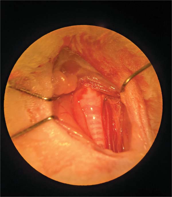
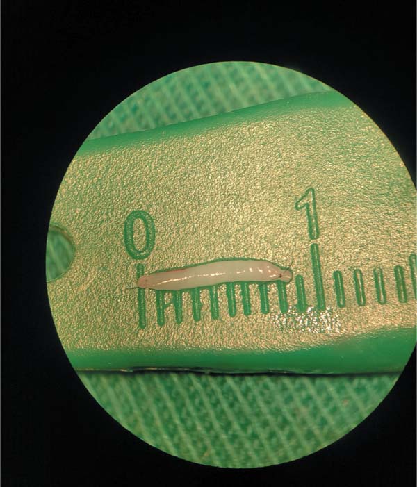
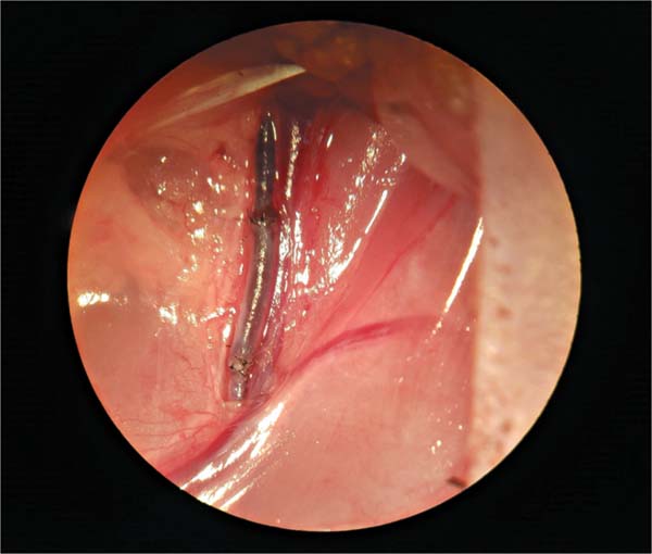
Using a surgical microscope at 10x magnification, dissection was initiated in the cervical region by isolating the external carotid vessel (►Fig. 1). Subsequently, a 9 mm graft was obtained from the external carotid artery. The rats were randomly divided into two groups: in the first (group C: 10 rats), the arterial graft was closed in the distal and proximal segments using 10-0 nylon (►Fig. 2) and introduced under the skin in the inguinal region. In the second one (group O: 10 rats, ►Fig. 3), an arterial graft was introduced under the skin (►Fig. 4) without occlusion of the extremities. To introduce the graft, an incision of about 15 mm was made in the inguinal region, dissecting the local subcutaneous tissue and allocating the stretchedgraft without tension, sothat the skinwas sutured above it. There was no graft exchange between the rats.
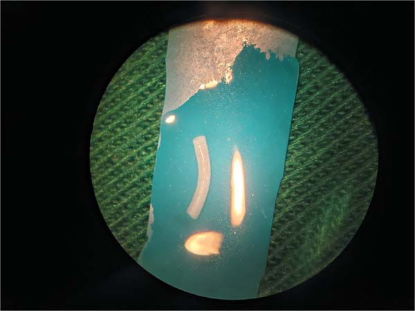
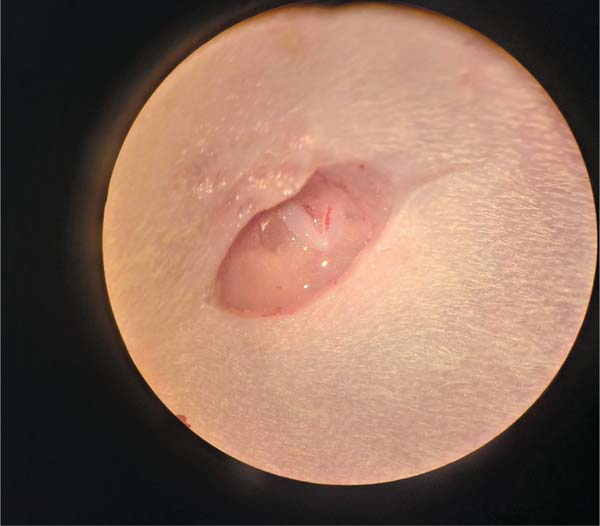
After surgery, the rats were provided with water and food ad libitum and allowed to rest for 7 days with the grafts lodged under the inguinal subcutaneous tissue.
Second Surgical Procedure
The animals were anesthetized via inhalation and received analgesics following the same procedure.
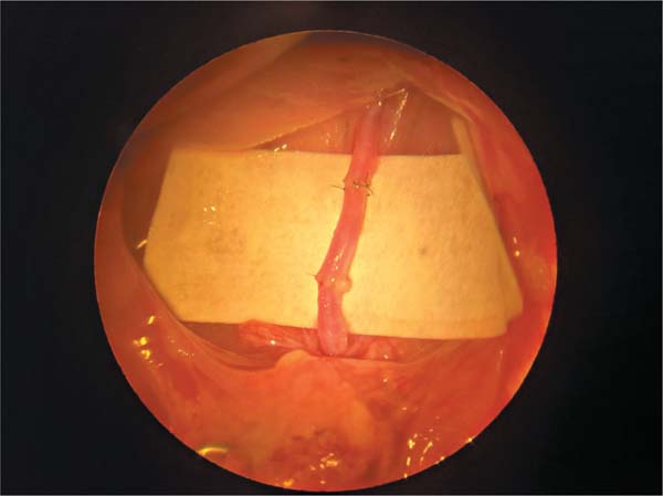
The 9 mm graft from the external carotid artery was surgically removed from the subcutaneous tissue (►Fig. 5). Then, the extremities were resected and it was interposed through an end-to-end anastomosis between the femoral artery stumps (►Fig. 6). For that, the femoral artery was dissected and the adventitia removed at the point chosen for the anastomosis, performed 10 mm from its origin under the inguinal ligament. Blood flow was obstructed with the aid of a microvascular clamp, and the artery was sectioned.
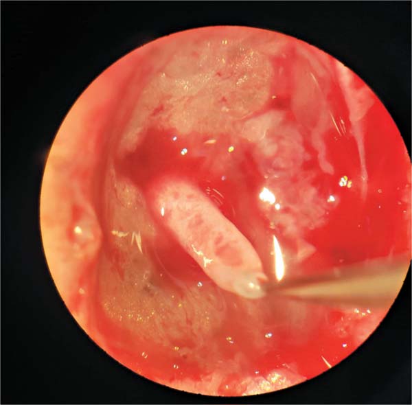
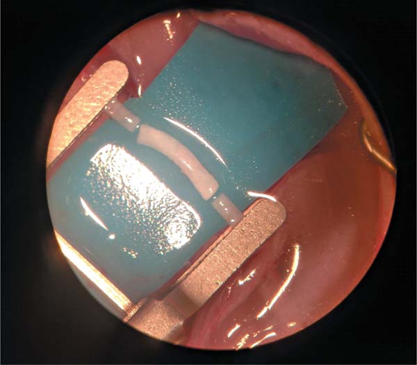
Each anastomosis, distal and proximal, was performed with 12 simple separate stitches, using 10-0 nylon and a cylindrical needle. A microscope (DF Vasconcelos) with optical magnification from 25 to 40x was used during the procedure.
In the first 20 minutes after the procedure, the graft’s patency was constantly monitored. For that, pulsation and the “milk test,” which consists of constricting the vessel with two microsurgery forceps and separating them, so that a segment of the vessel, distal to anastomosed graft, is bloodless. Then, the proximal forceps is released, and blood flow is reestablished. In cases where the graft did not present pulsation and the blood did not return after the “milk test”, the result was considered negative and patency absent, suggesting thrombosis.
After the graft anastomosis, the rats were kept in separate cages, with controlled temperature and light, sodium dipyrone (100 mg/kg) was administered subcutaneously every 6 hours for the next 24 hours, and the rats were provided drinking water and food ad libitum.
Permeability Test and Euthanasia
After 24 hours of the graft anastomosis, the rats underwent inhalation anesthesia again, as previously described, and the patency of the grafted artery was evaluated by the coloration, pulsation, and the “milk test”. In cases where the graft did not present light red coloration, pulsation, and the blood did not return after the “milk test”, patency was considered absent, suggesting thrombosis (►Fig. 7). In cases with light red color, pulsation, and positive “milk test”, the graft was considered patent (►Fig. 8).
Euthanasia was performed using an overdose of 120 mg/kg intravenous thiopental, immediately after permeability test.
Histological Analysis
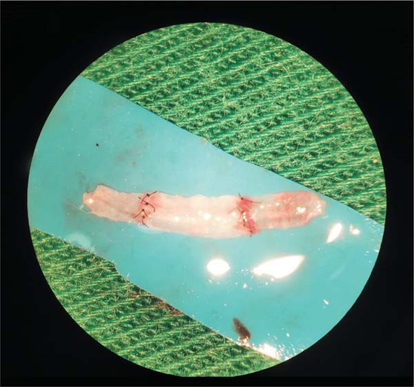
Grafts and anastomosis were cut into a rectangular shape (►Fig. 9), fixed in formol solution (10%) for 24 hours, embedded in paraffin. Then, 5 μm longitudinal sections were obtained and stained with hematoxylin and eosin (HE) solution for blinded histopathological evaluation. The slides were scanned and specimens evaluated for histological alterations (media muscle integrity and inflammatory infiltrate), according to the International Harmonization of Nomenclature and Diagnostic Criteria for Lesions in Rats and Mice,23 and compared to the femoral artery slides taken from rats 19 and 20 nonoperated limbs after euthanasia. The incidence of the observed alterations (in %) was calculated for each intervention.
Results
The rats were numbered and randomly assigned blind draw between the O and C groups, which were composed as follows:
►Table 1 provides information on graft patency in each rat immediately after anastomosis completion, in the first 20 minutes, and 24 hours after anastomosis completion.
| Rat | Group | Patency after 20 min After 24 hours | Final weight | |
|---|---|---|---|---|
| After 20 min | After 24 hours | |||
| 1 | O | Pervious | Thrombosis | 267 g |
| 2 | C | Pervious | Thrombosis | 269 g |
| 3 | O | Pervious | Thrombosis | 254 g |
| 4 | C | Pervious | Thrombosis | 251g |
| 5 | O | Pervious | Pervious | 280 g |
| 6 | O | Pervious | Thrombosis | 221 g |
| 7 | C | Thrombosis | Thrombosis | 290 g |
| 8 | O | Pervious | Thrombosis | 299 g |
| 9 | C | Pervious | Thrombosis | 323 g |
| 10 | C | Pervious | Thrombosis | 271 g |
| 11 | O | Thrombosis | Thrombosis | 276 g |
| 12 | O | Pervious | Thrombosis | 260 g |
| 13 | O | Pervious | Thrombosis | 302 g |
| 14 | C | Pervious | Thrombosis | 298 g |
| 15 | C | Thrombosis | Thrombosis | 287 g |
| 16 | C | Pervious | Thrombosis | 270 g |
| 17 | C | Pervious | Thrombosis | 276 g |
| 18 | O | Pervious | Thrombosis | 251g |
| 19 | C | Pervious | Thrombosis | 298 g |
| 20 | O | Pervious | Thrombosis | 299 g |
Abbreviations: C, closed group; O, open group.

Statistics
The immediate patency (first 20 min) in the O group was 9/10 (90%) and in the C one, it was 8/10 (80%). There was no statistically significant difference between the groups (p ¼ 0.99).
The late patency (after 24 h) in the O group was 1/10 (10%), and there were no cases in the C group (0%). There was no statistically significant difference between the groups (p ¼ 0.99).
There were no cases of infection in the inguinal area, where the arterial flap was buried, in any group.
Histological Analysis
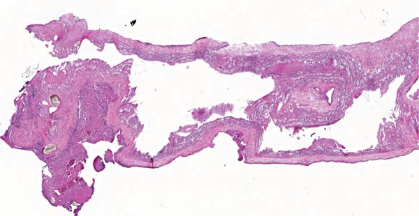
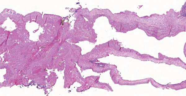
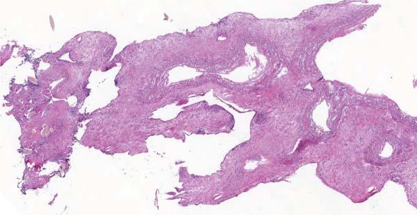
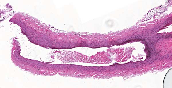
The histopathological evaluation showed that all graft samples from the open and closed groups (100%) presented intima and media degeneration, represented by loss of nuclear detail and fragmentation of endothelial and smooth muscle cells (►Figs. 10–12).



The analysis also showed that all graft samples from the open and closed groups (100%) presented foreign body reaction,24 in this case inflammatory mononuclear infiltrate, only at the anastomotic lines.
None of the graft samples from the open or closed groups (0%) showed endothelial hypertrophy, or intimal and mid-graft media inflammatory infiltration (►Figs. 10–12).
These findings were also observed for graft number 5 (►Fig. 10), which was patent after 24 hours of anastomosis. The evaluation of the control slides (►Fig. 13) shows an intima layer with no evidence of endothelial degeneration, nuclei detail loss, or hyperplasia. Additionally, it is possible to observe a media layer with no evidence of degeneration, no smooth muscle cells fragmentation nor loss of nuclei detail.
Discussion
Studies have shown that the vascular graft capacity for endothelization is related to thrombosis, this consists of the growth of neointima over the graft inner wall, close to the anastomosis.25,26 Additionally, repopulation of the media layer with new muscular cells has also been documented,27,28 resulting in an increase in graft functionality in the first few months after the procedure. Our findings indicate that the same process may occur with buried autologous grafts, with the advantage of being less immunogenic than non-autologous ones.28,29 Further studies are required to assess these possibilities.
Studies have shown that the glycerol preservation method also turns intima and media cells into nonviable cells. However, the extracellular matrix is composed of collagen III, elastin, proteoglycans, and glycoproteins, being responsible for an important part of the arterial structure.24,30 Thus, the functional integrity of the artery graft was not compromised. Our histological analysis showed that muscle cells in the media had shady nuclei, indicating that the muscle cells were nonviable and the cohesion among endothelial cells was reduced, as well as between them and nuclei with detail loss (►Figs. 10–12). Despite this, there were no cases of graft rupture, suggesting that the functional capacity of the artery was maintained.
We assessed that the inflammatory mononuclear infiltrate was only present at the anastomotic line,24 which was expected due to foreign body reaction.
Unfortunately, after 24 hours, only 10% patency was achieved in the open group and none in the closed group. However, the singular success case (►Fig. 8) reinforced the viability of the method. We believe that, in the case of histological abnormalities related to the storage method, all grafts would have failed and would not allow 90% patency in the O group and 80% patency in the C one after 20 minutes of anastomosis. We attribute the low final patency rate to aspects of the animal model used and absence of postsurgical thrombotic prophylaxis treatment. The overall diameter of the carotid graft was significantly larger compared to the femoral artery (►Fig. 6), and no antiplatelet or anticoagulation therapy31 was used.
The literature presents varied patency rates for microanastomosis in femoral arteries of Wistar rats.32
Additionally, the use of rabbits as animal models may be valid for future studies.
The low patency rate should not diminish the histological findings, which suggest the feasibility of an autologous artery graft subcutaneous bank. This technique has a simple execution, low cost, and low infection rate. It can potentially enhance the efficacy of artery grafts and reduce the need for additional vascular exploration.
Future studies with more detailed histological analyses, including transverse cuts of artery grafts could be performed to simultaneously evaluate the three layers of arterial tissue. Specific staining techniques can be employed to evaluate the elastic structure of grafts. Additionally, a longer recovery period would allow the evaluation of neointima formation, the possibility of stenosis, and the repopulation process.
The present project is innovative and pioneering. No studies have evaluated the possibility of creating subcutaneous banks for autologous arterial grafts. Owing to its novelty and great technical difficulty, a large percentage of patency in the arterial grafts was not expected.
All procedures were performed by the same trained researcher using the same technique.
A higher percentage of anastomosis might exhibit patency if methodological adaptations are implemented. The graft patency of rat number 5 encourages us to maintain our line of research and invest more time and studies in this new technique, which may benefit patient outcomes if improved.
The animal model and postsurgery antiplatelet or anticoagulation therapy can be improved and have played an important role in the graft patency outcomes. Histological analysis could aim for the extracellular matrix, responsible for maintaining the artery graft functionality. Findings from animal models should be interpreted cautiously when extrapolated for clinical use.
Conclusion
After the experiment, all graft samples from the O and C groups showed deletion of smooth muscle cell nuclei and reduced cohesion between endothelial cells. There was no statistical difference between groups in terms of immediate and late patency.
The histological alterations present appear to be similar to those observed in other techniques used to preserve arterial grafts. The present pilot study described a new, promising and low-cost method with no cases of infection.
Video 1
Arterial graft patent 24 hours after anastomosis. Online content including video sequences viewable at: https://www.thieme-connect.com/products/ejournals/html/10.1055/s-0045-1812995.
REFERENCES
1. Mavrogenis AF, Markatos K, Saranteas T, et al. The history of microsurgery. Eur J Orthop Surg Traumatol 2019;29(02): 247–254. Doi: 10.1007/s00590-019-02378-7
2. Wong CH, Wei FC. Anterolateral thigh flap. Head Neck 2010;32 (04):529–540. Doi: 10.1002/hed.21204
3. Gottlieb LJ. Radial Forearm. Clin Plast Surg 2018;45(03):391–398. Doi: 10.1016/j.cps.2018.03.009
4. Haynes WI, de Boutray M, Kennel T, Boetto J. Osteocutaneous radial forearm free flap for anterior cranial base reconstruction: Technical note. Neurochirurgie 2021;67(06):606–610. Doi: 10.1016/j.neuchi.2021.03.009
5. Jeong W, Lee S, Kim J. Meta-analysis of flap perfusion and donor site complications for breast reconstruction using pedicled versus free TRAM and DIEP flaps. Breast 2018;38:45–51. Doi: 10.1016/j. breast.2017.12.003
6. van Gijn DR, D’Souza J, King W, Bater M. Free Flap Head and Neck Reconstruction with an Emphasis on Postoperative Care. Facial Plast Surg 2018;34(06):597–604. Doi: 10.1055/s-0038-1676076
7. Pohlenz P, Klatt J, Schön G, Blessmann M, Li L, Schmelzle R. Microvascular free flaps in head and neck surgery: complications and outcome of 1000 flaps. Int J Oral Maxillofac Implants 2012;41 (06):739–743. Doi: 10.1016/j.ijom.2012.02.012
8. Squiers JJ, Mack MJ. Coronary artery bypass grafting-fifty years of quality initiatives since Favaloro. Ann Cardiothorac Surg 2018;7 (04):516–520. Doi: 10.21037/acs.2018.05.13
9. Maltais S, Widmer RJ, Bell MR, et al. Reoperation for Coronary Artery Bypass Grafting Surgery: Outcomes and Considerations for Expanding Interventional Procedures. Ann Thorac Surg 2017;103 (06):1886–1892. Doi: 10.1016/j.athoracsur.2016.09.097
10. Gallo M, Trivedi JR, Monreal G, Ganzel BL, Slaughter MS. Risk Factors and Outcomes in Redo Coronary Artery Bypass Grafting. Heart Lung Circ 2020;29(03):384–389. Doi: 10.1016/j.hlc.2019.02.008
11. Moreira RWC, Costa PVA, Carrilho DDR. Tratamento da isquemia crítica de membros inferiores usando uma técnica híbrida. J Vasc Bras 2014;13(03):257–261. Doi: 10.1590/jvb.2014.043
12. Aboyans V, Ricco JB, Bartelink MEL, et al. Editor’s Choice - 2017 ESC Guidelines on the Diagnosis and Treatment of Peripheral Arterial Diseases, in collaboration with the European Society for Vascular Surgery (ESVS). Eur J Vasc Endovasc Surg 2018;55(03): 305–368. Doi: 10.1016/j.ejvs.2017.07.018
13. Azadgoli B, Leland HA, Wolfswinkel EM, Bakhsheshian J, Russin JJ, Carey JN. Combined Direct and Indirect Cerebral Revascularization Using Local and Flow-Through Flaps [Revascularização cerebral direta e indireta combinada usando retalhos locais e de fluxo contínuo]. J Reconstr Microsurg 2018;34(02):103–107. Doi: 10.1055/s-0037–1606552
14. Godina M. Preferential use of end-to-side arterial anastomoses in free flap transfers. Plast Reconstr Surg 1979;64(05):673–682
15. Deppe AC, Liakopoulos OJ, Choi YH, et al. Endoscopic vein harvesting for coronary artery bypass grafting: a systematic review with meta-analysis of 27,789 patients. J Surg Res 2013;180(01): 114–124. Doi: 10.1016/j.jss.2012.11.013
16. Singla N, Singh SP, Gupta SK, Karthigeyan M, Radotra BD. Histopathology of subcutaneously preserved autologous bone flap after decompressive craniectomy: a prospective study. Acta Neurochir (Wien) 2014;156(07):1369–1373. Doi: 10.1007/s00701-014-2071-3
17. Baldo S, Tacconi L. Effectiveness and safety of subcutaneous abdominal preservation of autologous bone flap after decompressive craniectomy: a prospective pilot study. World Neurosurg 2010;73(05):552–556. Doi: 10.1016/j.wneu.2010.02.018
18. Kakodkar R, Nundy S, Soin AS. Use of banked cryopreserved veins from explanted recipient livers in living donor liver transplantation. Surgery 2008;144(01):93–95. Doi: 10.1016/j.surg.2007.10.008
19. Špaček M, Meřička P, Janoušek L, et al. Current vascular allograft
20. Garbe S, Zatschler B, Müller B, et al. Preservation of human artery function following prolonged cold storage with a new solution. J Vasc Surg 2011;53(04):1063–1070. Doi: 10.1016/j.jvs.2010. 10.093
21. Fahner PJ, Legemate DA, van der Wal AC, et al. Comparison of preserved vascular allografts using glycerol and University of Wisconsin solution in a goat carotid artery transplantation model. Eur Surg Res 2012;48(02):64–72. Doi: 10.1159/000334170
22. Carrel A. LATENT LIFE OF ARTERIES. J Exp Med 1910;12(04): 460–486. Doi: 10.1084/jem.12.4.460
23. Berridge BR, Mowat V, Nagai H, et al. Non-proliferative and Proliferative Lesions of the Cardiovascular System of the Rat and Mouse. J Toxicol Pathol 2016;29(03):1S–47S. Doi: 10.1293/tox.29.3s-1
24. Fahner PJ, Idu MM, van Gulik TM, van Wijk B, van der Wal AC, Legemate DA. Glycerol-preserved arterial allografts evaluated in the infrarenal rat aorta. Eur Surg Res 2009;42(02):78–86. Doi: 10.1159/000180114
25. Pashova A, Work LM, Nicklin SA. The role of extracellular vesicles in neointima formation post vascular injury. Cell Signal 2020; 76:109783. Doi: 10.1016/j.cellsig.2020.109783
26. Wang Y, Xu Y, Yan S, et al. Adenosine kinase is critical for neointima formation after vascular injury by inducing aberrant DNA hypermethylation. Cardiovasc Res 2021;117(02):561–575. Doi: 10.1093/cvr/cvaa040
27. Amensag S, Goldberg L, O’Malley KA, Rush DS, Berceli SA, McFetridge PS. Pilot assessment of a human extracellular matrix-based vascular graft in a rabbit model. J Vasc Surg 2017;65(03): 839–847.e1. Doi: 10.1016/j.jvs.2016.02.046
28. Massaro MS, Kochová P, Pálek R, et al. Decellularization of Porcine Carotid Arteries: From the Vessel to the High-Quality Scaffold in Five Hours. Front Bioeng Biotechnol 2022;10:833244. Doi: 10.3389/fbioe.2022.833244
29. Mangold S, Schrammel S, Huber G, et al. Evaluation of decellularized human umbilical vein (HUV) for vascular tissue engineering - comparison with endothelium-denuded HUV. J Tissue Eng Regen Med 2015;9(01):13–23. Doi: 10.1002/term.1603
30. Fahner PJ, Idu MM, Legemate DA, et al. Morphological and functional alterations in glycerol preserved rat aortic allografts. Int J Artif Organs 2004;27(11):979–989. Doi: 10.1177/039139880402701111
31. DelMauro MA, Chen K, Keller A. Reducing Length of Stay after Microsurgical Breast Reconstruction with a Standardized Postoperative Protocol. J Reconstr Microsurg 2019;35(08):557–567. Doi: 10.1055/s-0039-1687916
32. Menezes Neto BF, Oliveira Neto FV, Secanho MS, Carvalho LB, Moragas WR, Fernandes MS. Submerged vascular anastomosis. A technique for vascular suturing in experimental microsurgery. Acta Cir Bras 2021;36(08):e360807. Doi: 10.1590/ACB360807
1. Departamento de Cirurgia, Faculdade de Medicina, Universidade Estadual Paulista
(Unesp), Botucatu, SP, Brazil
Animal Ethics Approval The study was approved by the Animal Ethics Committee, under license number 1327/2019.
Data Availability Statement The research data of the present article is available from the corresponding author upon reasonable request (mat.scuracchio@gmail.com).
Financial Support The project was supported by the São Paulo Research Foundation (FAPESP), process No. 2021/04429-8, under the responsibility of Professor Fausto Viterbo.
Address for correspondence Matheus Scuracchio Fernandes, MD, Universidade Estadual Paulista (Unesp), Faculdade de Medicina, Departamento de Cirurgia, Av. Professor Montenegro, s/n, Botucatu, SP, 18618-687, Brasil (e-mail: mat.scuracchio@gmail.com).
Article received: April 25, 2025.
Article accepted: June 01, 2025.
Conflict of Interests The authors have no conflict of interests to disclose.
Editor-in-Chief: Dov Charles Goldenberg.
 Read in Portuguese
 Read in English
 PDF PT
 Print
Print
 Send this article by email
 How to Cite
 Mendeley
 Pocket
 Facebook
 Twitter