


Original Article - Year 2011 - Volume 26 -
Reconstruction of pelviperineal injuries with perforator flaps: clinical experience with 22 cases
Reconstruções pelveperineais com uso de retalhos cutâneos baseados em vasos perfurantes: experiência clínica com 22 casos
ABSTRACT
INTRODUCTION: Plastic surgery consultation is commonly sought for the treatment of pelviperineal injuries in general hospitals. The objective of this study was to present the experience acquired in the treatment of perineal, sacral, and hip injuries with the use of perforator flaps.
METHODS: Patients referred to the Plastic Surgery Division of the Clinical Hospital of Medicine College of Universidade de São Paulo for evaluation of pelviperineal and hip wounds from February to May 2009 were retrospectively evaluated. A total of 22 patients underwent reconstruction with skin and fasciocutaneous flaps based on the perforator vessels, according to the inclusion criteria. The average follow-up period was 6 months.
RESULTS: Pelviperineal injuries consisted of pressure ulcers in 20 cases (91%), deep infection in 1 case (45%), and perineal hidradenitis in 1 case (4.5%). The choice of flap for reconstruction was dependent on the local wound: 15 cases (68.2%) of sacral ulcers were repaired with a superior gluteal artery perforator flap; 3 cases (13.6%) of ischial ulcers were repaired with an inferior gluteal artery perforator flap; and 2 cases (9.1%) of trochanteric ulcers were repaired using a tensor fascia lata perforator flap. A fasciocutaneous gluteofemoral flap was selected for reconstruction of post Fournier' syndrome in 1 patient and was used after resection of perineal hidradenitis in 1 patient. A new suture for late primary closure was necessary in 3 (13.6%) cases in which the suture line dehiscence was < 10% of the injury perimeter during the first 15 post-operative days. There were no cases of > 3% necrosis of the flap surface. These results were maintained during the follow-up evaluation period.
CONCLUSIONS: The results of the study were satisfactory, and the utility of surgical flaps without the incorporation of muscle for pelviperineal reconstruction was demonstrated. This treatment alternative decreases donor site morbidity and preserves the muscular tissue for future interventions.
Keywords: Perineum. Wounds and injuries. Pressure ulcer. Surgical flaps. Plastic surgery.
RESUMO
INTRODUÇÃO: As lesões pelveperineais representam grande parcela das interconsultas para o cirurgião plástico em hospitais gerais. O objetivo do presente trabalho é apresentar a experiência obtida no tratamento de pacientes com lesões perineais, sacrais e de quadril com o uso de retalhos com vasos perfurantes.
MÉTODO: Foram estudados, retrospectivamente, os pacientes submetidos a avaliação pela equipe da Divisão de Cirurgia Plástica do Hospital das Clínicas da Faculdade de Medicina da Universidade de São Paulo, nos meses de fevereiro a maio de 2009, que apresentavam feridas em região pelveperineal e no quadril. No total, 22 pacientes foram submetidos a reconstrução com retalhos cutâneos e fasciocutâneos baseados em vasos perfurantes, de acordo com os critérios de inclusão. O período de seguimento médio foi de 6 meses.
RESULTADOS: A lesão pelveperineal foi úlcera por pressão em 20 (91%) casos, infecção profunda em 1 (4,5%), e hidradenite perineal em 1 (4,5%). A opção dos retalhos foi previamente estabelecida, dependendo do local da ferida: úlceras sacrais, retalho baseado nas perfurantes da artéria glútea superior em 15 (68,2%) casos; úlceras isquiáticas, retalho baseado nos vasos perfurantes da artéria glútea inferior em 3 (13,6%) casos; e úlceras trocantéricas, retalho tensor da fascia lata perfurante em 2 (9,1%) casos. Retalho fasciocutâneo inervado gluteofemoral foi a opção para a reconstrução pós-síndrome de Fournier em um paciente e após ressecção de hidradenite perineal em outro. Houve necessidade de nova sutura para fechamento primário tardio em deiscência < 10% do perímetro da lesão em 3 (13,6%) casos, durante os primeiros 15 dias de pós-operatório. Não houve casos de necrose > 3% da superfície do retalho. Os resultados foram mantidos no período de seguimento avaliado.
CONCLUSÕES: Os resultados obtidos no presente estudo foram satisfatórios e ficou demonstrada a utilidade de retalhos cirúrgicos sem incorporação de músculo para reconstruções pelveperineais. Essa alternativa para tratamento é menos mórbida para as áreas doadoras e preserva o tecido muscular para possível intervenção futura.
Palavras-chave: Períneo. Ferimentos e lesões. Úlcera de pressão. Retalhos cirúrgicos. Cirurgia plástica.
Plastic surgery consultation is commonly sought for the treatment of pelviperineal and hip injuries in general hospitals. The majority of these wounds are caused by pressure ulcers, although other etiologies are common, including perineal hidradenitis, deep infections, trauma, burns sequelae, neoplasias, and surgical trauma. The treatment usually consists of local care, surgical debridement, proper dressing, frequent position changes, and nutritional support1.
Certain patients with pelviperineal and hip wounds require reconstructive plastic surgery2. Primary closure, dermoepidermal grafts, and fasciocutaneous or musculocutaneous flaps are the best options for reconstruction with the objective of filling, covering, and protecting the flaps3. The use of myocutaneous flaps is very common, as described by Costa et al.4, who reported on a series of cases of pelviperineal reconstruction of pressure ulcers using myocutaneous flaps.
In the Plastic Surgery Division of the Clinical Hospital of the College of Medicine of Universidade de São Paulo (HCFMUSP), the use of perforator flaps is common practice. In 2004, Munhoz5 used a transverse abdominal skin flap based on the superficial epigastric artery perforator for breast reconstructions. Improved topographic localization of dominant perforator vessels in the pelvis and hip enabled the establishment of a protocol for pelviperineal reconstruction using cutaneous perforator flaps.
The objective of this study was to describe our experience with the use of perforator flaps for the treatment of perineal, sacral, and hip injuries.
METHODS
A total of 22 patients with perineal, sacral, and hip injuries were evaluated and followed up at the Plastic Surgery Division of HCFMUSP from February to May 2009. The Wound Group was formed by interconsultation with inpatients in other clinics of HCFMUSP.
Demographic data was collected, and an epidemiological inventory of each patient was created, including age, gender, and cause of injury. The indication criteria for surgical intervention were favorable clinical conditions, proper nutritional state, possibility of home care, prognosis, and underlying disease.
During the period included in the study, 22 patients underwent pelviperineal and hip reconstruction with local flaps, including 13 (59.1%) men and 9 (40.9%) women. The patients ranged in age from 17 to 46 years, and the average age was 32.6 years. The cause of perineal injury was pressure ulcers in 20 cases (91%; level III or IV ulcers in paraplegic or quadriplegic patients), deep infection in 1 (4.5%) case, and perineal hidradenitis in 1 case (4.5%).
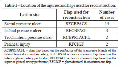
The general procedure consisted of surgical debridement and installation of a vacuum therapy device for 1 to 2 weeks. The reconstruction was carried out with local flaps, especially those based on perforator vessels. The flaps were planned and the isolation and dissection of the perforators were based on the anatomic repairs described6. In sacral ulcers (15 cases, 62.2%), a superior gluteal artery perforator flap was used7; ischial ulcers (3 cases, 13.6%) were repaired using an inferior gluteal artery perforator flap8 with V-Y advancement flaps; and trochanteric ulcers (2 cases, 9.1%) were treated with a skin flap based on the transverse branch of the lateral femoral circumflex artery9. The gluteofemoral fasciocutaneous flap10 based on the descending branch of the inferior gluteal artery was the choice for reconstruction in a patient with a perineal injury associated with Fournier syndrome and in 1 patient after resection of perineal hidradenitis (Table 1).
The post-operative follow-up period ranged from 4 to 9 months, with an average follow-up period of 6 months. The patients were evaluated after discharge in weekly outpatient consultations during the first month and subsequently every two months. The operated areas were evaluated and eventual necrosis, relapses, dehiscence, or infections were recorded. Other areas were also examined to detect possible new pressure ulcers.
RESULTS
All complications were considered to be minor. Total loss of the flap due to necrosis was not observed. A new suture for late primary closure was necessary in 3 (13.6%) cases in which the suture line dehiscence was <10% of the injury perimeter during the first 15 post-operative days. In these 3 cases, < 3% of the total flap surface showed necrosis, and the treatment involved debridement and direct suture without the development of new dehiscence.
All wounds showed complete closure. The flaps remained viable in all patients, without signs of relapse at the 2-month outpatient consultation. Patients were provided with instructions on recumbent positioning and appropriate maneuvers for position changes. Out of 15 patients with sacral ulcers, 3 (20%) presented level II11 ischial ulcers during the follow-up period, and the ulcers were treated conservatively.
Figures 1 to 3 illustrate some of the cases described in the present study.
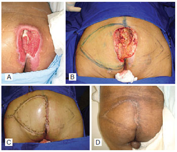
Figure 1 - Patient with sacral pressure ulcer. In A, pre-operative aspect. In B, pressure ulcer after debridement and marking of the fasciocutaneous flap based on the superior gluteal artery perforator. In C, immediate post-operative aspect. In D, late post-operative aspect.
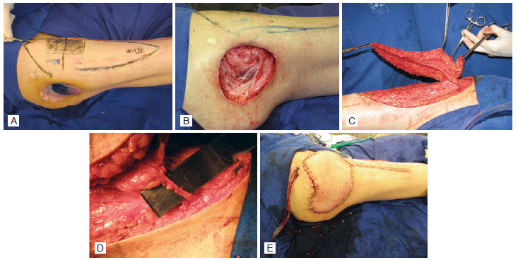
Figure 2 - Patient with trochanteric pressure ulcer. In A, pre-operative aspect. In B, pressure ulcer after debridement of the devitalized tissue. In C, skin flap based on the perforator of the transverse branch of the lateral femoral circumflex artery (tensor fascia lata perforator flap). In D, characteristics of the preserved perforator flaps. In E, immediate post-operative aspect.
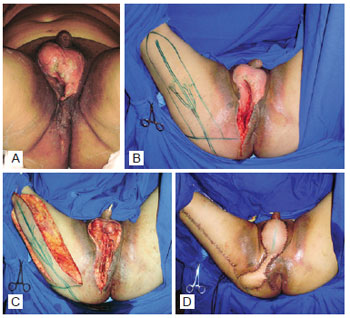
Figure 3 - Patient with perineal wound resulting from Fournier's syndrome. In A, pre-operative aspect. In B, appearance after debridement and marking of the flap. In C, dissected gluteofemoral fasciocutaneous flap. In D, immediate post-operative aspect.
DISCUSSION
Pelviperineal injuries represent a great challenge for the plastic surgeon. New pressure ulcers are common in general hospitals and are mostly caused by poor compliance with guidelines for position changes, mainly affecting intensive care patients. Flap reconstruction is often postponed until favorable clinical and local conditions are achieved. The initial treatment includes dressing changes and surgical debridement of the devitalized tissues in addition to orientation regarding posture, which is the responsibility of the nursing team in charge.
Surgical reconstruction is indicated in patients who were discharged from intensive therapy and have shown functional recovery, as well as in spinal cord trauma or stroke patients. The treatment of paralyzed patients involves the education of both, the patient and third parties in charge of patient care. In these patients, it is important to control neurogenic bladder spasticity and ensure regular position changes12.
To accelerate the preparation of the wound bed, our institution routinely uses the negative pressure (vacuum) method, which has been found to improve the results of surgery and prevent the need for multiple surgical interventions. During the reconstruction itself, the plastic surgeon should explore all the options for flaps whose dissection is based on anatomic repairs.
Musculocutaneous gluteus maximus flaps are commonly used for the repair of sacral and ischial pressure ulcers13. Hamstring muscle flaps have also been successfully applied for the treatment of ischial pressure ulcers14. Tensor fasciae latae muscle flaps are the treatment of choice for the repair of trochanteric pressure ulcers15. All these alternatives use the muscle as a vector for skin flap perfusion, compromising the donor site for future interventions. To diminish this type of morbidity, our institution began to use perforator flaps for cutaneous repairs, and these flaps do not require the inclusion of an underlying muscle.
Perforator flaps, which are based on the angiosome concept16, usually require more advanced knowledge of anatomy and surgical ability. Koshima et al.17 and Ao et al.18 were the pioneers in the use of perforator flaps based on gluteal, lumbar, and intercostal arteries for the repair of lumbosacral defects in the early 90's. In the present study, the flaps were designed to receive their blood supply through the reconstitution of well known vascular pedicles. Careful dissection keeping the perforator vessel anatomy in mind enabled the isolation of the perforators and the elevation of the fasciocutaneous flaps, including enough tissue to cover the injuries and allow primary closure of the donor areas.
The advantages of this type of flap are countless: it preserves the muscle, allows the inclusion of multiple components in the flap, and reduces pain and functional deficits at the donor site7,18. In comparison, musculocutaneous flaps are thick and well vascularized. Nevertheless, the muscular tissue is more sensitive to ischemia, and the removal of muscles from the donor area has greater functional morbidity19. The potential disadvantage of fasciocutaneous flaps based on perforator vessels is their reduced thickness, which limits their use to the reconstruction of wounds requiring greater filling7. In the patients included in this study, there was no need for volume addition besides that provided by the fasciocutaneous flap.
Approximately 80% of patients with paralysis are prone to recurrence of pressure ulcers or the development of new ones, depending on the source and follow-up period19. Even in these patients, in which the donor site functionality is not a priority, the use of perforator flaps preserves the underlying tissues, such as the muscle itself, which can be used as an alternative source in cases of relapse. Among the patients included in this study, there were no cases of relapse within a 6-month follow-up period, although 3 patients presented with new ulcers at other sites. These patients were paraplegic and had sacral pressure ulcers that were diagnosed and treated during their hospitalization for spinal cord trauma. Upon discharge, these patients used a wheelchair for mobility and developed level II11 ischial ulcers that were treated with regular dressings.
In the present study, no complications related to the viability of the flaps that could compromise the closure of lesions were observed, with the exception of 3 cases of partial dehiscence that were treated with debridement and direct suture. In patients with paralysis that present with pressure ulcers, the tendency towards recurrence or development of new wounds is common19,20, which emphasizes the importance of using flaps that enable the reuse of the donor site and local tissues.
CONCLUSIONS
The results obtained in this study were satisfactory, and the benefits of using surgical flaps that do not require the inclusion of an underlying muscle for pelviperineal reconstruction were demonstrated. This treatment alternative is associated with lower donor site morbidity and preserves the muscular tissue for future interventions.
REFERENCES
1. Conway H, Griffith BH. Plastic surgery for closure of decubitus ulcers in patients with paraplegia, based on experience with 1,000 cases. Am J Surg. 1956;91(6):946-75.
2. Sørensen JL, Jørgensen B, Gottrup F. Surgical treatment of pressure ulcers. Am J Surg. 2004;188(1A Suppl):42-51.
3. Rosen JM, Mo ST, Liu A. Experience with the island inferior gluteal thigh flap compared with other local flaps for the reconstruction of the pelvic area. Ann Plast Surg. 1990;24(6):498-509.
4. Costa MP, Sturtz G, Costa FPP, Ferreira MC, Barros Filho TEP Epidemiologia e tratamento das úlceras de pressão: experiência de 77 casos. Acta Ortop Bras. 2005;13(3):124-33.
5. Munhoz AM. Estudo crítico da anatomia arterial do retalho vascularizado pela artéria perfurante muscular da artéria epigástrica inferior [dissertação de mestrado]. São Paulo: Faculdade de Medicina da Universidade de São Paulo; 2004.
6. Hallock GG. A primer of schematics to facilitate the design of the preferred muscle perforator flaps. Plast Reconstr Surg. 2009;123(3):1107-15.
7. Coşkunfirat OK, Ozgentaş HE. Gluteal perforator flaps for coverage of pressure sores at various locations. Plast Reconstr Surg. 2004;113(7):2012-7.
8. Higgins JP, Orlando GS, Blondeel PN. Ischial pressure sore reconstruction using an inferior gluteal artery perforator (IGAP) flap. Br J Plast Surg. 2002;55(1):83-5.
9. Ishida LH, Munhoz AM, Montag E, Alves HR, Saito FL, Nakamoto HA, et al. Tensor fasciae latae perforator flap: minimizing donor-site morbidity in the treatment of trochanteric pressure sores. Plast Reconstr Surg. 2005;116(5):1346-52.
10. Hurwitz DJ, Swartz WM, Mathes SJ. The gluteal thigh flap: a reliable, sensate flap for the closure of buttock and perineal wounds. Plast Reconstr Surg. 1981;68(4):521-32.
11. Shea JD. Pressure sores: classification and management. Clin Orthop Relat Res. 1975;(112):89-100.
12. Bauer JD, Mancoll JS, Phillips LG. Pressure sores. In: Thorne CH, ed. Grabb & Smith's plastic surgery. 6a ed. New York: Lippincott Williams & Wilkins; 2007. p. 722-9.
13. Baek SM, Williams GD, McElhinney AJ, Simon BE. The gluteus maximus myocutaneous flap in the management of pressure sores. Ann Plast Surg. 1980;5(6):471-6.
14. James JH, Moir IH. The biceps femoris musculocutaneous flap in the repair of pressure sores around the hip. Plast Reconstr Surg. 1980;66(5):736-9.
15. Nahai F, Silverton JS, Hill HL, Vasconez LO. The tensor fascia lata musculocutaneous flap. Ann Plast Surg. 1978;1(4):372-9.
16. Taylor GI, Palmer JH. The vascular territories (angiosomes) of the body: experimental study and clinical applications. Br J Plast Surg. 1987;40(2):113-41.
17. Koshima I, Moriguchi T, Soeda S, Kawata S, Ohta S, Ikeda A. The gluteal perforator-based flap for repair of sacral pressure sores. Plast Reconstr Surg. 1993;91(4):678-83.
18. Ao M, Mae O, Namba Y, Asagoe K. Perforator-based flap for coverage of lumbosacral defects. Plast Reconstr Surg. 1998;101(4):987-91.
19. Bauer J, Phillips LG. MOC-PSSM CME article: Pressure sores. Plast Reconstr Surg. 2008;121(1 Suppl):1-10.
20. Seyhan T, Ertas NM, Bahar T, Borman H. Simplified and versatile use of gluteal perforator flaps for pressure sores. Ann Plast Surg. 2008;60(6):673-8.
1. Plastic surgeon, physician preceptor at the Plastic Surgery Division of the Clinical Hospital of the College of Medicine of the Universidade de São Paulo (HCFMUSP), São Paulo, SP, Brazil.
2. Resident physician of Plastic Surgery of HCFMUSP, São Paulo, SP, Brazil.
3. Master in Plastic Surgery, assistant physician of the Plastic Surgery Division of HCFMUSP, São Paulo, SP, Brazil.
4. Plastic surgeon, assistant physician of the Instituto do Câncer do Estado de São Paulo (Institute of Cancer of São Paulo State), São Paulo, SP, Brazil.
5. PhD in Plastic Surgery, assistant physician of the Plastic Surgery Division of HCFMUSP, São Paulo, SP, Brazil.
6. Titular professor of Plastic Surgery Discipline at Medicine College of Universidade de São Paulo, chief of Plastic Surgery Division of HCFMUSP, São Paulo, SP, Brazil.
Correspondence to:
Guilherme Cardinali Barreiro
Av. Dr. Enéas Carvalho de Aguiar, 255 - Cerqueira César
São Paulo, SP, Brazil - CEP 05403-000
E-mail: guilhermecardinali@hotmail.com
Paper submitted to SGP (Sistema de Gestão de Publicações/Manager Publications System) of RBCP (Revista Brasileira de Cirurgia Plástica/Brazilian Journal of Plastic Surgery).
Paper received: July 23, 2011
Paper accepted: October 28, 2011
Study conducted at the Plastic Surgery Division of the Clinical Hospital of the College of Medicine of Universidade de São Paulo, São Paulo, SP, Brazil.
Study presented at the Brazilian Congress of Plastic Surgery held in São Paulo, SP, Brazil, 2009.
 Read in Portuguese
 Read in English
 PDF PT
 Print
Print
 Send this article by email
 How to Cite
 Mendeley
 Pocket