


Original Article - Year 2023 - Volume 38 -
Facial anatomy applied to live models
Anatomia facial aplicada em modelos vivos
Ricardo Frota Boggio1,* ; Agnaldo Gonçalves de Castro1; Kazuyo Yamada1; Adriane Tartare1; Daniel Boro dos Santos1; Gladstone Eustáquio de Lima Faria1
ABSTRACT
Introduction: Anatomy is one of the foundations in medicine, and choosing a practical and dynamic teaching method is essential for better retention of your learning. The objective is to use facial anatomy applied to live models as an innovative teaching strategy and to evaluate the experience of the learning experience of students assigned to the method.
Method: The work analyzes the experience with body painting of 51 students from Instituto Boggio assigned this method (or instructed to use this method) during their classes. Different planes and anatomical structures were represented on live models' faces to simulate and teach the main injectable cosmetic procedures; syringes, needles, cannulas, and ultrasound gel stained with food inks were used. Overlapping latex layers were used for the anatomical study of the temple, middle third of the face, and nose, allowing the reproduction of fillers and biostimulators in these regions. The main muscle groups were represented for the discussion of high-precision botulinum toxin. After the entire demonstration, the students answered a questionnaire via "Google Forms" evaluating the methodology used.
Results: According to the answers to the questionnaires, most students considered body painting an innovative methodology that contributed to learning anatomical content and satisfactorily illustrating the demonstrated cosmetic procedures.
Conclusion: Practical learning through live models makes this new teaching method something innovative and unique that, in an enjoyable way, enables the study of anatomy and appropriately trains clinical skills.
Keywords: Anatomy; Anatomy, artistic; Models, anatomical; Face; Dermal fillers; Collagen; Botulinum toxins, type A
RESUMO
Introdução: A anatomia é um dos principais alicerces no exercício da medicina e a escolha de um método de ensino prático e dinâmico é fundamental para melhor retenção do seu aprendizado. O objetivo é utilizar a anatomia facial aplicada em modelos vivos como estratégia inovadora de ensino e avaliar a experiência do processo de aprendizagem dos alunos submetidos ao método.
Método: O trabalho analisa a experiência vivida com a pintura corporal por 51 alunos do Instituto Boggio submetidos ao método durante as aulas ministradas. Diferentes planos e estruturas anatômicas foram representados nas faces de modelos vivos. Para simulação e ensino dos principais procedimentos cosmiátricos injetáveis, seringas, agulhas, cânulas e gel de ultrassom corado com tintas alimentícias foram utilizados. Camadas de látex sobrepostas foram utilizadas para estudo anatômico da têmpora, terço médio da face e nariz, possibilitando a reprodução do uso de preenchedores e bioestimuladores nestas regiões. Os principais grupamentos musculares foram representados para discussão sobre toxina botulínica de alta precisão. Após toda a demonstração, os alunos responderam a um questionário via "Formulários Google" avaliando a metodologia utilizada.
Resultados: De acordo com as respostas dos questionários, a maioria dos alunos considerou a pintura corporal como uma metodologia inovadora e que contribui no aprendizado do conteúdo anatômico, bem como ilustra satisfatoriamente os procedimentos cosmiátricos demonstrados.
Conclusão: A aprendizagem prática por meio dos modelos vivos faz deste novo método de ensino algo inovador e único que, de maneira lúdica, possibilita o estudo da anatomia e o treinamento de habilidades clínicas adequadamente.
Palavras-chave: Anatomia; Anatomia artística; Modelos anatômicos; Face; Preenchedores dérmicos; Colágeno; Toxinas botulínicas tipo A
INTRODUCTION
Considered one of the foundations of learning, anatomy is of fundamental importance in medicine1. Traditionally, its teaching takes place through expository theoretical classes, followed by laboratory practice. Unfortunately, the study of anatomy has faced obstacles over the years, such as religious beliefs, shortage of anatomists, lack of available cadaver specimens, and the high cost of commercial anatomical models2,3,4.
Goodwin5, in the United Kingdom between 1995 and 2000, registered a 7-fold increase in medicolegal litigation related to surgical negligence due to anatomical lack of knowledge. Cahill et al.6 reported that a significant percentage of the 80,000 preventable deaths per year in the United States of America (USA) could be related to errors secondary to a lack of knowledge of anatomy.
Faced with numerous cases of malpractice related to insufficient anatomical knowledge, Kumar & Singh7 defend the teaching of anatomy based on science and art to provide accurate, sufficient, and understandable knowledge for the proper exercise of clinical practice. Thus, the authors defend the development of pedagogical models for teaching anatomy in medical education.
To keep up with the transformative trends observed in current history, anatomists must explore innovative, stimulating, engaging, intentional, and multimodal means to encourage proactive and indepth anatomy learning. New teaching strategies must emerge, especially with the incorporation of art and technology, in such a way as to create effective and multidimensional solutions capable of meeting the questioning spirits of contemporary times8,9,10,11,12,13.
Body painting is an art form that started in prehistoric times and persists today. In this type of art, the artist uses the human body as a real canvas to express beauty and creativity in a unique way. Science and art have always gone hand in hand; the human body, its forms, and its functioning mechanisms were designed and painted by the most distinguished artists over the years14.
Op Den Akker (2002) first described body painting as a method of medical education by replicating internal structures on the body’s surface. When using body painting to teach clinical skills, McLachlan (2004) and McMenamin (2008) reinforced the concepts suggested by Op Den Akker. Currently, many medical schools have replaced traditional anatomy study methods with live anatomy, using body painting and increasingly realistic models15,16,17,18,19,20,21. Developing active and engaging teaching strategies is undoubtedly a major challenge for educators and training centers.
OBJECTIVE
The primary objective of this study is to use facial anatomy applied to live models as an innovative teaching strategy and to evaluate the experience of the learning process of students assigned to the method.
METHOD
The present work descriptively analyzes the experience with body painting of 51 students from the Instituto Boggio de Ensino e Pesquisa, located in the city of São Paulo-SP, submitted to the method during classes taught at the institution. All students were either residents or specialists in dermatology or plastic surgery. The participants signed the Informed Consent Form (TCLE).
Live models were selected based on their characteristics and the project’s development needs. The models participated in the study voluntarily and signed terms of consent and authorization for using their images.
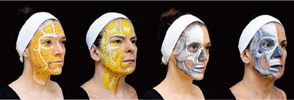
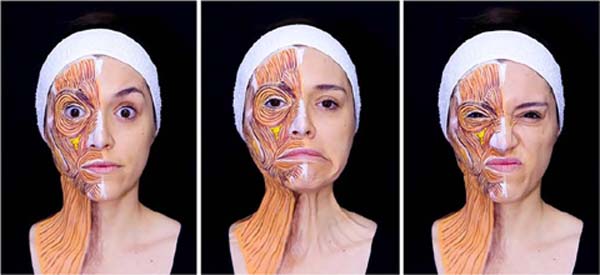
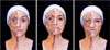
Initially, to represent the changes brought about by the aging process, two models were selected, one younger (39 years old) and the other older (59 years old). In both, the fatty layer and the facial skeleton were represented.
Comparing the two models through Figure 1, in the images on the left, it is possible to observe the differences in the pattern of organization of the fat pads, as well as the changes in the thickness of the subcutaneous tissue (lipoatrophy), with the consequent impairment of the contours and installation of facial skeletonization. As for the facial bone skeleton, when comparing the models, one can see changes in the frontal and temporal bones’ presentation, characterizing the upper third’s structural aging. Concerning the orbital rim, its elongation in the inferolateral direction can be noted, observable in the older model. The thinning and rarefaction of the zygomatico-malar bone, changes in the piriform aperture, maxillary and mandibular alterations, and different presentations of the chin are evidenced in the images on the right.

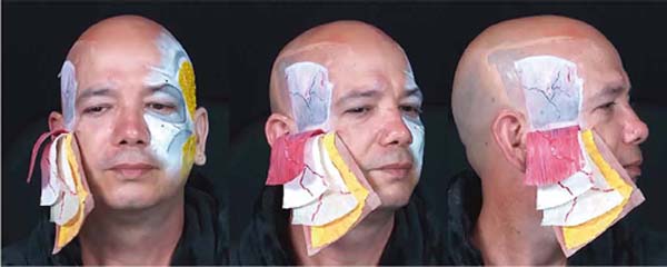
For the study of the temple, five layers were superimposed using latex and inks in varied colors and textures. A bald male live model was selected as he adequately fit the project’s needs. A first layer of latex was used to represent the skin and superficial fat pads. Another two layers were used to represent the superficial and deep temporal fascia. A fourth layer of latex, suitably colored, was used to rather highlight the temporal muscle. To represent the temporal bone, the painting was performed directly on the skin of the model’s temple (Figure 2).

To enhance representation of the treatment of the temple, ultrasound gel in different colors (food dyes) was used and injected in different anatomical planes. Figure 3 demonstrates the treatment of the temporal and zygomatico-malar regions. The image on the left shows the treatment of the upper compartments of the superficial fat layer of the temple, with a red-stained gel injected in a single bolus through a 22G, 50mm cannula.
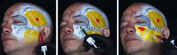

In addition to treating the temporal region, the treatment of the zygomatico-malar region was demonstrated in the present study using a gel stained in green and injected with a 27G needle in small boluses and the juxtaperiosteal plane (central image). The volumization of the medial cheek fat compartment, as well as the deep compartments of the region, was represented through the injection, in multiple boluses, of a red-colored gel (image on the right).
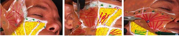
The use of biostimulators is a common practice in the offices of plastic surgeons and dermatologists. For a better study of the injection techniques, a latex film was applied over the face of the model, representing the cutaneous layer. Directly on the skin of the model’s face and immediately below the latex film, the superficial fat compartments of the face were represented, as well as some relevant bone structures.
Figure 4 demonstrates different forms of biostimulation that can be used in the daily clinical practice of the injector. With the use of a 22G cannula, the latex film was transfixed, with the tip of the cannula initially positioned over the zygomatic arch, where small boluses of green-colored gel were injected, simulating the focal stimulus points, as recommended in the technique of use of biostimulators (left image). As a complement, linear retroinjections were represented in the middle third of the face, in a fan and subdermal plane, to reproduce the maneuvers used for the global facial stimulus (central image). In order to represent the linear biostimulus, parallel retroinjections were performed in the subdermal plane next to the mandibular contour, with small aliquots of ultrasound gel stained in green being deposited along the segment (image on the right).

For the reproduction of facial muscles, different colors and textures were used in order to represent the muscular anatomy adequately. The main muscles’ origins, insertions, and positions were painted regarding the model’s anatomical parameters (Figure 5)21. With the use of a 1cc syringe, 30G needle, and colored ultrasound gel, a simulation of the toxin injection was carried out considering the exact position of the application points, the distribution of the points according to the muscle area, and the halo of action of the drug, as well as the proper angulation and grip of the syringe given the anatomical requirements of each muscle.
The nose is one of the most emblematic regions of the face and is represented in Figure 6. In the present study, a layer of latex was used to represent the skin and a second underlying layer to represent the nasal SMAS, with bones, cartilage, and deep fat represented directly on the skin of the model’s nose.
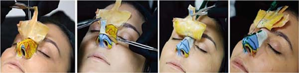
The first three images further to the left illustrate the described layers. A syringe containing green-colored ultrasound gel was attached to a 22G cannula for the simulation of advanced nose filling. The cannula, through an entrance port made of latex layers, was introduced until it reached the nasal dorsum, and the gel was injected into the bony and cartilaginous portions. To reproduce the technique used in the treatment of the nasal tip, injections were made on the domus, the lower margin of the lateral cross of the alar cartilages, and the columella, as shown in the image on the right.
After all this demonstration in live models, the students answered a questionnaire elaborated through the “Google Forms” platform about the applied teaching methodology. The effectiveness of body painting was evaluated using the following questions:
Do you consider that the body painting adequately demonstrated the facial anatomical structures?
Regarding cadaveric anatomical pieces, do you consider that the body painting facilitated the perception of anatomical structures, facilitating your spatial perception?
Have you ever participated in a class that used the body painting methodology as a learning tool?
Do you believe that the body painting methodology facilitated learning the technique demonstrated in class?
Do you consider that the body painting methodology has better learning retention when compared to the use of cadaveric anatomical parts?
Do you consider that the body painting methodology facilitates better learning retention when compared to the use of drawings of anatomical structures, such as those represented in books?
The responses were tabulated and submitted for analysis using simple descriptive statistics.
RESULTS
Skin, fat, SMAS, aponeuroses, cartilage, bones, and vessels were represented on the faces of live models, taking into account the anatomical individuality of each. Injectable procedures were also simulated on these models during classes at the Boggio Institute.
A total of 51 students who had already gone through the experience with body painting then filled out a form evaluating the teaching methodology via the “Google Forms” platform. The answers to the questions raised are described below:
Do you believe that the body painting adequately demonstrated the facial anatomical structures?
- Yes: 96.1% / No: 3.9%
Regarding cadaveric anatomical pieces, do you consider that the body painting presented facilitated the perception of anatomical structures, facilitating their spatial perception?
- Yes: 94.1% / No: 5.9%
Have you ever participated in a class that used the body painting methodology as a learning tool?
- Yes: 9.8% / No: 90.2%
Do you consider that the body painting methodology facilitated learning the technique demonstrated in class?
- Yes: 100% / No: 0%
Do you consider that the body painting methodology facilitates better learning retention when compared to the use of cadaveric anatomical parts?
- Yes: 51.0% / No: 49.0%
Do you consider that the body painting methodology facilitates better learning retention when compared to the use of drawings of anatomical structures, such as those represented in books?
- Yes: 90.2% / No: 9.8%
DISCUSSION
Considered one of the foundations of knowledge, anatomy has historically been referenced as a cornerstone in medical education. Although the importance of anatomy is undeniable, currently there is a debate about how it is taught. The traditional study of anatomy, based on lectures and cadaveric dissections, has been progressively replaced by various new methods, including curriculum integration, problem-based learning, computer-assisted learning (CAL), 3D printing, embalming, plastination, and body painting22,23,24,25,26,27,28.
When asked if they had already participated in a class that used the body painting methodology as a learning tool, 90.2% answered no, demonstrating that this technique is up-to-date and innovative even for students who already have some experience, as they are specialists in dermatology or plastic surgery.
Increasingly, body painting has stood out as a teaching method, enabling the study of living anatomy in a dynamic and easily reproducible way29. McLachlan & Regan de Bere20 used body painting in teaching anatomy, allowing their students to paint each other during the course. The authors observed that the students who were being painted, when receiving the tactile stimulus, found it easier to memorize what was being taught, while the students who were painting received the information in a kinesthetic way, thus memorizing the knowledge through another stimulus pathway. The authors also reported the tendency of students to choose strong and vivid colors, which, according to them, facilitated the memorization of painted structures and, consequently, the learning of anatomy.
In line with the findings of McLachlan & Regan de Bere20, McMenamin21 reinforced in his studies the effectiveness of using body painting in learning anatomy and developing clinical skills. When he asked the students of the Boggio Institute of Teaching and Research whether body painting adequately demonstrated the anatomical structures of the face, 96.1% answered yes, reinforcing the conclusion of the cited works even though in this study, the painting was only demonstrated and not performed by the students themselves. In addition, 94.1% of students responded that body painting facilitated the spatial perception of anatomical structures compared to cadaveric parts, which may be related to the choice of strong colors for painting and the dynamics of a live model.
It is worth mentioning that in the classes held within the Institute, in addition to the use of body painting in all its fullness in an unprecedented and totally innovative way, latex films were used to reproduce the different layer and facial anatomical structures. Asking the students whether body painting facilitated learning the technique demonstrated in class, 100% said yes.
By allowing the sequential and dynamic detachment of each of the anatomical planes represented, the overlap of layers of the temple allows a better understanding of the structural organization of the temporal region. In addition, the gel injection ultrasound scans with different colors and in different anatomical planes made it possible to discuss the most varied techniques to restore the region’s balance.
Regarding the representation of facial mimicry, the individualization of the design to the characteristics of the model allows the adequate reproduction of muscle dynamics on the skin surface and, consequently, a better understanding of the action of each of the muscles of the face. It is also possible to correlate each of the application points with muscle function and the presence of hyperkinetic wrinkles.
In this way, the plasticity and three-dimensionality of this new teaching method enabled a clear understanding of the applied anatomy and provided adequate conditions for the simulation of injectable cosmetic procedures, thus standing out as an important medical training tool30.
According to Finn31,32, body painting has many educational benefits, from acquiring anatomical knowledge to developing greater body awareness. Identifying, palpating, and reproducing anatomical structures give the student a unique learning experience, thus clearly translating the science and art binomial.
The conclusions obtained after the experience of the Boggio Institute students with body painting follow the same line of reasoning when it comes to learning: when questioning the students if they consider that this methodology presents better retention of content when compared to the use of cadaveric pieces, 51.0% answered yes. Compared to drawings of anatomical structures represented in books, 90.2% of the students stated that the body painting technique is also superior considering learning.
With these results, it is clear that the more dynamic the methodology, the greater the students’ satisfaction. In addition, although the percentage of students who reported better retention of content with the body painting was not so expressive, there was a very positive result concerning the facilitation of the spatial perception of the anatomical structures and learning of the technique demonstrated in class.
Unfortunately, the literature on this topic by national authors is scarce, and more studies are needed to assess better the effectiveness of the body painting method in student learning. The comparison of students’ performance in tests given after classes using body painting and older techniques (corpse pieces or book images), for example, is a possibility to be analyzed. In this way, we would be able to measure the gain in content retention more objectively with the body painting methodology.
CONCLUSION
Facial anatomy applied to live models is an innovative teaching method that students accept well, as it makes it possible to study anatomy and train clinical skills efficiently and in an enjoyable way.
1. Instituto Boggio, Cosmiatria, São Paulo, São Paulo, SP, Brazil.
Corresponding author: Ricardo Frota Boggio Instituto Boggio Rua Cincinato Braga, 37, 8° andar, Bela Vista, São Paulo, SP, Brazil. Zip code: 01333-010 E-mail: drboggio@clinicaboggio.com.br
 Read in Portuguese
 Read in English
 PDF PT
 Print
Print
 Send this article by email
 How to Cite
 Mendeley
 Pocket