


Case Reports - Year 1999 - Volume 14 -
Multiple Cylindromas - Turban Tumor Case Report
Cilindromas Múltiplos - Tumor em Turbante - Relato de Caso
ABSTRACT
"Turban tumor" corresponds to multiple cylindromas in almost the entire extension of the scalp. The disease is benign, autosomal dominant, and has a controversial histogenesis. A case of a 51 years old man is presented, in which some tumors were initially resected. Then, a large excision of the scalp was acomplished, followed by graft with skin from the thigh. Thirteen years later, the patient is in good conditions, with a satisfactory aesthetic result.
Keywords: Cylindroma; turban tumor; cutaneous appendage tumors.
RESUMO
O "tumor em turbante" corresponde a múltiplos cilindromas em praticamente toda a extensão do couro cabeludo. A doença é benigna, autossômica dominante e tem histogênese controvertida. Descreve-se um caso em homem de 51 anos, do qual, de início, se retiraram alguns tumores; depois se fez exérese ampla de couro cabeludo, seguida de enxerto de pele de coxa. Treze anos depois, o paciente se acha em boas condições, com resultado estético satisfatório.
Palavras-chave: Cilindroma; tumor em turbante; tumor de anexos cutâneos
Cylindroma is an uncommon benign neoplasia of the cutaneous appendages, which frequently manifests as a unique, small, pink-reddish, usually painless lesion, of sporadic occurence, located, in 90% of the cases, in the scalp, face or neck. Cases with multiple lesions are inherited, with autosomal dominant transmission and incomplete penetrance, described as smooth nodules, with round contour and variable sizes, located in the scalp and occasionally, in the face, rarely in the chest or extremities; when large portions of the scalp are in volved it is called "turban tumor"(l, 2, 3).
Elder et al.(2) states that the lesions of the "turban tumor" begin to appear in adulthood and gradually in crease in number and size.
Surgery is the preferential treatment with scalp excision followed by free skin graft.
CASE REPORT
A 51 year old caucasian man, farmer, looked for the Plastic Surgery Unit of the Uberlandia Federal University complaining of painful "lumps" in the head, which had been developing for approximately 30 years. Initially they were small but had grown steadily. He is a social etilist and former chronic smoker. Two paternal uncles and his father had had similar disease.
Physical examination revealed several tumors with variable sizes and shapes, confluent, smooth, with firm consistence and pink color, with telangiectasias in the surface, located in the scalp and face (Fig. 1).
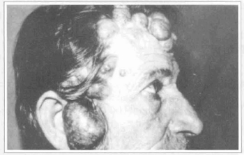
Fig.1 - Multiple tumoration in the face and scalp, with variable sizes, some of which with telangiectasias.
The smaller and isolated tumors underwent excision with direct approximation of the marges of the surgical wound, when feasible, or skin graft in the sites of larger extension. The coalescent tumoral mass was treated by large resection of the scalp, above the galea aponeurotica, involving the frontal, temporal and parieto-occipital regions (Fig. 2), followed by free skin graft in the crude resultant area. The histological analysis of the tumors revealed multiple cylindromas (Fig. 3) and one trichoepithelioma.
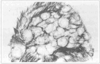
Fig. 2 - Segment of the resected scalp showing the "turban tumor".
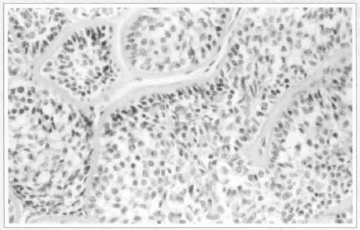
Fig. 3 -Cylindroma. Lobules with small and dark epithelial cells in the periphery and big and light ones in the center, surrounded by thick hyalin sheath. Note the small masses of hyalin material inside the lobules. H-E, original augmentation of 400x.
The patient evolved well in the postoperative period, with total integration of the grafts to the receptor bed, without significant clinical intercurrences. The appearance of the head in the grafted regions is considered aesthetically satisfactory (Figs. 4 & 5).
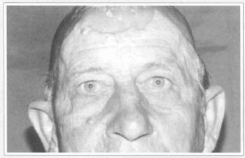
Fig. 4 - Late postoperative period (13 years) -Frontal view.
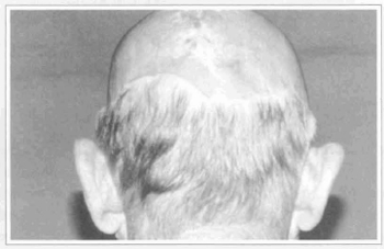
Fig. 5 - Late postoperative period (13 years) -Posterior view.
DISCUSSION
Cylindromas are more common in females and caucasians, beginning to appear more frequently in the third or fourth decade(l). According to our patient, his first tumors arised at age 20.
Cylindroma, histologicaly speaking, represents an epithelial neoplasia, W1encapsulated, composed of lobules of variable shapes and sizes, located in the upper dermis and surrounded by a usually thick hyalin sheath. The cells in the periphery of the lobules have small and dark-staining nuclei and often have a palisade arrangement, the central ones have large and light-staining nuclei; among the cells one may see globules of PAS-positive hyalin material, diastase-resistant, and tubular structures with amorphous material within the lumina(2) . Inside the tumor, areas with features of eccrine spiradenoma, trichoepithelioma and syringoma may be found (1, 4) . In several instances, multiple cylindromas are associated to trichoepithelioma (as in the present report) and eccrine spiradenomas(4) .
Its coexistence with structuraly similar tumor of the salivary gland (especially the parotid)(2, 5) is exceptionally described.
The histogenesis and differentiation of the tumor are controversial. Many authors postulate eccrine, apoccrine or pilar differentiation(3). The current opinion about the histogenesis, based on ultra-structural and imunohistochemical data, is that the cylindroma is originary from the coiled portion of the eccrine ductiles(6) .
The cylindroma seldom becomes malignant(2, 3). Cooper et al.(7) refer to the report of at least 14 cases. Such occurence is more common arising from "turban tumor"(3, 7). Usually the malignization is of a sole tumor(2). The malignant cylindromas are aggressive, with disseminated metastases to lymphonodus, viscera and bones(3). Death is caused by visceral metastases or, in some cases, due to cranian cavity invasion, with hemorrhage and meningitis(2). As the majority of the malignant cylindromas arises from "turban tumor", patients with this modality of cylindroma should be cautiously followed through, which is what we have been doing in this case, regarding the possibility of new tumors in the ramaining scalp which might not have been substituted by the graft.
The proposed therapy is usually the surgery, specially for aesthetic reasons(8); other indications are the ulceration, infection, suspected malignization, functional impairment (i.g, auditive) and eventually pain(4, 9). Irwin et al.(8) recommends individual excision of the lesions with less than 1cm in diameter or located in cosmetically important areas. Large resection of the scalp, followed by cutaneous graft, is restricted to the cases of "turban tumours". Other forms of treatrnent includes eletrocoagulation, radiotherapy and dermoabbrasion(8) .
The recidive after exeresis of isolated tumors is common, reaching 42% in the Crain and'Helwing series(1O). Maybe this is due to the multifocal growth or to the multinodularity of the cylindromas.
REFERENCES
1. BATSAKIS JG. Dermal eccrine cylindroma. Ann. Otol. Rhinol. Laryngol. 1989; 98:991-992.
2. ELDER D, ELENITSAS R, RAGSDALE BD. Tumors of the epidermal appendages. In: ELDER D, ELENITSAS R, JAWORSKY C, JOHNSON B Jr.- Lever's Histopathology of the skin, Lippincott-Raven, Philadelphia, USA, 8th ed. 1997; 747-803.
3. SANTA CRUZ DJ. Tumors of sweat gland differentiation. In: FARMER ER, HOOD AF.-Pathology of the skin, Appleton-Lange, Norwalk, USA, 1990; 624-662.
4. GUZZO C, JOHNSON B. Unusual abdominal location of a dermal cylindroma. Cutis. 1995; 56:239-240.
5. BATSAKIS JG, BRANNON RB. Dermal analogue tumours of major salivary glands. J. Laryngol. Otol. 1981; 95:155-164.
6. COTTON DWK, BRAYE SG. Dermal cylindromas originate from the eccrine sweat gland. Brit. J. Dermatol. 1984; 1ll: 53-61.
7. COOPER PH, FRIERSON HF, MORRISON AG. Malignant transformation of eccrine spiradenoma. Arch. Dermatol. 1985; 121: 1445-1448.
8. IRWIN LR, BAINBRIDGE LC, REID CA, PIGOTT TA, BROWN HG. Dermal eccrine cylindroma (turban tumour). Brit. J. Plast. Surg. 1990; 43: 702-705.
9. FREEDMAN AM, WOODS JE. Total scalp excision and auricular resurfacing for dermal cylindroma (turban tumor). Ann. Plast. Surg. 1989; 22: 50-57.
10. CRAIN RC, HELWIG EB. Dermal cylindroma (dermal eccrine cylindroma). Am. J. Clin. Pathol. 1961; 35: 505-515.
I - Ex-Resident, Plastic Surgery Service of Universidade Federal de Uberlândia.
II - Resident, Plastic Surgery Service of Universidade Federal de Uberlândia.
III - Chief, Plastic Surgery Service of Universidade Federal de Uberlândia.
IV - Titular Professor, Pathology Department of Universidade Federal de Uberlândia.
V - Associate Professor, Plastic Surgery Service of Universidade Federal de Uberlândia.
Address for correspondence:
Anderson Gonça1ves de Freitas Jr, MD
R. Francisco Antonio Fernandes, 209
38400-119 - Uberlândia - MG Brazil
 Read in Portuguese
 Read in English
 PDF PT
 Print
Print
 Send this article by email
 How to Cite
 Mendeley
 Pocket